

Patterns of gray matter atrophy in genetic frontotemporal dementia: results from the GENFI study
- PMID: 29172163
- PMCID: PMC5759893
- DOI: 10.1016/j.neurobiolaging.2017.10.008
Patterns of gray matter atrophy in genetic frontotemporal dementia: results from the GENFI study
Abstract
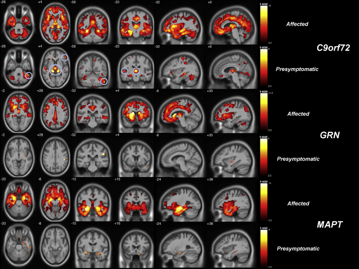
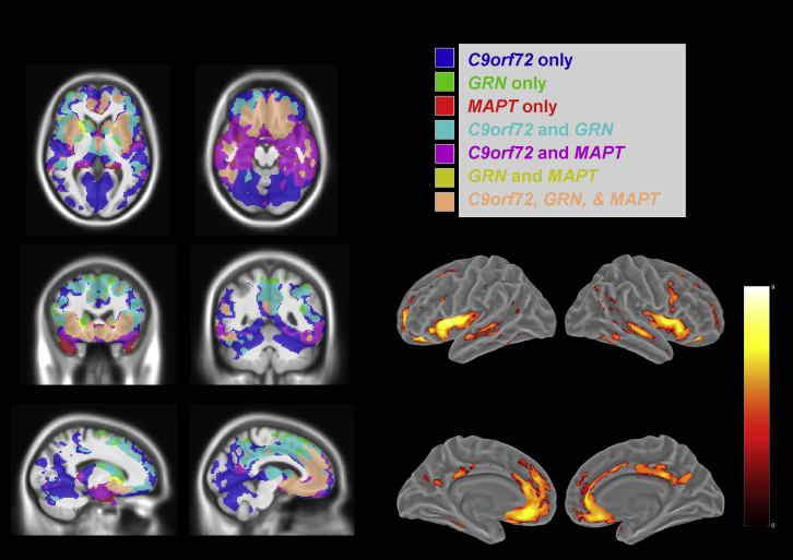
Frontotemporal dementia (FTD) is a highly heritable condition with multiple genetic causes. In this study, similarities and differences of gray matter (GM) atrophy patterns were assessed among 3 common forms of genetic FTD (mutations in C9orf72, GRN, and MAPT). Participants from the Genetic FTD Initiative (GENFI) cohort with a suitable volumetric T1 magnetic resonance imaging scan were included (319): 144 nonmutation carriers, 128 presymptomatic mutation carriers, and 47 clinically affected mutation carriers. Cross-sectional differences in GM volume between noncarriers and carriers were analyzed using voxel-based morphometry. In the affected carriers, each genetic mutation group exhibited unique areas of atrophy but also a shared network involving the insula, orbitofrontal lobe, and anterior cingulate. Presymptomatic GM atrophy was observed particularly in the thalamus and cerebellum in the C9orf72 group, the anterior and medial temporal lobes in MAPT, and the posterior frontal and parietal lobes as well as striatum in GRN. Across all presymptomatic carriers, there were significant decreases in the anterior insula. These results suggest that although there are important differences in atrophy patterns for each group (which can be seen presymptomatically), there are also similarities (a fronto-insula-anterior cingulate network) that help explain the clinical commonalities of the disease.
Keywords: Atrophy; Frontotemporal dementia; Magnetic resonance imaging; Preclinical dementia; Voxel-based morphometry.
Copyright © 2017 The Authors. Published by Elsevier Inc. All rights reserved.
Figures
References
-
- Ashburner J., Friston K.J. Unified segmentation. Neuroimage. 2005;26:839–851. - PubMed
-
- Caroppo P., Habert M.O., Durrleman S., Funkiewiez A., Perlbarg V., Hahn V., Bertin H., Gaubert M., Routier A., Hannequin D., Deramecourt V., Pasquier F., Rivaud-Pechoux S., Vercelletto M., Edouart G., Valabregue R., Lejeune P., Didic M., Corvol J.C., Benali H., Lehericy S., Dubois B., Colliot O., Brice A., Le Ber I. Lateral temporal lobe: an early imaging marker of the presymptomatic GRN disease? J. Alzheimers Dis. 2015;47:751–759. - PMC - PubMed
-
- Filippi M., Agosta F., Scola E., Canu E., Magnani G., Marcone A., Valsasina P., Caso F., Copetti M., Comi G., Cappa S.F., Falini A. Functional network connectivity in the behavioral variant of frontotemporal dementia. Cortex. 2013;49:2389–2401. - PubMed
Publication types
MeSH terms
Substances
Grants and funding
- 103838/WT_/Wellcome Trust/United Kingdom
- MC_UU_00024/1/MRC_/Medical Research Council/United Kingdom
- MC_U105597119/MRC_/Medical Research Council/United Kingdom
- MR/J009482/1/MRC_/Medical Research Council/United Kingdom
- MR/M023664/1/MRC_/Medical Research Council/United Kingdom
- WT_/Wellcome Trust/United Kingdom
- MC_UU_00005/12/MRC_/Medical Research Council/United Kingdom
- MR/M008525/1/MRC_/Medical Research Council/United Kingdom
- MR/M009106/1/MRC_/Medical Research Council/United Kingdom
- MR/M009041/1/MRC_/Medical Research Council/United Kingdom
- MR/M024873/1/MRC_/Medical Research Council/United Kingdom
LinkOut - more resources
Full Text Sources
Other Literature Sources
Miscellaneous
