Review
. 2020 Jul;11(4):256-270.
doi: 10.1080/21541248.2017.1405773. Epub 2017 Dec 31.

Cortactin function in invadopodia

Affiliations
Review

Cortactin function in invadopodia

Pauline Jeannot et al. Small GTPases. 2020 Jul.

Abstract

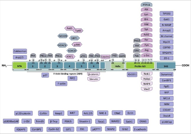
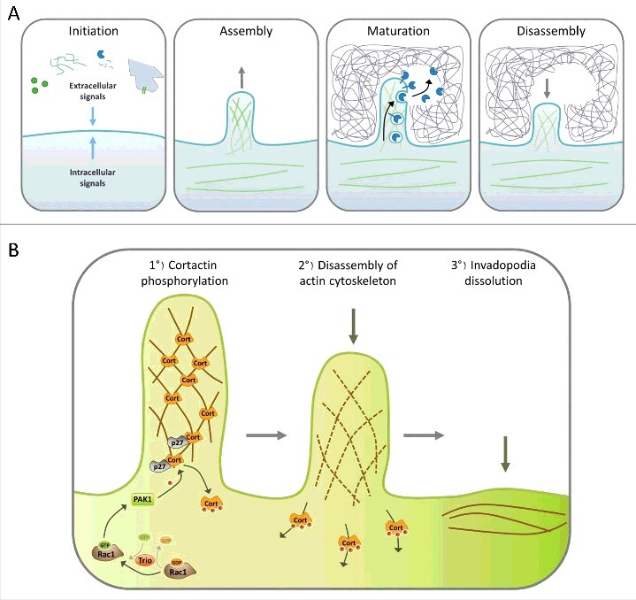
Actin remodeling plays an essential role in diverse cellular processes such as cell motility, vesicle trafficking or cytokinesis. The scaffold protein and actin nucleation promoting factor Cortactin is present in virtually all actin-based structures, participating in the formation of branched actin networks. It has been involved in the control of endocytosis, and vesicle trafficking, axon guidance and organization, as well as adhesion, migration and invasion. To migrate and invade through three-dimensional environments, cells have developed specialized actin-based structures called invadosomes, a generic term to designate invadopodia and podosomes. Cortactin has emerged as a critical regulator of invadosome formation, function and disassembly. Underscoring this role, Cortactin is frequently overexpressed in several types of invasive cancers. Herein we will review the roles played by Cortactin in these specific invasive structures.

Keywords: ARP2/3; Cortactin; Rac1; Rho GTPases; actin cytoskeleton; invadopodia; invasion; migration; podosome.


Figures

Figure 1.
Figure 1.
Domain structure of Cortactin and interacting partners. Cortactin is composed of an N-terminal acidic domain that allows the interaction with Arp2/3, followed by six and a half F-actin binding repeats of 32 amino acids that mediate binding to F-actin and can be acetylated on lysines or phosphorylated on serines. On its C-terminal part, Cortactin has a helical domain, a proline-rich domain that is extensively regulated by phosphorylation and whose tyrosines are targeted, among others, by Src-family kinases. Finally, Cortactin has an SH3 domain at its C-terminal end, which binds many different proteins mostly involved in the regulation of actin cytoskeleton dynamics, including N-WASP. The known binding partners of Cortactin are indicated close to the domain to which they bind to or target, when known, or in the lower part of the figure when unknown. Lysines targeted for acetylation are indicated by @ (amino acids 87, 124, 144, 161, 181, 198, 235, 272, 295, 304, 309, 346). The amino acid numbering refers to the mouse protein (NP_031829.2).
Figure 2.
Figure 2.
Steps of invadopodia formation. A. The invadopodia lifecycle can be divided in four steps: Initiation, assembly, maturation and disassembly. Cells begin to form invadopodia in response to activating signals transmitted by growth factor or matrix receptors, MMP activity, heterotypic cell interaction, EMT or oncogenic transformation. These signals activate different signaling pathways that induce branched actin polymerization and formation of a cellular protrusion. Once invadopodia are mature, different proteases are secreted, allowing degradation of the surrounding matrix. The final step consists in the dissolution of the invadopodia, which includes branched actin disassembly. B. Role of Cortactin in invadopodia disassembly. Invadopodia dissolution is triggered by the activation of Rac1 by its GEF Trio; in turn Rac1 activates PAK1, which is recruited by p27 on Cortactin. Then, PAK1 phosphorylates Cortactin on S113, S150 and/or S282, which probably induces the release of Cortactin from branched F-actin, destabilizing the branched actin network which disassembles, allowing the return to a basal situation.

References

    1. Goley ED, Welch MD.. The ARP2/3 complex: an actin nucleator comes of age. Nat Rev Mol Cell Biol. 2006;7:713–26. doi: 10.1038/nrm2026. - DOI - PubMed
    1. Nürnberg A, Kitzing T, Grosse R. Nucleating actin for invasion. Nat Rev Cancer. 2011;11:177–87. doi: 10.1038/nrc3003. - DOI - PubMed
    1. Pollard TD. Actin and Actin-Binding Proteins. Cold Spring Harb Perspect Biol. 2016;8(8):a018226. doi: 10.1101/cshperspect.a018226. - DOI - PMC - PubMed
    1. Weaver AM, Karginov AV, Kinley AW, et al. Cortactin promotes and stabilizes Arp2/3-induced actin filament network formation. Curr Biol CB. 2001;11:370–4. doi: 10.1016/S0960-9822(01)00098-7. - DOI - PubMed
    1. Wu H, Reynolds AB, Kanner SB, et al. Identification and characterization of a novel cytoskeleton-associated pp60src substrate. Mol Cell Biol 1991;11:5113–24. doi: 10.1128/MCB.11.10.5113. - DOI - PMC - PubMed

Publication types

LinkOut - more resources