. 2017 Dec 12;9(6):1765-1779.
doi: 10.1016/j.stemcr.2017.10.024. Epub 2017 Nov 22.

A COUP-TFII Human Embryonic Stem Cell Reporter Line to Identify and Select Atrial Cardiomyocytes

Affiliations

A COUP-TFII Human Embryonic Stem Cell Reporter Line to Identify and Select Atrial Cardiomyocytes

Verena Schwach et al. Stem Cell Reports. .

Abstract

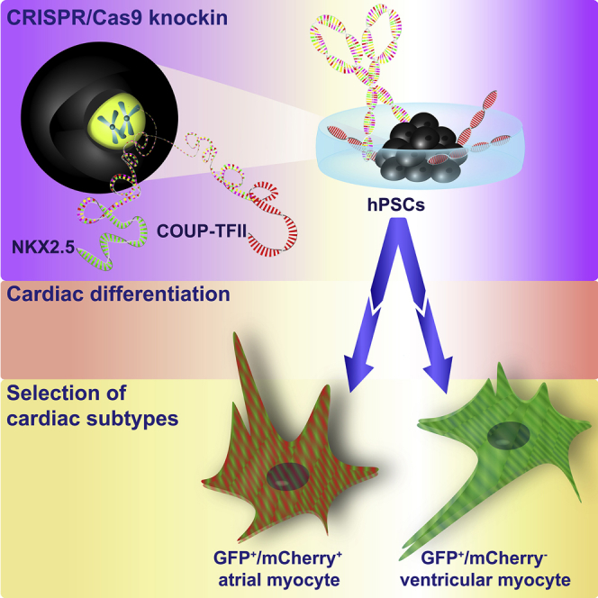
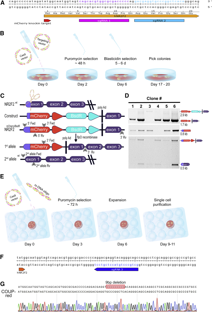
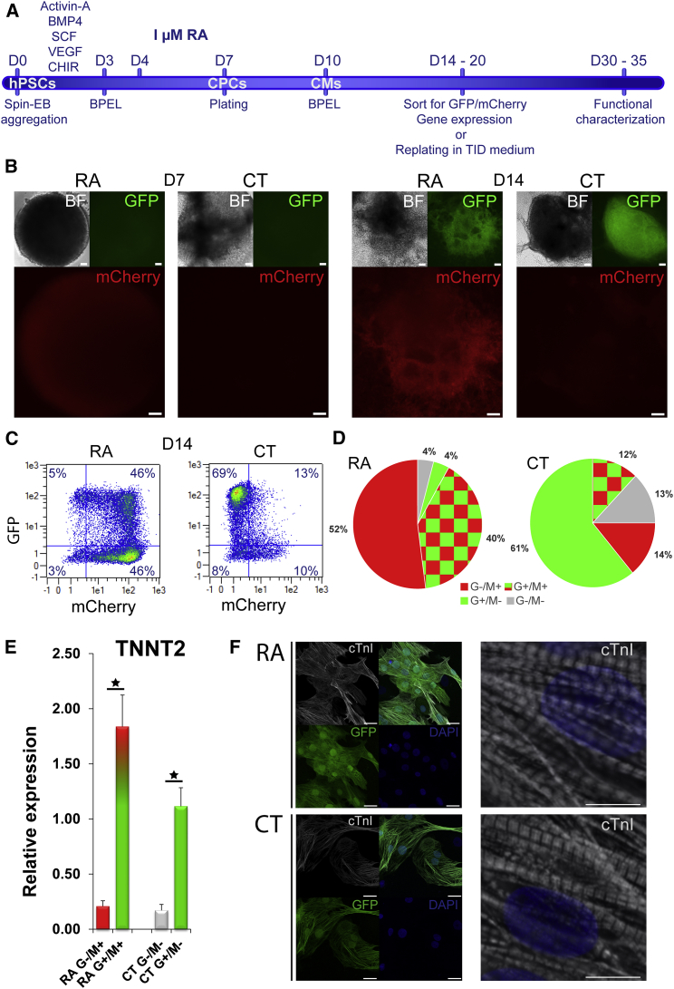
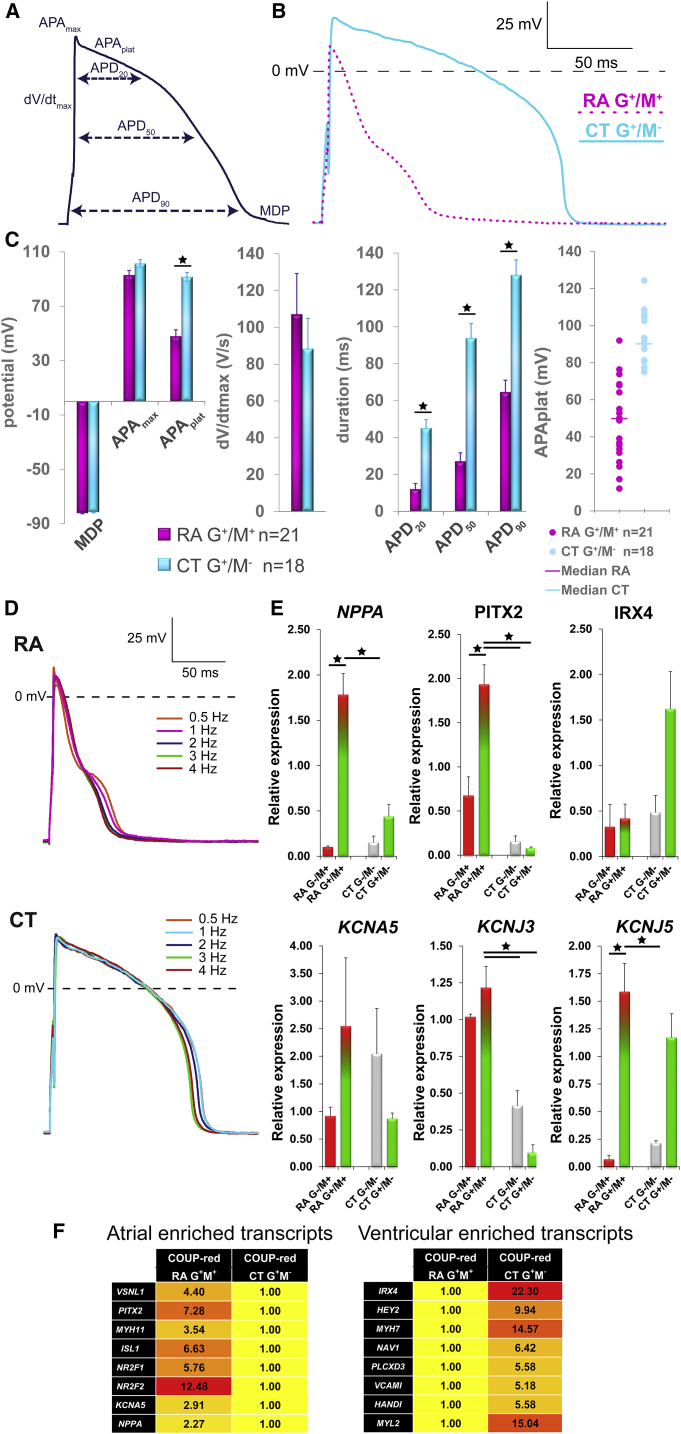
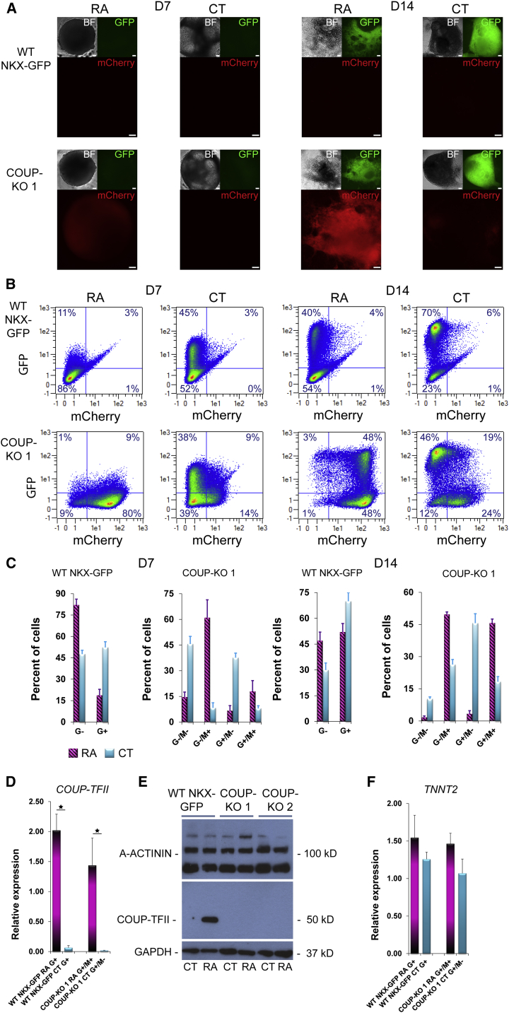
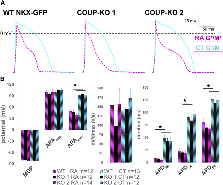
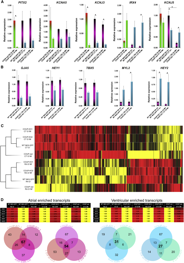
Reporter cell lines have already proven valuable in identifying, tracking, and purifying cardiac subtypes and progenitors during differentiation of human pluripotent stem cells (hPSCs). We previously showed that chick ovalbumin upstream promoter transcription factor II (COUP-TFII) is highly enriched in human atrial cardiomyocytes (CMs), but not ventricular. Here, we targeted mCherry to the COUP-TFII genomic locus in hPSCs expressing GFP from the NKX2.5 locus. This dual atrial NKX2.5EGFP/+-COUP-TFIImCherry/+ reporter line allowed identification and selection of GFP+ (G+)/mCherry+ (M+) CMs following cardiac differentiation. These cells exhibited transcriptional and functional properties of atrial CMs, whereas G+/M- CMs displayed ventricular characteristics. Via CRISPR/Cas9-mediated knockout, we demonstrated that COUP-TFII is not required for atrial specification in hPSCs. This new tool allowed selection of human atrial and ventricular CMs from mixed populations, of relevance for studying cardiac specification, developing human atrial disease models, and examining distinct effects of drugs on the atrium versus ventricle.

Keywords: COUP-TFII-knockout; COUP-TFII-mCherry fluorescent stem cell reporter; CRISPR/Cas9; atrial specification; cardiac differentiation; human embryonic stem cells.


Figures

None
Graphical abstract
Figure 1
Figure 1
CRISPR/Cas9-Mediated Knockin of mCherry into COUP-TFII (A) Location and sequence of single-guide RNAs (sgRNAs) 1 and 2 in the COUP-TFII locus. (B) Schematic representation of the CRISPR/Cas9 plasmid-based targeting and dual-selection protocol. (C) Schematic overview of wild-type (WT) COUP-TFII, COUP-TFII-mCherry targeting construct, and the resulting targeted (first allele) and WT COUP-TFII (second allele) alleles with forward (FWD) and reverse (RV) primer binding sites for screening. (D) PCR screen to identify targeted clones: Upper panel: 3′ end screen and confirmation of excision of the blasticidin-resistance cassette in 1, 2, 4, 5, and 6, but not 3 (targeted, but blasticidin-resistance cassette still present). Middle panel: 5′ end screen shows that clones 1–3, and 5 and 6, were targeted at the 5′ end. Lower panel: screen to determine if heterozygous (clones 2, 3, 4, and 6) or homozygous (clones 1 and 5) mCherry knockin occurred. (E) Schematic representation of the CRISPR/Cas9-mediated correction using a WT COUP-TFII single-stranded oligonucleotide (ssDNA oligo). (F) Sequence corresponding to the 9 bp deletion in the COUP-TFII locus along with the annealing sites for sgRNA 3 designed to repair the deletion. (G) Sanger sequencing confirming correction of the second COUP-TFII allele from the COUP-red clone after correction. See also Figures S1 and S2.
Figure 2
Figure 2
Characterization of the Dual Human NKX2.5EGFP/+-COUP-TFIImCherry/+ Stem Cell Line (COUP-red) as Atrial Reporter (A) Atrial-directed pin-embryoid body (spin-EB) protocol and treatment with retinoic acid (RA). (B) mCherry expression together with GFP and bright-field (BF) images at D7 and D14 of differentiation in RA and control (CT) differentiations. Scale bar, 100 μm. (C) Representative flow cytometry plots depicting the percentage of GFP-positive (G+) or mCherry (M+) cells at D14 of differentiation in RA or CT differentiations. (D) Averaged G/M percentage calculated from four independent differentiations (n = 4): RA condition (G+/M+: 41% ± 2%, G/M+: 52% ± 3%, G+/M: 4% ± 1%, G/M: 4% ± 1%) versus CT (G+/M+: 12% ± 1%, G/M+: 14% ± 2%, G+/M: 61% ± 4%, G/M: 13% ± 2%). Data are displayed as means ± SEM (E) Relative mRNA expression of cardiac troponin (TNNT2) in sorted G/M populations at day 20 of differentiation. Data are displayed as means ± SEM; p < 0.05. (F) Immunostaining of cardiac troponin (cTnI) together with endogenous GFP expression and DAPI as nuclear staining in unsorted cultures after dissociation and re-plating. Scale bars, 25 μm (left), 2.5 μm (right). See also Figure S3.
Figure 3
Figure 3
M+ COUP-red CMs Exhibit Atrial Properties (A) Analyzed AP parameters. (B) Representative AP of G+/M+ CMs generated from RA-treated and G+/M CMs from CT differentiations at 1 Hz stimulation. (C) Averaged maximum diastolic potential (MDP), maximum AP amplitude (APAmax) and AP plateau amplitude (APAplat), maximum upstroke velocity (dV/dtmax), and AP duration at 20%, 50%, and 90% repolarization (APD20, APD50, and APD90). Scatterplot depicting the APAplat of single CMs and calculated median from RA and CT conditions. n = 21 cells for RA G+/M+ and n = 18 cells for CT G+/M; from 3 independent differentiations. Data are displayed as means ± SEM; p < 0.05. (D) Typical AP traces stimulated at different frequencies. (E) Transcriptional profiling of selected atrial or ventricular-specific genes by qPCR of G+/M+ and G/M+ fractions from RA differentiations compared with G+/M and G/M fractions from CT differentiations at D20 of differentiations (n = 4). Data are displayed as means ± SEM; p < 0.05. (F) Genome-wide transcriptional profiling by microarray. Heatmaps to display the fold difference of selected atrial- and ventricular-enriched transcripts in RA-treated G+/M+ and CT G+/M from COUP-red at a threshold of 2-fold difference. See also Figure S3.
Figure 4
Figure 4
Characterization of COUP-KO Lines (A) mCherry overlapping with GFP and bright-field (BF) images at D7 and D14 of differentiation in RA or CT differentiations from WT NKX-GFP or COUP-KO cells from subclone COUP-KO 1. Scale bars, 100 μm. (B) Representative flow cytometry plots depicting the percentage of GFP-positive (G+) and mCherry (M+) cells at D7 and D14 of differentiation in RA or CT samples from WT NKX-GFP or COUP-KO 1. (C) Averaged G/M percentage calculated from three independent differentiations (n = 3) at day 7 of differentiation in RA (G+/M+, 18% ± 6%; G/M+, 61% ± 10%; G+/M, 7% ± 3%; G/M, 15% ± 3%) and CT (G+/M+, 8% ± 2%; G/M+, 8% ± 3%; G+/M, 38% ± 3%; G/M, 46% ± 4%) and at day 14 in RA (G+/M+, 46% ± 2%; G/M+, 50% ± 1%; G+/M, 4% ± 2%; G/M, 2% ± 1%) and CT (G+/M+, 18% ± 2%; G/M+, 26% ± 2%; G+/M, 46% ± 2%; G/M, 10% ± 1%), together with WT NKX-GFP cells (D7, CT G+/M+, 5% ± 3%; G+/M 65% ± 3%; G/M 30% ± 3%; G/M+, 1% ± 1%; RA G+/M+, 4% ± 1%; G+/M 48% ± 4%; G/M 45% ± 4%; G/M+, 2% ± 1%; D14, CT G+/M+, 3% ± 1%; G+/M 49% ± 3%; G/M 48% ± 3%; G/M+, 0% ± 0%; RA G+/M+, 4% ± 1%; G+/M 15% ± 4%; G/M 81% ± 4%; G/M+, 1% ± 1%). n = 3 independent differentiations; mean ± SEM. (D) mRNA expression of COUP-TFII in COUP-KO 1 cells at day 14 of differentiation (n = 4 independent differentiations; mean ± SEM; p < 0.05). (E) Validation of complete knock out of COUP-TFII expression by western blot in unpurified differentiated CMs from RA and CT differentiations from COUP-KO cells compared with WT NKX-GFP cells. (F) mRNA expression of cardiac troponin (TNNT2) in differentiated CMs from RA and CT differentiations from COUP-KO 1 and WT NKX-GFP cells (n = 4 independent differentiations). See also Figures S4, S5, and S8.
Figure 5
Figure 5
COUP-KO-derived CMs Functionally Resemble their WT Counterparts (A) Representative action potentials (APs) of G+/M+ CMs generated from RA-treated and G+/M CMs from CT differentiations of WT NKX-GFP, COUP-KO1, and COUP-KO 2 cells. (B) Averaged maximum diastolic potential (MDP), maximum AP amplitude (APAmax) and AP plateau amplitude (APAplat), maximum upstroke velocity (dV/dtmax), and AP duration at 20%, 50%, and 90% repolarization (APD20, APD50, and APD90). n = 12 cells for WT NKX-GFP RA G+, n = 12 cells for COUP-KO 1 RA G+/M+, and n = 14 cells for COUP-KO 2 RA G+/M+; n = 13 cells for WT NKX-GFP CT G+, n = 12 cells for COUP-KO 1 CT G+/M, and n = 12 cells for COUP-KO 2 CT G+/M; from three independent differentiations. Data are displayed as means ± SEM; p < 0.05. See also Figure S6.
Figure 6
Figure 6
G+M+ CMs Lacking Functional COUP-TFII Exhibit Atrial Characteristics on Transcriptional Level (A and B) qPCR analysis of selected atrial or ventricular transcripts of sorted COUP-KO 1 derived G+/M+ fractions from RA and G+/M populations from CT compared with G+ populations of WT NKX-GFP RA and CT differentiations at D14 (n = 4 independent differentiations). Data are displayed as means ± SEM; p < 0.05. (C) Hierarchical clustering of G+/M+ RA fractions and G+/M CT fractions of two independent subclones COUP-KO 1 and 2 revealed high similarity to their G+ WT NKX-GFP or G+/M+ and G+/M COUP-red equivalents. (D) Heatmaps: fold difference of atrial- and ventricular-enriched transcripts with Venn diagrams to display the overlap of differentially expressed genes between RA (atrial transcripts, left panel) and CT (ventricular transcripts, right panel) in COUP-KO 1, 2, and WT NKX-GFP or COUP-red at a threshold of 2-fold difference. See also Figures S6 and S7 and Table S3.

Similar articles

Cited by

References

    1. Achatz G., Hölzl B., Speckmayer R., Hauser C., Sandhofer F., Paulweber B. Functional domains of the human orphan receptor ARP-1/COUP-TFII involved in active repression and transrepression. Mol. Cell. Biol. 1997;17:4914–4932. - PMC - PubMed
    1. Al Turki S., Manickaraj A.K., Mercer C.L., Gerety S.S., Hitz M.-P., Lindsay S., D’Alessandro L.C.A., Jawahar Swaminathan G., Bentham J., Arndt A.-K. Rare variants in NR2F2 cause congenital heart defects in humans. Am. J. Hum. Genet. 2014;94:574–585. - PMC - PubMed
    1. Beqqali A., Van Eldik W., Mummery C., Passier R. Human stem cells as a model for cardiac differentiation and disease. Cell. Mol. Life Sci. 2009;66:800–813. - PMC - PubMed
    1. Birket M.J., Ribeiro M.C., Kosmidis G., Ward D., Leitoguinho A.R., van de Pol V., Dambrot C., Devalla H.D., Davis R.P., Mastroberardino P.G. Contractile defect caused by mutation in MYBPC3 revealed under conditions optimized for human PSC-cardiomyocyte function. Cell Rep. 2015;13:1–13. - PMC - PubMed
    1. Braam S.R., Tertoolen L., van de Stolpe A., Meyer T., Passier R., Mummery C.L. Prediction of drug-induced cardiotoxicity using human embryonic stem cell-derived cardiomyocytes. Stem Cell Res. 2010;4:107–116. - PubMed

Publication types

MeSH terms