. 2018 Jan:7:45-56.
doi: 10.1016/j.molmet.2017.11.004. Epub 2017 Nov 11.

Fatty acid oxidation is required for active and quiescent brown adipose tissue maintenance and thermogenic programing

Affiliations

Fatty acid oxidation is required for active and quiescent brown adipose tissue maintenance and thermogenic programing

Elsie Gonzalez-Hurtado et al. Mol Metab. 2018 Jan.

Abstract

Objective: To determine the role of fatty acid oxidation on the cellular, molecular, and physiologic response of brown adipose tissue to disparate paradigms of chronic thermogenic stimulation.

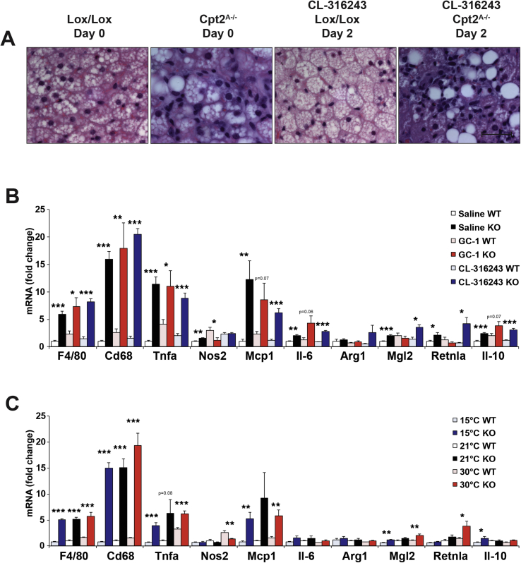
Methods: Mice with an adipose-specific loss of Carnitine Palmitoyltransferase 2 (Cpt2A-/-), that lack mitochondrial long chain fatty acid β-oxidation, were subjected to environmental and pharmacologic interventions known to promote thermogenic programming in adipose tissue.

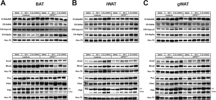
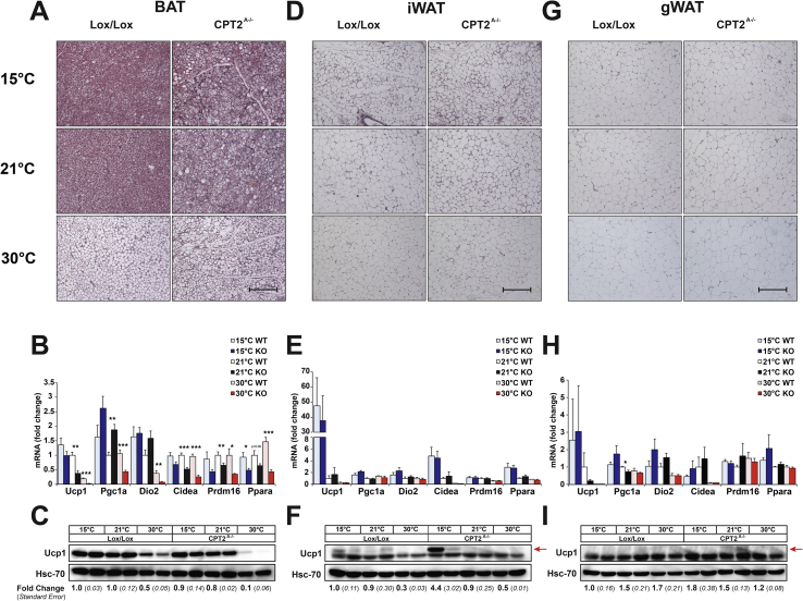
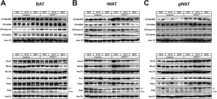
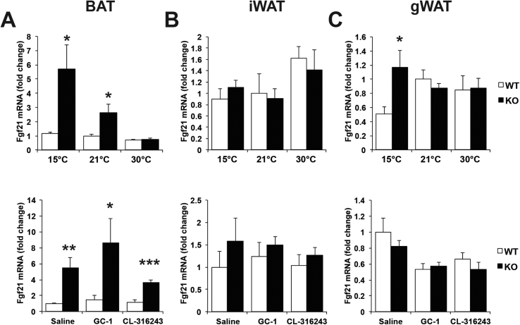
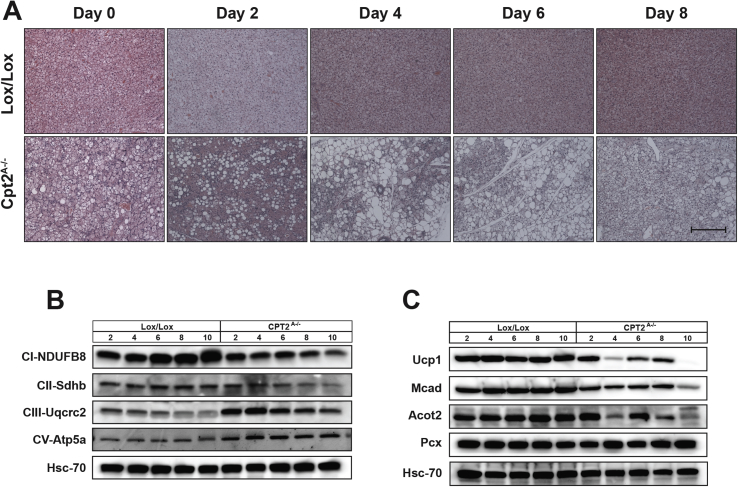
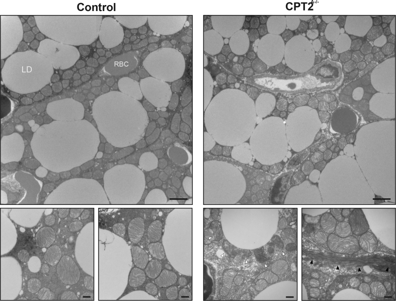
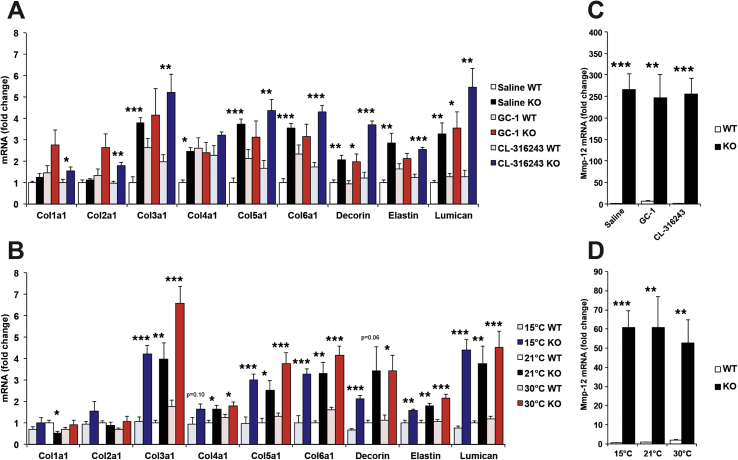
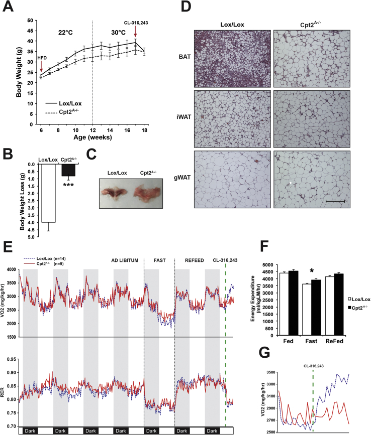
Results: Chronic administration of β3-adrenergic (CL-316243) or thyroid hormone (GC-1) agonists induced a loss of BAT morphology and UCP1 expression in Cpt2A-/- mice. Fatty acid oxidation was also required for the browning of white adipose tissue (WAT) and the induction of UCP1 in WAT. In contrast, chronic cold (15 °C) stimulation induced UCP1 and thermogenic programming in both control and Cpt2A-/- adipose tissue albeit to a lesser extent in Cpt2A-/- mice. However, thermoneutral housing also induced the loss of UCP1 and BAT morphology in Cpt2A-/- mice. Therefore, adipose fatty acid oxidation is required for both the acute agonist-induced activation of BAT and the maintenance of quiescent BAT. Consistent with this data, Cpt2A-/- BAT exhibited increased macrophage infiltration, inflammation and fibrosis irrespective of BAT activation. Finally, obese Cpt2A-/- mice housed at thermoneutrality exhibited a loss of interscapular BAT and were refractory to β3-adrenergic-induced energy expenditure and weight loss.

Conclusion: Mitochondrial long chain fatty acid β-oxidation is critical for the maintenance of the brown adipocyte phenotype both during times of activation and quiescence.

Keywords: Adipose macrophage; Adrenergic signaling; Brown adipose tissue; Cold induced thermogenesis; Fatty acid oxidation.


Figures

Figure 1
Figure 1
Effect of CL-316243 and GC-1 on adipose tissue of Cpt2A−/−mice. A) H&E staining of BAT from 12-week old Cpt2A−/− and Cpt2lox/lox mice injected with Saline, 0.3 mg/kg GC-1, or 1 mg/kg CL-316243 for 10 days. Scale bar, 200 um. B) mRNA expression in BAT from 12-week old Cpt2A−/− and Cpt2lox/lox mice injected with Saline, 0.3 mg/kg GC-1, or 1 mg/kg CL-316243 for 10 days (n = 6). C) UCP1 protein expression in BAT from 12-week old Cpt2A−/− and Cpt2lox/lox mice injected with Saline, 0.3 mg/kg GC-1, or 1 mg/kg CL-316243 for 10 days. D) H&E staining of iWAT from 12-week old Cpt2A−/− and Cpt2lox/lox mice injected with Saline, 0.3 mg/kg GC-1, or 1 mg/kg CL-316243 for 10 days. Scale bar, 200 um. E) mRNA expression in iWAT from 12-week old Cpt2A−/− and Cpt2lox/lox mice injected with Saline, 0.3 mg/kg GC-1, or 1 mg/kg CL-316243 for 10 days (n = 6). F) UCP1 protein expression in iWAT from 12-week old Cpt2A−/− and Cpt2lox/lox mice injected with Saline, 0.3 mg/kg GC-1, or 1 mg/kg CL-316243 for 10 days. Arrow indicates Ucp1 band. G) H&E staining of gWAT from 12-week old Cpt2A−/− and Cpt2lox/lox mice injected with Saline, 0.3 mg/kg GC-1, or 1 mg/kg CL-316243 for 10 days. Scale bar, 200 μm H) mRNA expression in gWAT from 12-week old Cpt2A−/− and Cpt2lox/lox mice injected with Saline, 0.3 mg/kg GC-1, or 1 mg/kg CL-316243 for 10 days (n = 6). I) UCP1 protein expression in gWAT from 12-week old Cpt2A−/− and Cpt2lox/lox mice injected with Saline, 0.3 mg/kg GC-1, or 1 mg/kg CL-316243 for 10 days. Arrow indicates Ucp1 band. Western blot quantification is expressed as fold change +/− SE (n = 3). Data are expressed as mean ± SEM. *p < 0.05; **p < 0.01; ***p < 0.001.
Figure 2
Figure 2
Effect of CL-316243 and GC-1 on mitochondrial proteins in adipose tissue of Cpt2A−/−mice. A) Western blots of mitochondrial proteins in BAT from 12-week old Cpt2A−/− and Cpt2lox/lox mice injected with Saline, 0.3 mg/kg GC-1, or 1 mg/kg CL-316243 for 10 days. B) Western blots of mitochondrial proteins in iWAT from 12-week old Cpt2A−/− and Cpt2lox/lox mice injected with Saline, 0.3 mg/kg GC-1, or 1 mg/kg CL-316243 for 10 days. C) Western blots of mitochondrial proteins in gWAT from 12-week old Cpt2A−/− and Cpt2lox/lox mice injected with Saline, 0.3 mg/kg GC-1, or 1 mg/kg CL-316243 for 10 days.
Figure 3
Figure 3
Effect of ambient temperature on adipose tissue of Cpt2A−/−mice. A) H&E staining of BAT from 12-week old Cpt2A−/− and Cpt2lox/lox mice acclimated to 15 °C, 21 °C, or 30 °C for 10 days. Scale bar, 200 um. B) mRNA expression in BAT from 12-week old Cpt2A−/− and Cpt2lox/lox mice acclimated to 15 °C, 21 °C, or 30 °C for 10 days (n = 6). C) UCP1 protein expression in BAT from 12-week old Cpt2A−/− and Cpt2lox/lox mice acclimated to 15 °C, 21 °C, or 30 °C for 10 days. D) H&E staining of iWAT from 12-week old Cpt2A−/− and Cpt2lox/lox mice acclimated to 15 °C, 21 °C, or 30 °C for 10 days. Scale bar, 200 um. E) mRNA expression in iWAT from 12-week old Cpt2A−/− and Cpt2lox/lox mice acclimated to 15 °C, 21 °C, or 30 °C for 10 days (n = 6). F) UCP1 protein expression in iWAT from 12-week old Cpt2A−/− and Cpt2lox/lox mice acclimated to 15 °C, 21 °C, or 30 °C for 10 days. Arrow indicates Ucp1 band. G) H&E staining of gWAT from 12-week old Cpt2A−/− and Cpt2lox/lox mice acclimated to 15 °C, 21 °C, or 30 °C for 10 days. Scale bar, 200 um. H) mRNA expression in gWAT from 12-week old Cpt2A−/− and Cpt2lox/lox mice acclimated to 15 °C, 21 °C, or 30 °C for 10 days (n = 6). I) UCP1 protein expression in gWAT from 12-week old Cpt2A−/− and Cpt2lox/lox mice acclimated to 15 °C, 21 °C, or 30 °C for 10 days. Arrow indicates Ucp1 band. Western blot quantification is expressed as fold change +/− SE (n = 3). Data are expressed as mean ± SEM. *p < 0.05; **p < 0.01; ***p < 0.001.
Figure 4
Figure 4
Effect of ambient temperature on mitochondrial proteins in adipose tissue of Cpt2A−/−mice. A) Western blots of mitochondrial proteins in BAT from 12-week old Cpt2A−/− and Cpt2lox/lox mice acclimated to 15 °C, 21 °C, or 30 °C for 10 days. B) Western blots of mitochondrial proteins in iWAT from 12-week old Cpt2A−/− and Cpt2lox/lox mice acclimated to 15 °C, 21 °C, or 30 °C for 10 days. C) Western blots of mitochondrial proteins in gWAT from 12-week old Cpt2A−/− and Cpt2lox/lox mice acclimated to 15 °C, 21 °C, or 30 °C for 10 days.
Figure 5
Figure 5
Loss of fatty acid oxidation induces Fgf21 in BAT. A) Fgf21 expression in BAT of 12-week old Cpt2A−/− and Cpt2lox/lox mice acclimated to different ambient temperature or thermogenic agonists for 10 days (n = 6). B) Fgf21 expression in iWAT of 12-week old Cpt2A−/− and Cpt2lox/lox mice acclimated to different ambient temperature or thermogenic agonists for 10 days (n = 6). C) Fgf21 expression in gWAT of 12-week old Cpt2A−/− and Cpt2lox/lox mice acclimated to different ambient temperature or thermogenic agonists for 10 days (n = 6). Data are expressed as mean ± SEM. *p < 0.05; **p < 0.01; ***p < 0.001.
Figure 6
Figure 6
Time course of CL-316243 mediated dysfunction in Cpt2A−/−BAT. A) H&E staining of BAT from Cpt2A−/− and Cpt2lox/lox mice after injection with 1 mg/kg CL-316243 for 0, 2, 4, 6, or 8 days. Scale bar, 200 um. B) Western blots of representative proteins in oxidative phosphorylation complexes I, II, III, and V in BAT from 12-week old Cpt2A−/− and Cpt2lox/lox mice injected with 1 mg/kg CL-316243 for 0, 2, 4, 6, 8, and 10 days. C) Mitochondrial protein expression in BAT from 12-week old Cpt2A−/− and Cpt2lox/lox mice injected with 1 mg/kg CL-316243 for 0, 2, 4, 6, 8, and 10 days.
Figure 7
Figure 7
Loss of adipose fatty acid oxidation causes macrophage infiltration and inflammation in BAT. A) H&E staining of BAT from Cpt2A−/− and Cpt2lox/lox mice after injection with 1 mg/kg CL-316243 for 0, 2, 4, 6, or 8 days. B) mRNA expression of macrophage & inflammatory genes in BAT from 12-week old Cpt2A−/− and Cpt2lox/lox mice injected with Saline, 0.3 mg/kg GC-1, or 1 mg/kg CL-316243 for 10 days (n = 6). C) mRNA expression of macrophage & inflammatory genes in BAT from 12-week old Cpt2A−/− and Cpt2lox/lox mice acclimated to 15 °C, 21 °C, or 30 °C for 10 days (n = 6). Data are expressed as mean ± SEM. *p < 0.05; **p < 0.01; ***p < 0.001.
Figure 8
Figure 8
Transmission Electron Micrographs of control and Cpt2A−/−BAT. TEM of BAT from 16-week old Cpt2A−/− and Cpt2lox/lox mice. Scale bars, 2 um and 500 nm, respectively. White arrowheads pointing to dysmorphic mitochondria. Black arrowheads pointing to fibrin deposition. Lipid Droplet (LD), Red Blood Cell (RBC).
Figure 9
Figure 9
Loss of adipose fatty acid oxidation induces genes in extracellular matrix. A) mRNA expression of collagen and extracellular matrix genes in BAT from 12-week old Cpt2A−/− and Cpt2lox/lox mice injected with Saline, 0.3 mg/kg GC-1, or 1 mg/kg CL-316243 for 10 days (n = 6). B) mRNA expression of collagen and extracellular matrix genes in BAT from 12-week old Cpt2A−/− and Cpt2lox/lox mice acclimated to 15 °C, 21 °C, or 30 °C for 10 days (n = 6). C) Mmp-12 mRNA expression in BAT from 12-week old Cpt2A−/− and Cpt2lox/lox mice injected with Saline, 0.3 mg/kg GC-1, or 1 mg/kg CL-316243 for 10 days (n = 6). D) Mmp-12 mRNA expression in BAT from 12-week old Cpt2A−/− and Cpt2lox/lox mice acclimated to 15 °C, 21 °C, or 30 °C for 10 days (n = 6). Data are expressed as mean ± SEM. *p < 0.05; **p < 0.01; ***p < 0.001.
Figure 10
Figure 10
Adipose fatty acid oxidation is required for acute β3-adrenergic-induced weight loss. A) Body weights male Cpt2A−/− and Cpt2lox/lox littermate control mice on HFD for 6-weeks at 22 °C and 6 weeks at 30 °C. Arrows indicate the start of high-fat diet feeding and injections with 1 mg/kg CL-316243 (n = 10–14). B) Loss in body weight of male Cpt2A−/− and Cpt2lox/lox littermate control mice after injections with 1 mg/kg CL-316243 for 10 days at 30 °C (n = 10–14). C) Gross interscapular BAT morphology of male Cpt2A−/− and Cpt2lox/lox littermate control mice housed at 30 °C on high-fat diet plus 1 mg/kg CL-316243 injections for 10 days. D) H&E staining of BAT, iWAT, and gWAT from male Cpt2A−/− and Cpt2lox/lox littermate control mice housed at 30 °C on high-fat diet plus 1 mg/kg CL-316243 injections for 10 days. Scale bar, 200 um. E) VO2 consumption and RER of male Cpt2A−/− and Cpt2lox/lox littermate control mice housed at 30 °C on high-fat diet plus 1 mg/kg CL-316243 injections for 10 days (n = 9–14). Data were collected during a 96 h ad libitum period, 24 h fast, 24 h reefed, and 3 h after injection with 10 mg/kg CL-316243. F) Energy expenditure of male Cpt2A−/− and Cpt2lox/lox littermate control mice housed at 30 °C on high-fat diet plus 1 mg/kg CL-316243 injections for 10 days under fed, fast, and re-fed conditions (n = 9–14). G) VO2 consumption of male Cpt2A−/− and Cpt2lox/lox littermate control mice housed at 30 °C on high-fat diet plus 1 mg/kg CL-316243 injections for 10 days. VO2 consumption was measured for 3 h after injection with 10 mg/kg CL-316243 (n = 9–14). Data are expressed as mean ± SEM. *p < 0.05; **p < 0.01; ***p < 0.001.

Similar articles

Cited by

References

    1. Guerra C., Koza R.A., Walsh K., Kurtz D.M., Wood P.A., Kozak L.P. Abnormal nonshivering thermogenesis in mice with inherited defects of fatty acid oxidation. The Journal of Clinical Investigation. 1998;102(9):1724–1731. - PMC - PubMed
    1. Tolwani R.J. Medium-chain acyl-CoA dehydrogenase deficiency in gene-targeted mice. PLoS Genetics. 2005;1(2):e23. - PMC - PubMed
    1. Schuler A.M., Gower B.A., Matern D., Rinaldo P., Vockley J., Wood P.A. Synergistic heterozygosity in mice with inherited enzyme deficiencies of mitochondrial fatty acid beta-oxidation. Molecular Genetics and Metabolism. 2005;85(1):7–11. - PubMed
    1. Fedorenko A., Lishko P.V., Kirichok Y. Mechanism of fatty-acid-dependent UCP1 uncoupling in brown fat mitochondria. Cell. 2012;151(2):400–413. - PMC - PubMed
    1. Hankir M.K. Dissociation between brown adipose tissue 18F-FDG uptake and thermogenesis in uncoupling protein 1-deficient mice. Journal of Nuclear Medicine. 2017;58(7):1100–1103. - PubMed

Publication types