. 2018 Feb:192:30-45.
doi: 10.1016/j.trsl.2017.10.007. Epub 2017 Nov 3.

Peripheral vascular atherosclerosis in a novel PCSK9 gain-of-function mutant Ossabaw miniature pig model

Affiliations

Peripheral vascular atherosclerosis in a novel PCSK9 gain-of-function mutant Ossabaw miniature pig model

Ahmad F Hedayat et al. Transl Res. 2018 Feb.

Abstract

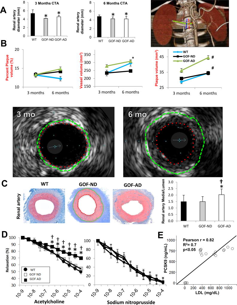
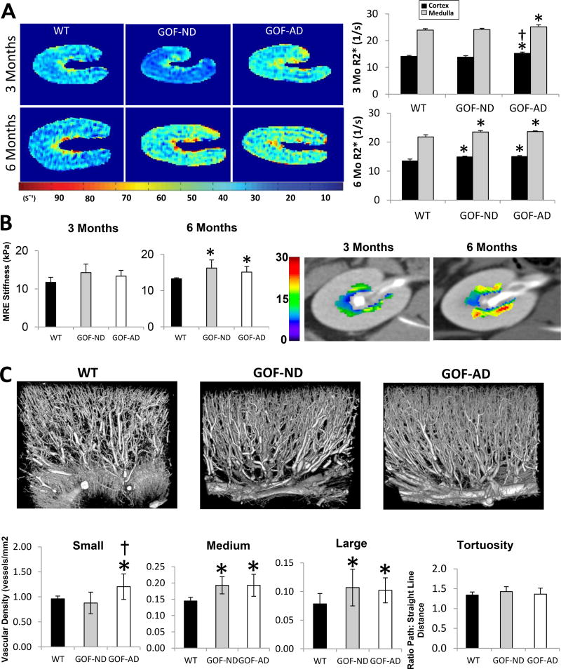
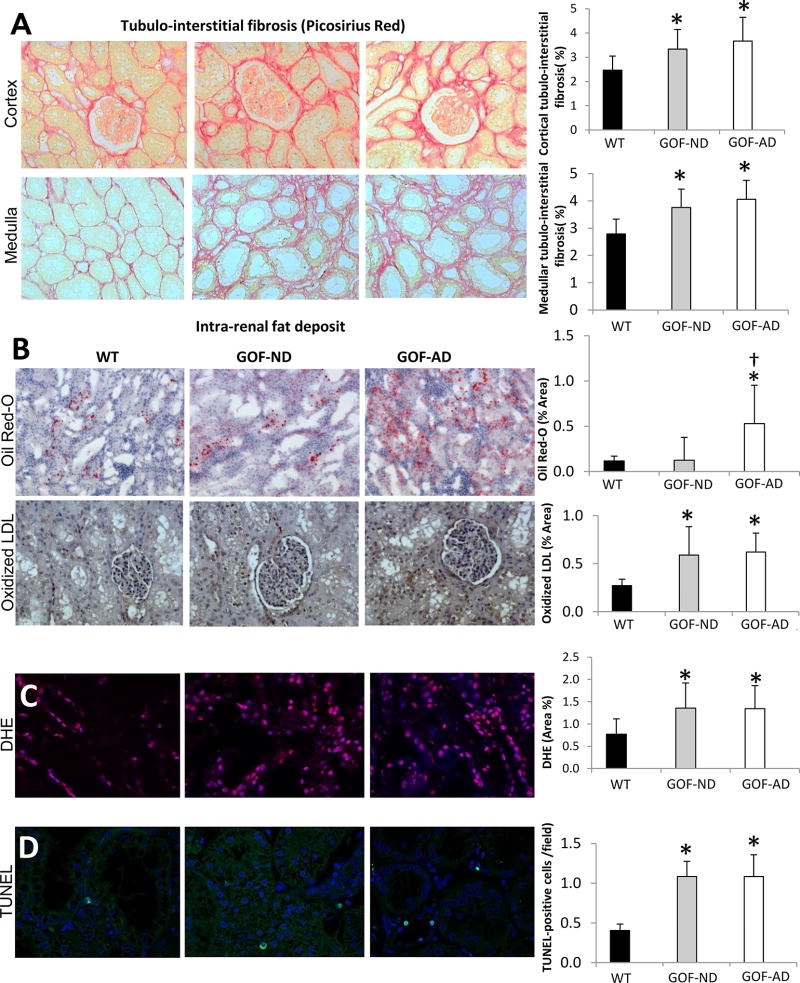
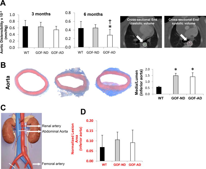
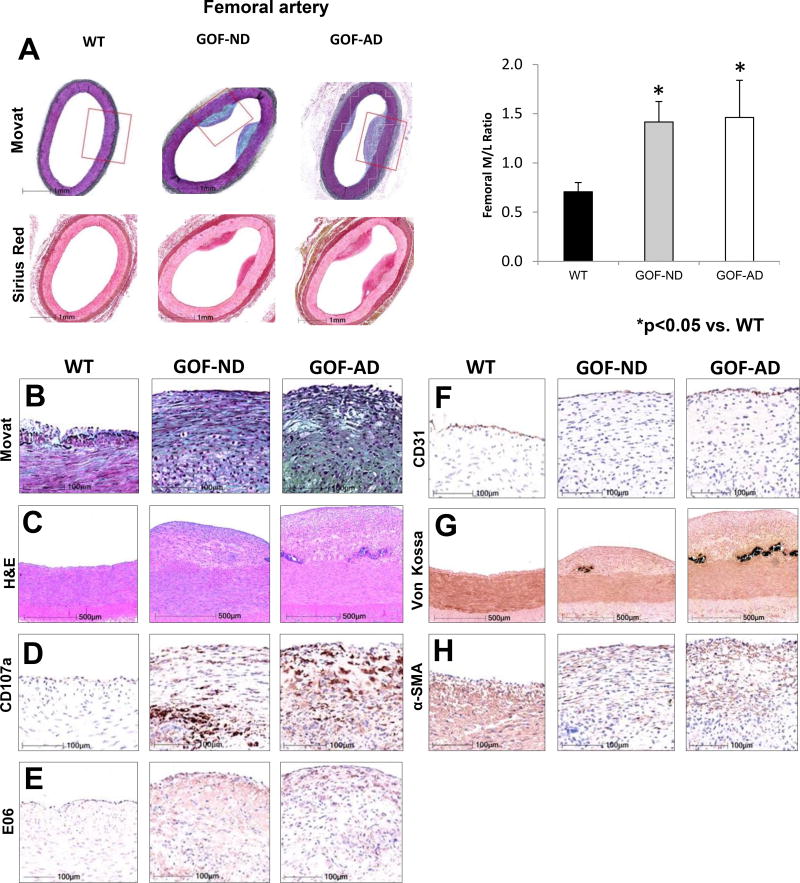
Hypercholesterolemia is a major risk factor for atherosclerosis. Remaining challenges in the management of atherosclerosis necessitate development of animal models that mimic human pathophysiology. We characterized a novel mutant pig model with DNA transposition of D374Y gain-of-function (GOF) cDNA of chimp proprotein convertase subtilisin/kexin type-9 (PCSK9), and tested the hypothesis that it would develop peripheral vascular remodeling and target organ injury in the kidney. Wild-type or PCSK9-GOF Ossabaw miniature pigs fed a standard or atherogenic diet (AD) (n = 7 each) were studied in vivo after 3 and 6 months of diet. Single-kidney hemodynamics and function were studied using multidetector computed tomography and kidney oxygenation by blood oxygen level-dependent magnetic resonance imaging. The renal artery was evaluated by intravascular ultrasound, aortic stiffness by multidetector computed tomography, and kidney stiffness by magnetic resonance elastography. Subsequent ex vivo studies included the renal artery endothelial function and morphology of abdominal aorta, renal, and femoral arteries by histology. Compared with wild type, PCSK9-GOF pigs had elevated cholesterol, triglyceride, and blood pressure levels at 3 and 6 months. Kidney stiffness increased in GOF groups, but aortic stiffness only in GOF-AD. Hypoxia, intrarenal fat deposition, oxidative stress, and fibrosis were observed in both GOF groups, whereas kidney function remained unchanged. Peripheral arteries in GOF groups showed medial thickening and development of atheromatous plaques. Renal endothelial function was impaired only in GOF-AD. Therefore, the PCSK9-GOF mutation induces rapid development of atherosclerosis in peripheral vessels of Ossabaw pigs, which is exacerbated by a high-cholesterol diet. This model may be useful for preclinical studies of atherosclerosis.


Conflict of interest statement

Conflicts of Interest: All authors have read the journal’s policy on disclosure of potential conflicts of interest.

Figures

Figure 1
Figure 1
(A) Representative computed tomography angiography (CTA) of the renal artery (green), showing proximal (red) and distal (blue) markings of a 5mm renal artery segment, and quantification showing a consistent decrease in the diameter of the renal artery, suggesting diffuse narrowing (n=7/group). (B) Intravascular ultrasound (IVUS) images obtained in a PCSK9 GOF-AD pig after 3 versus 6 months of diet, and their quantification in the groups. The images show the vessel wall (green dashed line: external elastic lamina) and lumen (red dashed line) areas. The area enclosed between them illustrates diffuse concentric neointimal hyperplasia at 6 compared to 3 months. (C) The renal artery in GOF-AD showed an increase in media thickness (enclosed between the green and red outlines) relative to the lumen area (n=7/group). (D) Endothelial-dependent (left) and independent (right) relaxation responses to acetylcholine of renal arterial segments from Wild-type (WT) and GOF pigs, showing impaired relaxation response in GOF-AD (n=7/group). (E) LDL-C and PCSK9 levels showed a direct correlation. *p<0.05 vs. WT; #p<0.05 vs. 3mo; †p<0.05 vs. GOF-ND.
Figure 2
Figure 2
(A) Single-kidney BOLD-MRI illustrating hypoxic regions (yellow-red). R2* in the cortex and medulla increased in GOF-AD by 3-months on diet, and in GOF-ND by 6 months (n=7/group). (B) MRE showed in-vivo elevated renal medullary stiffness (yellow-red) at 6-month in both GOF groups (n=5/group). (C) Representative three-dimensional micro-CT images of kidney segments and quantification of renal microvascular spatial density and tortuosity, showing an increase in the microvascular density in GOF groups unchanged tortuosity. *p<0.05 vs. WT; †p<0.05 vs. GOF-ND.
Figure 3
Figure 3
(A) Cortical and medullary tubulointerstitial Picrosirius red staining showing increased fibrosis in GOF groups. (B) Intra-renal fat deposits (Oil-red-O) increased in GOF-AD, while tubulointerstitial Oxidized LDL (brown) accumulated in both GOF groups. (C) Renal production of superoxide anion (DHE, magenta) and (D) Apoptosis (TUNEL, cyan) increased in both GOF-ND and GOF-AD (n=7/group). *p<0.05 vs. WT; †p<0.05 vs. GOF-ND.
Figure 4
Figure 4
(A) CT-determined distensibility showed a fall in aortic elasticity at 6 months. (B) Media-to-lumen ratio of the aorta increased in both GOF-ND and GOF-AD (n=7/group). (C) Diagram showing the harvested vessels location, marked with white line segments. (D) Histomorphometrical assessments of normalized lesion area in the inferior aorta were not significantly increased in the GOF groups. *p<0.05 vs. WT.
Figure 5
Figure 5
Femoral arterial early lesions in GOF pigs. (A) Images and media-to-lumen ratio. (B) Movat, (C) H&E, (D) CD107a, (E) E06 (oxidized-LDL), (F) surface endothelium (CD31/ PECAM-1), (G) Von Kossa (calcium), (H) and α-smooth muscle cell actin (a-SMA), showed increased staining in GOF-ND and GOF-AD (n=7/group). The higher power magnifications H&E and von Kossa represent regions corresponding to the red box in the low power Movat pentachrome stain. The minimal lesions in the WT mainly consisted of smooth muscle cells and neointimal hyperplasia. Contrarily, fibroatheromatous plaques with oxidized LDL accumulation were noted in GOF-ND and GOF-AD. *p<0.05 vs. WT.

References

    1. Lusis AJ. Atherosclerosis. Nature. 2000;407:233–41. - PMC - PubMed
    1. Celermajer DS, Chow CK, Marijon E, Anstey NM, Woo KS. Cardiovascular disease in the developing world: prevalences, patterns, and the potential of early disease detection. J Am Coll Cardiol. 2012;60:1207–16. - PubMed
    1. Sillesen H, Falk E. Why not screen for subclinical atherosclerosis? Lancet. 2011;378:645–6. - PubMed
    1. Breslow JL. Mouse models of atherosclerosis. Science. 1996;272:685–8. - PubMed
    1. Mohler ER, 3rd, Sarov-Blat L, Shi Y, Hamamdzic D, Zalewski A, Macphee C, et al. Site-specific atherogenic gene expression correlates with subsequent variable lesion development in coronary and peripheral vasculature. Arterioscler Thromb Vasc Biol. 2008;28:850–5. - PubMed

Publication types

Substances