. 2018 Apr:14:656-668.
doi: 10.1016/j.redox.2017.11.012. Epub 2017 Nov 16.

Chicoric acid prevents PDGF-BB-induced VSMC dedifferentiation, proliferation and migration by suppressing ROS/NFκB/mTOR/P70S6K signaling cascade

Affiliations

Chicoric acid prevents PDGF-BB-induced VSMC dedifferentiation, proliferation and migration by suppressing ROS/NFκB/mTOR/P70S6K signaling cascade

Qing-Bo Lu et al. Redox Biol. 2018 Apr.

Abstract

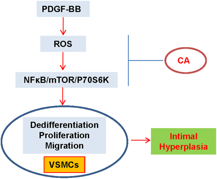
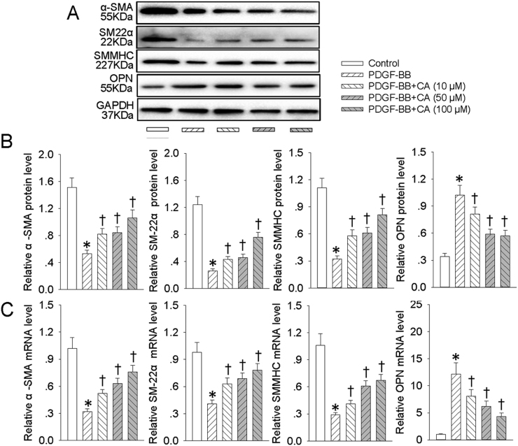
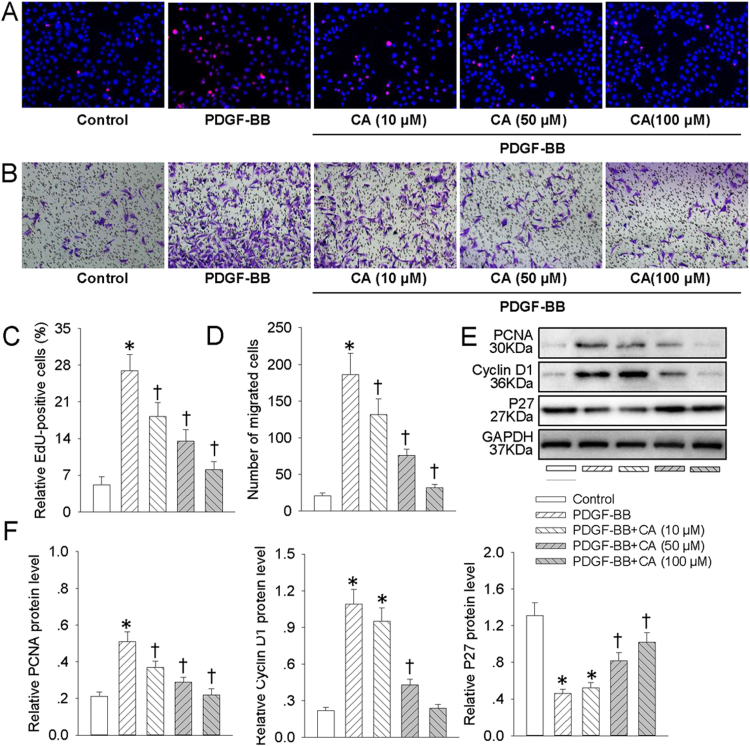
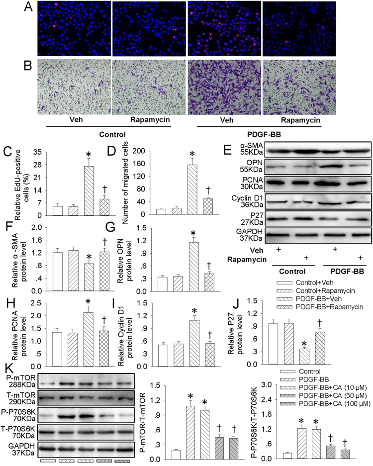
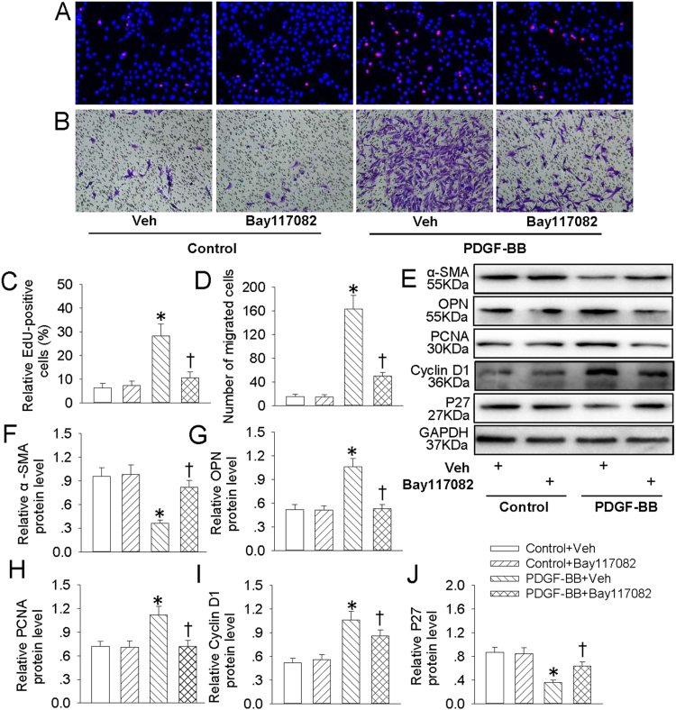
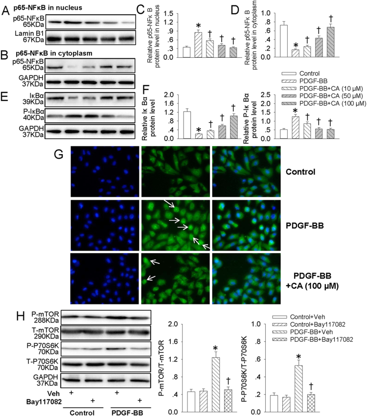
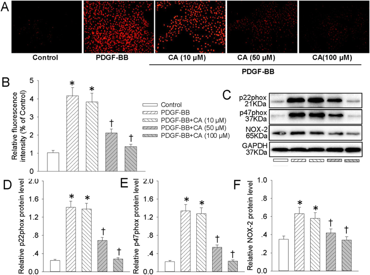
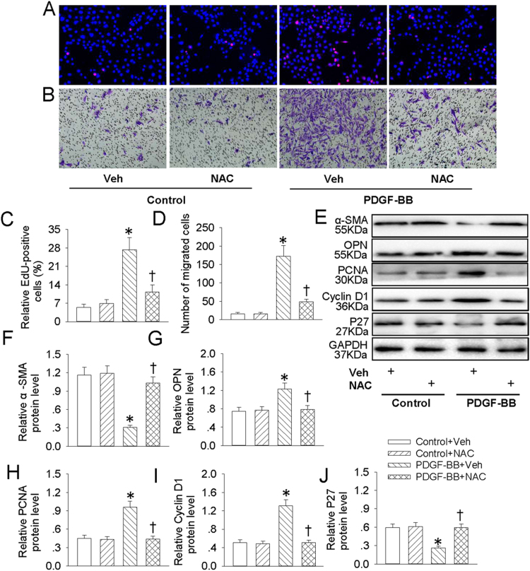
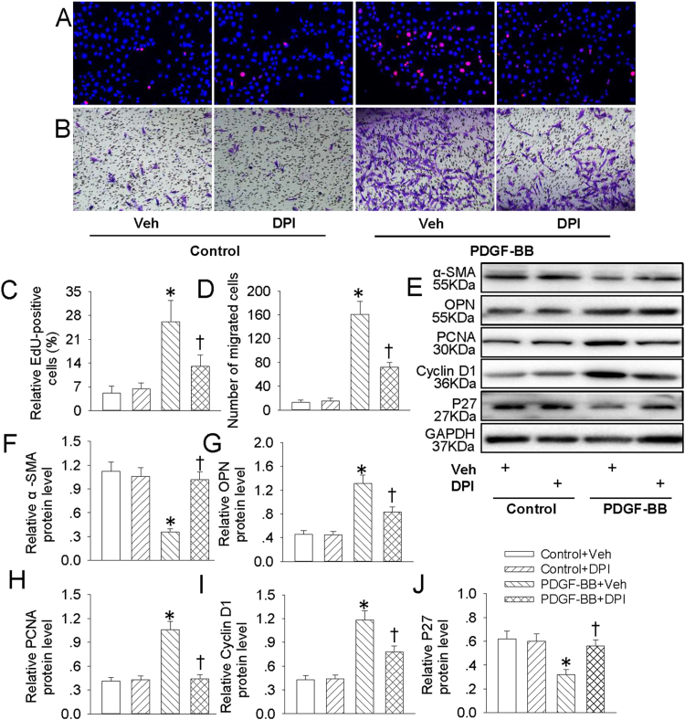
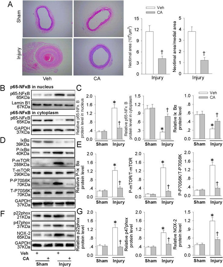
Phenotypic switch of vascular smooth muscle cells (VSMCs) is characterized by increased expressions of VSMC synthetic markers and decreased levels of VSMC contractile markers, which is an important step for VSMC proliferation and migration during the development and progression of cardiovascular diseases including atherosclerosis. Chicoric acid (CA) is identified to exert powerful cardiovascular protective effects. However, little is known about the effects of CA on VSMC biology. Herein, in cultured VSMCs, we showed that pretreatment with CA dose-dependently suppressed platelet-derived growth factor type BB (PDGF-BB)-induced VSMC phenotypic alteration, proliferation and migration. Mechanistically, PDGF-BB-treated VSMCs exhibited higher mammalian target of rapamycin (mTOR) and P70S6K phosphorylation, which was attenuated by CA pretreatment, diphenyleneiodonium chloride (DPI), reactive oxygen species (ROS) scavenger N-acetyl-l-cysteine (NAC) and nuclear factor-κB (NFκB) inhibitor Bay117082. PDGF-BB-triggered ROS production and p65-NFκB activation were inhibited by CA. In addition, both NAC and DPI abolished PDGF-BB-evoked p65-NFκB nuclear translocation, phosphorylation and degradation of Inhibitor κBα (IκBα). Of note, blockade of ROS/NFκB/mTOR/P70S6K signaling cascade prevented PDGF-BB-evoked VSMC phenotypic transformation, proliferation and migration. CA treatment prevented intimal hyperplasia and vascular remodeling in rat models of carotid artery ligation in vivo. These results suggest that CA impedes PDGF-BB-induced VSMC phenotypic switching, proliferation, migration and neointima formation via inhibition of ROS/NFκB/mTOR/P70S6K signaling cascade.

Keywords: Chicoric acid; Migration; PDGF-BB; Proliferation; VSMCs.


Figures

fx1
Graphical abstract
Fig. 1
Fig. 1
CA abrogated PDGF-BB-induced VSMC dedifferentiation. VSMCs were pretreated with various concentrations (10, 50 and 100 μM) of CA for 6 h followed by stimulation with PDGF-BB (20 ng/mL) for 24 h. (A) Western blot was employed to quantitate the expression levels of contractile protein α-SMA, SMMHC, SM22α and synthetic proteins OPN. (B) Bar graph showing the relative protein level of α-SMA, SMMHC, SM22α and OPN. (C) Bar graph showing the relative mRNA level ofα-SMA, SMMHC, SM22α and OPN. Values are mean±SE. * P < 0.05 vs. Control, † P < 0.05 vs. PDGF-BB. n = 6 for each group.
Fig. 2
Fig. 2
CA abated PDGF-BB-induced VSMC proliferation and migration. VSMCs were pretreated with various concentrations (10, 50 and 100 μM) of CA for 6 h followed by stimulation with PDGF-BB (20 ng/mL) for 24 h. (A) DNA synthesis in VSMCs determined with EdU incorporation assay. Blue fluorescence (Hoechst 33342) shows cell nuclei and green fluorescence (Edu) stands for cells with DNA synthesis. (B) Transwell assay was performed to determine the migration of VSMCs. (C) The ratio of EdU-positive cells to total cells. (D) Bar graph showing the number of migrated VSMCs. (E) Represented images showing the protein expressions of PCNA, cyclin D1 and P27. (F) Bar graph showing the relative protein level of PCNA, cyclin D1 and P27. Values are mean±SE. * P < 0.05 vs. Control, † P < 0.05 vs. PDGF-BB. n = 6 for each group.
Fig. 3
Fig. 3
CA inhibited PDGF-BB-induced VSMC dedifferentiation, proliferation and migration via suppressing mTOR/P70S6K signaling. VSMCs were pretreated with rapamycin (100 nM) for 6 h followed by stimulation with PDGF-BB (20 ng/mL) for 24 h. (A) EdU assay. (B) Transwell assay was performed to determine the migration of VSMCs. (C) The ratio of EdU-positive cells to total cells. (D) Bar graph showing the number of migrated VSMCs. (E) Represented images showing the protein expressions of α-SMA, OPN, PCNA, cyclin D1 and P27. The relative protein expressions of α-SMA (F), OPN (G), PCNA (H), cyclin D1 (I) and P27 (J) were quantified. (K) VSMCs were pretreated with various concentrations (10, 50 and 100 μM) of CA for 6 h followed by stimulation with PDGF-BB (20 ng/mL) for 24 h. The phosphorylated and total mTOR and P70S6K protein levels were measure by western blot. Values are mean±SE. * P < 0.05 vs. Control + Vehicle (Veh) or Control, † P < 0.05 vs. PDGF-BB + Vehicle (Veh) or PDGF-BB. n = 6 for each group.
Fig. 4
Fig. 4
Bay117082 retarded PDGF-BB-induced VSMC dedifferentiation, proliferation and migration. VSMCs were pretreated with Bay117082 (10 μM) for 6 h followed by stimulation with PDGF-BB (20 ng/mL) for 24 h. (A) EdU assay. (B) Transwell assay was performed to determine the migration of VSMCs. (C) The ratio of EdU-positive cells to total cells. (D) Bar graph showing the number of migrated VSMCs. (E) Represented images showing the protein expressions of α-SMA, OPN, PCNA, cyclin D1 and P27. The relative protein expressions of α-SMA (F), OPN (G), PCNA (H), cyclin D1 (I) and P27 (J) were quantified. Values are mean ± SE. * P < 0.05 vs. Control + Vehicle (Veh), † P < 0.05 vs. PDGF-BB + Vehicle (Veh). n = 6 for each group.
Fig. 5
Fig. 5
CA retarded PDGF-BB-induced NFκB signaling activation. VSMCs were pretreated with various concentrations (10, 50 and 100 μM) of CA for 6 h followed by stimulation with PDGF-BB (20 ng/mL) for 24 h. (A) Represented images showing the protein expressions of p65-NFκB in nucleus. (B) Represented images showing the protein expressions of p65-NFκB in cytoplasm. (C) Bar graph showing the relative protein expressions of p65-NFκB in nucleus. (D) Bar graph showing the relative protein expressions of p65-NFκB in cytoplasm. (E) Represented images showing the protein expressions of IκBα and phosphorylated IκBα. (F) Bar graph showing the relative protein expressions of IκBα and phosphorylated IκBα. (G) The translocation of p65-NFκB from cytoplasm to nucleus was measure by immunofluorescence, white arrow showing the nuclear localization of p65-NFκB. (H) VSMCs were pretreated with Bay117082 (10 μM) for 6 h followed by stimulation with PDGF-BB (20 ng/mL) for 24 h. The phosphorylated and total mTOR and P70S6K protein levels were measure by western blot. Values are mean ± SE. * P < 0.05 vs. Control or Control + Vehicle (Veh), † P < 0.05 vs. PDGF-BB or PDGF-BB + Vehicle (Veh). n = 6 for each group.
Fig. 6
Fig. 6
CA prevented PDGF-BB-induced ROS production in VSMCs. VSMCs were pretreated with various concentrations (10, 50 and 100 μM) of CA for 6 h followed by stimulation with PDGF-BB (20 ng/mL) for 24 h. (A) ROS generation was evaluated by DHE fluorescence. (B) Relative ROS fluorescence intensity. (C) Represented images showing the protein expressions of NAD(P)H oxidase subunits p22phox, p47phox, NOX-2. The relative protein expressions of NAD(P)H oxidase subunits p22phox (D), p47phox (E), NOX-2 (F) were quantified. Values are mean ± SE. * P < 0.05 vs. Control, † P < 0.05 vs. PDGF-BB. n = 6 for each group.
Fig. 7
Fig. 7
NAC retarded PDGF-BB-induced VSMC dedifferentiation, proliferation and migration. VSMCs were pretreated with NAC (1 mM) for 6 h followed by stimulation with PDGF-BB (20 ng/mL) for 24 h. (A) EdU assay. (B) Transwell assay was performed to determine the migration of VSMCs. (C) The ratio of EdU-positive cells to total cells. (D) Bar graph showing the number of migrated VSMCs. (E) Represented images showing the protein expressions of α-SMA, OPN, PCNA, cyclin D1 and P27. The relative protein expressions of α-SMA (F), OPN (G), PCNA (H), cyclin D1 (I) and P27 (J) were quantified. Values are mean ± SE. * P < 0.05 vs. Control + Vehicle (Veh), † P < 0.05 vs. PDGF-BB + Vehicle (Veh). n = 6 for each group.
Fig. 8
Fig. 8
DPI retarded PDGF-BB-induced VSMC dedifferentiation, proliferation and migration. VSMCs were pretreated with DPI (10 μM) for 6 h followed by stimulation with PDGF-BB (20 ng/mL) for 24 h. (A) EdU assay. (B) Transwell assay was performed to determine the migration of VSMCs. (C) The ratio of EdU-positive cells to total cells. (D) Bar graph showing the number of migrated VSMCs. (E) Represented images showing the protein expressions of α-SMA, OPN, PCNA, cyclin D1 and P27. The relative protein expressions of α-SMA (F), OPN (G), PCNA (H), cyclin D1 (I) and P27 (J) were quantified. Values are mean ± SE. * P < 0.05 vs. Control + Vehicle (Veh), † P < 0.05 vs. PDGF-BB + Vehicle (Veh). n = 6 for each group.
Fig. 9
Fig. 9
CA repressed intimal hyperplasia and suppressed ROS/NFκB/mTOR/P70S6K signaling cascade in vivo. Three days after vascular injury, CA (50 mg/kg/day) was administered gastric gavage to rats for a total of 8 weeks. (A) Representative HE staining of carotid arteries in sham and injury rats. (B) Represented blots showing the protein expressions of p65-NFκB in nucleus or in cytoplasm. (C) Bar graph showing the relative protein expressions of p65-NFκB in nucleus, p65-NFκB in cytoplasm and IκBα. (D) Represented images showing the protein expressions of IκBα, phosphorylated IκBα, phosphorylated and total mTOR and P70S6K. (E) Bar graph showing the relative protein expressions of phosphorylated IκBα, mTOR and P70S6K. (F) Represented images showing the protein expressions of NAD(P)H oxidase subunits p22phox, p47phox, NOX-2. (G) Bar graph showing the relative protein expressions of NAD(P)H oxidase subunits p22phox, p47phox, NOX-2. Values are mean ± SE. * P < 0.05 vs. Sham, † P < 0.05 vs. Veh. n = 6 for each group.

References

    1. Liao X.H., Wang N., Zhao D.W., Zheng D.L., Zheng L., Xing W.J., Ma W.J., Bao L.Y., Dong J., Zhang T.C. STAT3 protein regulates vascular smooth muscle cell phenotypic switch by interaction with myocardin. J. Biol. Chem. 2015;290:19641–19652. - PMC - PubMed
    1. Cao T., Zhang L., Yao L.L., Zheng F., Wang L., Yang J.Y., Guo L.Y., Li X.Y., Yan Y.W., Pan Y.M., Jiang M., Chen L., Tang J.M., Chen S.Y., Jia N. S100B promotes injury-induced vascular remodeling through modulating smooth muscle phenotype. Biochim. Biophys. Acta. 2017 - PubMed
    1. Zhu L.H., Huang L., Zhang X., Zhang P., Zhang S.M., Guan H., Zhang Y., Zhu X.Y., Tian S., Deng K., Li H. Mindin regulates vascular smooth muscle cell phenotype and prevents neointima formation. Clin. Sci. 2015;129:129–145. - PubMed
    1. Bennett M.R., Sinha S., Owens G.K. Vascular smooth muscle cells in atherosclerosis. Circ. Res. 2016;118:692–702. - PMC - PubMed
    1. Chistiakov D.A., Orekhov A.N., Bobryshev Y.V. Vascular smooth muscle cell in atherosclerosis. Acta Physiol. 2015;214:33–50. - PubMed

MeSH terms