

Traumatic pneumocephaly: trapped air from where?
- PMID: 29175906
- PMCID: PMC5720322
- DOI: 10.1136/bcr-2017-219420
Traumatic pneumocephaly: trapped air from where?
Abstract
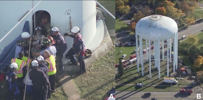
Traumatic pneumocephaly is literally defined as 'air in the head' after trauma. While this phenomenon has been well described in the literature, our case report is unique in describing diffuse pneumocephalus in the subaponeurotic space, subdural space, subarachnoid space, brain and ventricles without a break in the cranial vault: a 26-year-old man fell from a =9 meter scaffolding in a water tower. Following an arduous and delayed extrication, the patient was unresponsive with loss of pulse requiring intubation, cardiopulmonary resuscitation and release of tension pneumothorax with bilateral thoracostomy tubes. Examination remained poor with a Glasgow Coma Scale of 3. Immediate exploratory laparotomy was performed for a small right retroperitoneal haematoma on Focused Assessment with Sonography for Trauma. Postoperative imaging revealed diffuse pneumocephaly without facial fractures. This case presentation explores unusual causes of fistulous connections with the atmosphere that may lead to air trapped in and around the cranial vault.
Keywords: accidents, injuries; interventional radiology; trauma.
© BMJ Publishing Group Ltd (unless otherwise stated in the text of the article) 2017. All rights reserved. No commercial use is permitted unless otherwise expressly granted.
Conflict of interest statement
Competing interests: None declared.
Figures


References
-
- Vinken P, Bruyn G. Handbook of clinical neurology. 1969.
-
- Garland LH, Mottram ME. Traumatic pneumocephalus. Radiology 1945;44:237–40. 10.1148/44.3.237 - DOI
Publication types
MeSH terms
LinkOut - more resources
Full Text Sources
Other Literature Sources
Medical