. 2017 Nov 22;9(11):2411-2435.
doi: 10.18632/aging.101328.

TNFα-senescence initiates a STAT-dependent positive feedback loop, leading to a sustained interferon signature, DNA damage, and cytokine secretion

Affiliations

TNFα-senescence initiates a STAT-dependent positive feedback loop, leading to a sustained interferon signature, DNA damage, and cytokine secretion

Renuka Kandhaya-Pillai et al. Aging (Albany NY). .

Abstract

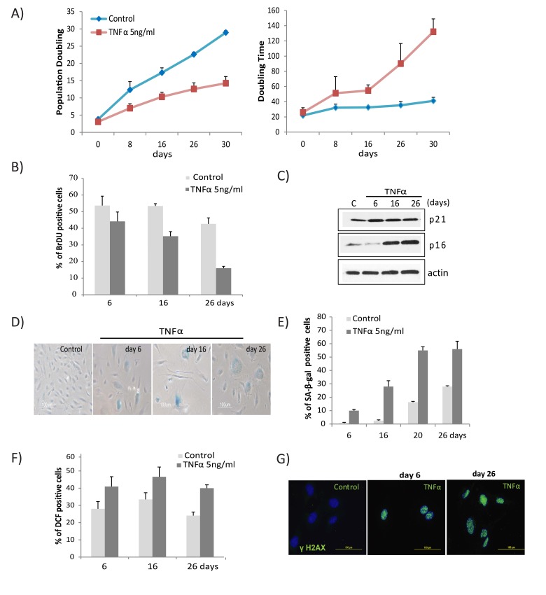
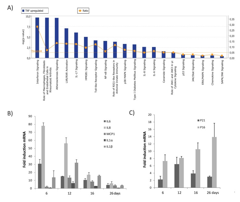
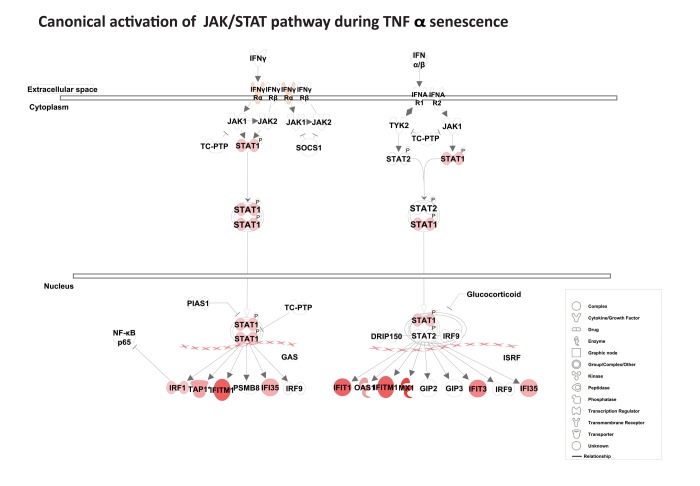
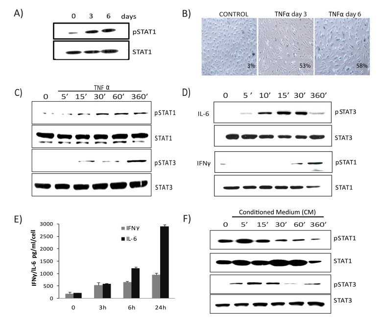
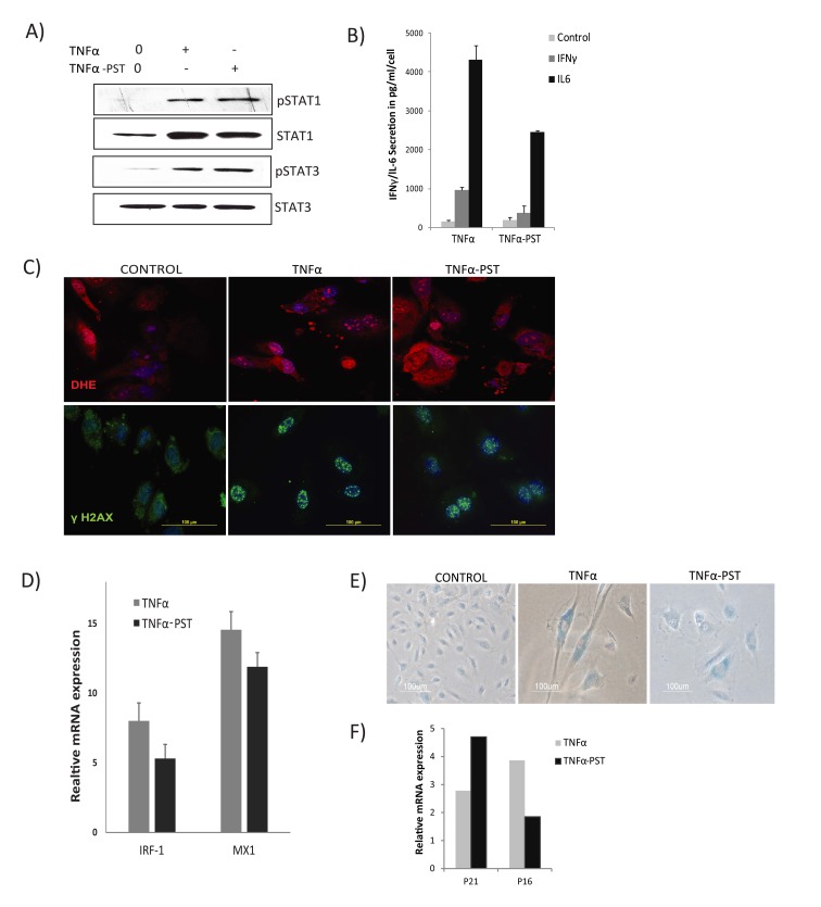
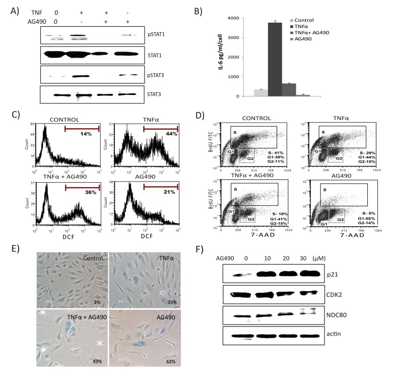
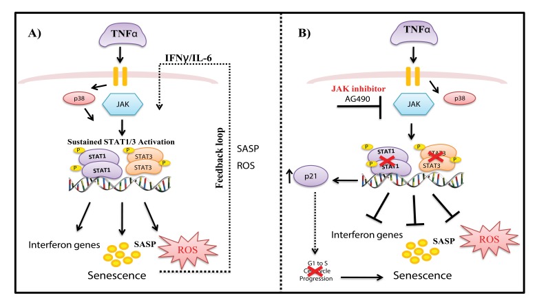
Cellular senescence is a cell fate program that entails essentially irreversible proliferative arrest in response to damage signals. Tumor necrosis factor-alpha (TNFα), an important pro-inflammatory cytokine secreted by some types of senescent cells, can induce senescence in mouse and human cells. However, downstream signaling pathways linking TNFα-related inflammation to senescence are not fully characterized. Using human umbilical vein endothelial cells (HUVECs) as a model, we show that TNFα induces permanent growth arrest and increases p21CIP1, p16INK4A, and SA-β-gal, accompanied by persistent DNA damage and ROS production. By gene expression profiling, we identified the crucial involvement of inflammatory and JAK/STAT pathways in TNFα-mediated senescence. We found that TNFα activates a STAT-dependent autocrine loop that sustains cytokine secretion and an interferon signature to lock cells into senescence. Furthermore, we show STAT1/3 activation is necessary for cytokine and ROS production during TNFα-induced senescence. However, inhibition of STAT1/3 did not rescue cells from proliferative arrest, but rather suppressed cell cycle regulatory genes and altered TNFα-induced senescence. Our findings suggest a positive feedback mechanism via the STAT pathway that sustains cytokine production and reveal a reciprocal regulatory role of JAK/STAT in TNFα-mediated senescence.

Keywords: DNA-damage; JAK/STAT pathway; inflammation; interferon response genes; senescence.


Conflict of interest statement

CONFLICTS OF INTEREST

The authors declare no conflicts of interest.

Figures

Figure 1
Figure 1. TNFα induces senescence and DNA damage in HUVECs
(A) Long-term growth curve of cells exposed to recombinant human TNFα (5ng/ml). Untreated cells were used as controls. Population doubling and doubling times were calculated based on cell density at confluence. Data represent mean values from 3 independent experiments. (B) The percentage of BrdU-positive cells was determined by FACS analysis in cells untreated or chronically treated with TNFα at the concentration indicated. (C) Western blot analysis of p21, p16, and actin in cells treated with TNFα 5ng/ml for the indicated times. (D) SA-β-gal activity in TNFα (5ng/ml)-treated or control cells for the indicated number of days. (E) Percentages of SA-β-gal-positive cells in control or TNFα-treated cultures. The data represent 2 independent counts of 200 cells from 3 independent experiments. (F) Intracellular ROS levels were monitored by 2′,7′-dichlorodihydrofluorescein diacetate staining followed by flow cytometry. Bar graph represents percentage of DCFDA-positive cells treated with TNFα or medium alone. (G) Immunofluorescence detection of γH2AX foci in controls or cells treated with TNFα (5ng/ml) for indicated days. Data in A, B, E, and F represent mean value ± standard deviation (s.d.) from n=3, 2, 3, and 2 independent experiments, respectively.
Figure 2
Figure 2. Top canonical pathways identified by IPA analysis in TNFα-induced senescence
(A) Bar chart represents the top canonical signaling pathways that were influenced during TNFα-induced senescence. p-values were determined using Fisher's exact test with a threshold value of >0.05. Ratios represent the number of genes that mapped to a specific canonical pathway divided by the total number of genes that make up the respective pathway. (B) Relative mRNA expression of SASP components in cells treated with TNFα at 6, 16, or 26 days compared to untreated cells. (C) Relative mRNA expression of p16 and p21 in cells treated with TNFα (5ng/ml) compared to untreated cells. Results are mean ± standard deviation of n=2 independent experiments.
Figure 3
Figure 3. Activation of the canonical JAK/ STAT pathway
Pathway analysis using IPA reveals canonical activation of STAT1 acts as a central regulator for JAK/ STAT-mediated interferon response genes during TNFα-induced senescence. The red symbols represent up-regulated genes, with the intensity of node color indicating degree of up-regulation and white indicating genes absent from the list. Shape of nodes denotes functions of gene products.
Figure 4
Figure 4. Prolonged activation of JAK/ STAT signaling in TNFα-induced senescence
(A) Immunoblot detection of p-Ser727-STAT1 and total STAT1 in cells exposed to TNFα 20ng/ml for the indicated times. (B) SA-β-gal activity in TNFα (20ng/ml)-treated or control cells for 3 or 6 days. (C) Immunoblot detection of p-Ser727-STAT1, total STAT1, p-Tyr705-STAT3, and total STAT3 in cells exposed to TNFα (5ng/ml) for the indicated intervals. (D) Immunoblot detection of pSTAT1, total STAT1, pSTAT3, and total STAT3 in cells stimulated with IL6 (10ng/ml) or IFNγ (1ng/ml) for the indicated intervals. (E) Secretion of IFNγ/IL6 quantified by ELISA in conditioned medium collected in the presence or absence of TNFα. (F) Immunoblot detection of p-Ser727-STAT1, p-Tyr705-STAT3, STAT1, and STAT3 in cells treated with conditioned medium (CM) (cell free-culture supernatants from control and cells stimulated with TNFα for 3 days transferred after 1:4 dilution with fresh culture medium) from TNFα-induced senescent cells or from non-senescent cells for the indicated times.
Figure 5
Figure 5. Persistent activation of STAT1/3
Cells were exposed to TNFα (20ng/ml) for 3 days, then washed to remove the residual TNFα and cultured for 3 days in the absence of exogenous TNFα (TNFα-PST). Parallel cultures were exposed to exogenous TNFα throughout the experiment. (A) Levels of p-Ser727-STAT1, p-Tyr705-STAT3, and total STAT3 proteins were quantified by immunoblot. (B) Secretion of IL-6/IFNγ was assessed in culture supernatants from cells treated with TNFα as indicated. (C) Immunodetection of ROS production and γH2AX foci in control or cells treated with TNFα or TNFα-post-stimulated (PST), as indicated. (D) Real-time gene expression of IRF1 and MX1 in cells exposed to TNFα or TNFα-PST for 3 days. Results were normalized to internal control TBP and are shown relative to untreated cells. (E) SA-β-gal activity in TNFα-treated cells for 3 days or in cells treated with TNFα (20ng/ml) for three days, then washed to remove residual TNFα and left untreated for another 3 days. (F) mRNA expression of p21 and p16 quantified by real-time PCR in cells exposed to TNFα or TNFα-PST for 3 days. Data in D and F represent mean value of ± sd from 2 independent experiments.
Figure 6
Figure 6. JAK2 inhibitor decreases TNFα-mediated inflammation, ROS levels, and interferon signature
Cells were exposed to TNFα alone, TNFα in combination with AG490 (30μM), or AG490 alone for 3 days. (A) Immunoblot detection of p-Ser727-STAT1, p-Tyr705-STAT3, and total STAT1 and STAT3. (B) Secretion of IL6 was estimated by ELISA in culture supernatants from cells treated with TNFα alone, or in combination with AG490, or AG490 alone for 3 days. (C) FACS analysis of ROS levels in cells stimulated either with TNFα, TNFα along with AG490, or AG490 alone for 3 days using DCFDA staining 2′,7′-dichlorofluorescein (DCF) positive cells were analyzed. Inhibition of STAT signals modulates senescence. (D) Cell cycle analysis using BrdU and 7- aminoactinomycin D (7-AAD) staning in cells exposed to TNFα, TNFα in combination with AG490, or AG490 alone for 3 days. (E) Percentage of SA-β-gal-positive cells. Quantification of SA-β-gal activity in cells stimulated with TNFα 20ng/ml, TNFα in combination with AG490, or AG490 alone for 3 days. The data represent means of 3 independent counts of 200 cells from 2 independent experiments. (F) Effect of AG490 on cell cycle regulatory proteins. Western analysis performed using cells treated with AG490 for 3 days and blotted against anti-p21, CDK2, and NDC80. Actin serves as loading control.
Figure 7
Figure 7. Model of mechanisms involved in TNFα-induced senescence of HUVECs
(A) TNFα activates JAK/STAT and p38 signaling pathways, which mediate increased expression of STAT1/3 phosphorylation. Activation of the JAK pathway leads to persistent phosphorylation of STAT1/3 signaling, which together with ROS, interferon genes, and other SASP components, drives a positive auto-regulatory loop, leading to sustained inflammation and stable senescence. (B) Inhibition of STAT1/3 with the JAK inhibitor AG490 decreased ROS and IL-6 production and decreased expression of interferon response genes. On the other hand, blockade of STAT1/3 expression decreased S phase entry of cells and increased p21 expression, leading to senescence.

References

    1. Tchkonia T, Zhu Y, van Deursen J, Campisi J, Kirkland JL. Cellular senescence and the senescent secretory phenotype: therapeutic opportunities. J Clin Invest. 2013;123:966–72. https://doi.org/10.1172/JCI64098 - DOI - PMC - PubMed
    1. Hayflick L, Moorhead PS. The serial cultivation of human diploid cell strains. Exp Cell Res. 1961;25:585–621. https://doi.org/10.1016/0014-4827(61)90192-6 - DOI - PubMed
    1. Sahin E, Colla S, Liesa M, Moslehi J, Müller FL, Guo M, Cooper M, Kotton D, Fabian AJ, Walkey C, Maser RS, Tonon G, Foerster F, et al. Telomere dysfunction induces metabolic and mitochondrial compromise. Nature. 2011;470:359–65. https://doi.org/10.1038/nature09787 - DOI - PMC - PubMed
    1. von Zglinicki T, Saretzki G, Ladhoff J, d'Adda di Fagagna F, Jackson SP. Human cell senescence as a DNA damage response. Mech Ageing Dev. 2005;126:111–17. https://doi.org/10.1016/j.mad.2004.09.034 - DOI - PubMed
    1. Herbig U, Jobling WA, Chen BP, Chen DJ, Sedivy JM. Telomere shortening triggers senescence of human cells through a pathway involving ATM, p53, and p21(CIP1), but not p16(INK4a) Mol Cell. 2004;14:501–13. https://doi.org/10.1016/S1097-2765(04)00256-4 - DOI - PubMed

MeSH terms