Diffusion tensor optical coherence tomography
- PMID: 29176039
- PMCID: PMC5773385
- DOI: 10.1088/1361-6560/aa9cfe
Diffusion tensor optical coherence tomography
Abstract
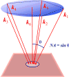
In situ measurements of diffusive particle transport provide insight into tissue architecture, drug delivery, and cellular function. Analogous to diffusion-tensor magnetic resonance imaging (DT-MRI), where the anisotropic diffusion of water molecules is mapped on the millimeter scale to elucidate the fibrous structure of tissue, here we propose diffusion-tensor optical coherence tomography (DT-OCT) for measuring directional diffusivity and flow of optically scattering particles within tissue. Because DT-OCT is sensitive to the sub-resolution motion of Brownian particles as they are constrained by tissue macromolecules, it has the potential to quantify nanoporous anisotropic tissue structure at micrometer resolution as relevant to extracellular matrices, neurons, and capillaries. Here we derive the principles of DT-OCT, relating the detected optical signal from a minimum of six probe beams with the six unique diffusion tensor and three flow vector components. The optimal geometry of the probe beams is determined given a finite numerical aperture, and a high-speed hardware implementation is proposed. Finally, Monte Carlo simulations are employed to assess the ability of the proposed DT-OCT system to quantify anisotropic diffusion of nanoparticles in a collagen matrix, an extracellular constituent that is known to become highly aligned during tumor development.
Figures


Similar articles
-
Imaging Extracellular Matrix Remodeling In Vitro by Diffusion-Sensitive Optical Coherence Tomography.Biophys J. 2016 Apr 26;110(8):1858-1868. doi: 10.1016/j.bpj.2016.03.014. Biophys J. 2016. PMID: 27119645 Free PMC article.
-
Probing biological nanotopology via diffusion of weakly constrained plasmonic nanorods with optical coherence tomography.Proc Natl Acad Sci U S A. 2014 Oct 14;111(41):E4289-97. doi: 10.1073/pnas.1409321111. Epub 2014 Sep 29. Proc Natl Acad Sci U S A. 2014. PMID: 25267619 Free PMC article.
-
Diffusion tensor of water in model articular cartilage.Eur Biophys J. 2011 Jan;40(1):81-91. doi: 10.1007/s00249-010-0629-4. Epub 2010 Oct 23. Eur Biophys J. 2011. PMID: 20972563
-
Diffusion tensor imaging in musculoskeletal disorders.Radiographics. 2014 May-Jun;34(3):E56-72. doi: 10.1148/rg.343125062. Radiographics. 2014. PMID: 24819802 Review.
-
The role of diffusion tensor imaging in the evaluation of ischemic brain injury - a review.NMR Biomed. 2002 Nov-Dec;15(7-8):561-9. doi: 10.1002/nbm.786. NMR Biomed. 2002. PMID: 12489102 Review.
Cited by
-
In situ pulmonary mucus hydration assay using rotational and translational diffusion of gold nanorods with polarization-sensitive optical coherence tomography.J Biomed Opt. 2024 Apr;29(4):046004. doi: 10.1117/1.JBO.29.4.046004. Epub 2024 Apr 30. J Biomed Opt. 2024. PMID: 38690122 Free PMC article.
-
A noninvasive fluorescence imaging-based platform measures 3D anisotropic extracellular diffusion.Nat Commun. 2021 Mar 26;12(1):1913. doi: 10.1038/s41467-021-22221-0. Nat Commun. 2021. PMID: 33772014 Free PMC article.
-
Label-free metabolic and structural profiling of dynamic biological samples using multimodal optical microscopy with sensorless adaptive optics.Sci Rep. 2022 Mar 2;12(1):3438. doi: 10.1038/s41598-022-06926-w. Sci Rep. 2022. PMID: 35236862 Free PMC article.
-
Genetically Encodable Contrast Agents for Optical Coherence Tomography.ACS Nano. 2020 Jul 28;14(7):7823-7831. doi: 10.1021/acsnano.9b08432. Epub 2020 Feb 10. ACS Nano. 2020. PMID: 32023037 Free PMC article.
-
Optical coherence tomography for noninvasive monitoring of drug delivery.Adv Drug Deliv Rev. 2025 May;220:115571. doi: 10.1016/j.addr.2025.115571. Epub 2025 Mar 24. Adv Drug Deliv Rev. 2025. PMID: 40139506 Free PMC article. Review.
References
-
- Anderson AW. Theoretical analysis of the effects of noise on diffusion tensor imaging. Magnetic Resonance in Medicine. 2001;46(6):1174–1188. - PubMed
-
- Basser PJ, Mattiello J, LeBihan D. Mr diffusion tensor spectroscopy and imaging. Biophysical Journal. 1994;66(1):259–267. URL: http://dx.doi.org/10.1016/S0006-3495(94)80775-1. - DOI - PMC - PubMed
Publication types
MeSH terms
Substances
Grants and funding
LinkOut - more resources
Full Text Sources
Other Literature Sources
