. 2018 Jan;41(1):19-31.
doi: 10.1097/CJI.0000000000000199.

Preclinical Optimization of a CD20-specific Chimeric Antigen Receptor Vector and Culture Conditions

Affiliations

Preclinical Optimization of a CD20-specific Chimeric Antigen Receptor Vector and Culture Conditions

Sang Yun Lee et al. J Immunother. 2018 Jan.

Abstract

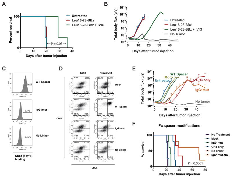
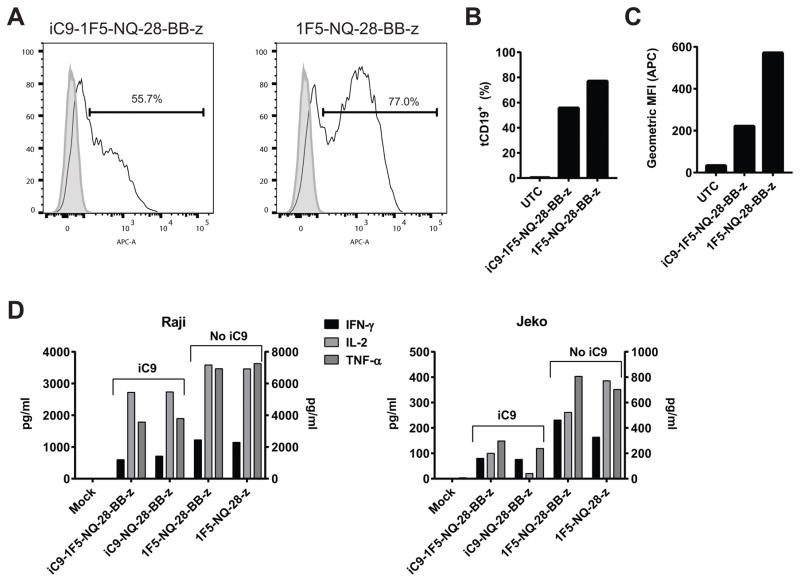
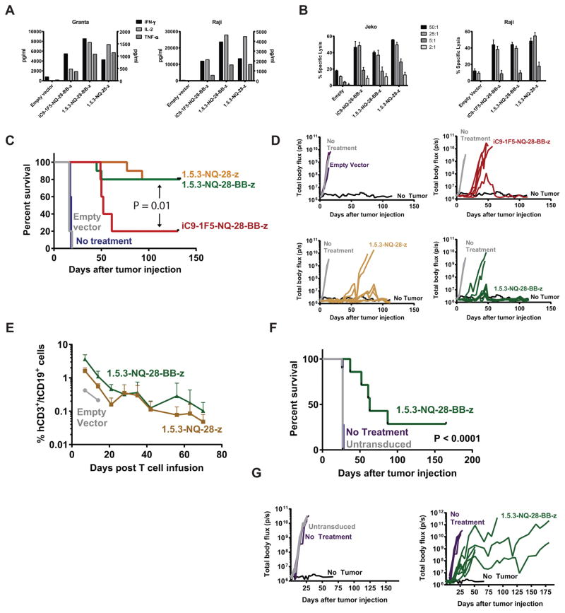
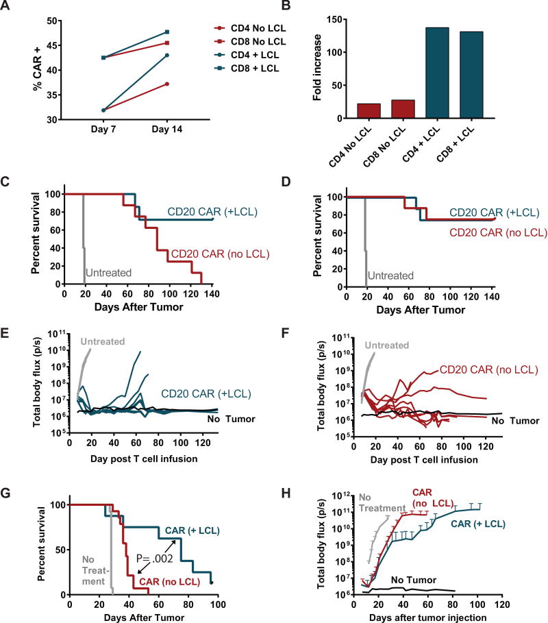
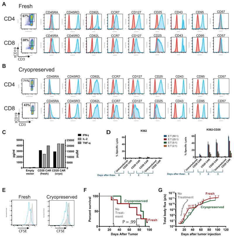
Chimeric antigen receptor (CAR)-based adoptive T-cell therapy is a highly promising treatment for lymphoid malignancies, and CD20 is an ideal target antigen. We previously developed a lentiviral construct encoding a third generation CD20-targeted CAR but identified several features that required additional optimization before clinical translation. We describe here several improvements, including replacement of the immunogenic murine antigen-binding moiety with a fully human domain, streamlining the transgene insert to enhance lentiviral titers, modifications to the extracellular IgG spacer that abrogate nonspecific activation resulting from binding to Fc receptors, and evaluation of CD28, 4-1BB, or CD28 and 4-1BB costimulatory domains. We also found that restimulation of CAR T cells with an irradiated CD20 cell line boosted cell growth, increased the fraction of CAR-expressing cells, and preserved in vivo function despite leading to a reduced capacity for cytokine secretion in vitro. We also found that cryopreservation of CAR T cells did not affect immunophenotype or in vivo antitumor activity compared with fresh cells. These optimization steps resulted in significant improvement in antitumor activity in mouse models, resulting in eradication of established systemic lymphoma tumors in 75% of mice with a single infusion of CAR T cells, and prolonged in vivo persistence of modified cells. These results provide the basis for clinical testing of a lentiviral construct encoding a fully human CD20-targeted CAR with CD28 and 4-1BB costimulatory domains and truncated CD19 (tCD19) transduction marker.


Figures

Figure 1
Figure 1. Modified extracellular spacer domain abrogates FcγR binding and improves in vivo CAR T cell function
(A & B) NSG mice were inoculated i.v. with Raji-ffLuc tumors followed 2 days later by infusion of 5 × 10⁶ CD20-targeted CAR+ T cells (TCM-enriched healthy donor T cells transduced with Leu16-28-BBz and re-stimulated with CD20+ LCL cells), with or without i.p. injection of 30 mg i.v immune globulin (IVIG) per mouse 4 hours prior to T cell infusion. Survival is shown in (A), with the curves compared using a log-rank (Mantel-Cox) test. Tumor burden as measured by bioluminescence imaging (mean ± SD of 3 mice per group) is shown in (B). Results are representative of 2 independent experiments. (C) CD64 (FcγRI) binding was tested using Jurkat cells transduced with a wild-type IgG1 Fc spacer CAR (iC9-SP163-1F5-IgG1-28-BB-z; “WT spacer”), CAR with spacer replacement of IgG1 hinge-linker by IgG2 hinge linker a.a. sequence (iC9-SP163-1F5-IgG1mut-28-BB-z; “IgG1mut”), and a CAR construct with deletion of the IgG1 hinge-linker (“No linker”). Transduced cells were identified by anti-CD19 antibody staining to detect the tCD19 transduction marker, and cells were stained with CD64, 3 days after transduction. The CD19+ population was gated, and frequencies of gated cells are shown in the histograms. (D) TCM T cells were transduced with Mock, WT spacer, IgG1mut, and No Linker constructs, and co-cultured with K562 or K562/CD64 in serum-free medium. To analyze T cell activation, cells were collected after 24 hours of co-culture and stained with CD3, CD19, CD25, and CD69. Frequencies of CD69+ and CD25+cells (gated on CD3+ CD19+ cells) are listed in the quadrants. (E) NSG mice were inoculated i.v. with Raji-ffLuc tumors followed 2 days later by infusion of 5 × 10⁶ CD20-targeted CAR+ T cells transduced with the following 1F5-based 3rd generation lentiviral vectors: WT spacer, the same vector but with CH2 domain deleted (“CH3 only”), or IgG1mut, or mokc-transduced T cells, and tumor burden was measured by bioluminescence over time (n=3 mice/group). (F) Mice were treated as in (E) but also included the No linker CAR and the IgG1mut CAR with N297Q mutation (iC9-SP163-1F5-NQ-28-BB-z; “IgGmut-NQ”). Kaplan-Meier survival curves are shown (n=5 mice/group) and compared using a log-rank test. Tumor burden curves for this experiment are shown in Figure S2 (Supplemental Digital Content).
Figure 2
Figure 2. Absence of iC9 gene improves transduction efficiency and function of CD20 CAR T cells
Healthy donor PBMC enriched for TCM were activated with anti-CD3/CD28 beads and transduced with a lentiviral vector containing either the iC9-1F5-NQ-28-BB-z construct or the same construct lacking the iC9 gene. Transgene expression was assessed by staining cells with anti-CD19 antibody to detect the tCD19 marker and analyzed by flow cytometry. (A) Histograms representing tCD19 expression of transduced cells (solid line) or untransduced control cells (filled histogram) are shown. The percent transduced cells is quantified in (B) and the geometric mean fluorescence intensity (MFI) of tCD19 expression is shown in (C). These results are representative of 3 independent experiments. (D) To measure cytokine secretion, TCM cells transduced with 1F5-NQ-28-BB-z or 1F5-NQ-28-z, with or without iC9 included, were co-incubated at a 1:1 ratio with irradiated Raji cells (left panel) or Jeko cells (right panel); supernatants were harvested 24 hours later and the indicated cytokines were measured by Luminex assay. IFN-γ and TNF-α are shown on the left y-axes and IL-2 is shown on the right y-axes. These results are representative of 2 independent experiments.
Figure 3
Figure 3. Fully human CD20 CAR exhibits potent in vitro and in vivo anti-tumor activity
Healthy donor central memory T cells (TCM) were stimulated with anti-CD3/CD28 beads and transduced the next day with iC9-1F5-NQ-28-BB-z, 1.5.3-NQ-28-BB-z, or 1.5.3-NQ-28-z lentiviral vectors, or a vector encoding only iC9 and tCD19 (“empty vector”), and expanded in vitro. (A) CAR-expressing cells were co-cultured in a 1:1 ratio with irradiated Granta-519 cells (left) or Raji-ffLuc cells (right), and supernatants were harvested 24 hours later and analyzed for the indicated cytokines using a Luminex assay. IL-2 and TNF-α (left axis) and IFN-γ (right axis) levels are shown. (B) Cytotoxicity was measured by using these cells as effectors in a standard 4-hour 51Cr-release assay against either Jeko (left) or Raji-ffLuc (right) cell lines. (C and D) NSG mice were injected i.v. with 5 × 10⁵ Raji-ffLuc cells followed 2 days later by 5 × 10⁶ cells transduced with the vectors described in (A). A Kaplan-Meier curve for overall survival is shown in (C) and tumor burden over time of individual mice as measured by bioluminescence is shown in (D). The survival curves for iC9_1F5-NQ-28-BB-z, 1.5.3-NQ-28-BB-z, and 1.5.3-NQ-28-z were compared using a log-rank (Mantel-Cox) test. (E) Retroorbital blood samples of mice treated with T cells transduced with 1.5.3-NQ-28-z, 1.5.3-NQ-28-BB-z, or an empty vector in the experiment described in C and D were analyzed by flow cytometry for circulating infused CAR T cells. The percentage of cells in the mCD45 hCD3+ hCD19+ gate over time are shown (n = 2 mice per group). (F) NSG mice were injected i.v. with 5 × 10⁵ Granta-ffLuc cells followed 2 days later by either 5 × 10⁶ tCD19+ cells transduced with the 1.5.3-NQ-28-BB-z or an equal number of untransduced cells. A Kaplan-Meier curve for overall survival is shown in (F), with curves compared using a log-rank test, and tumor burden over time of individual mice as measured by bioluminescence is shown in (G).
Figure 4
Figure 4. Re-stimulation with CD20+ cells in culture improves CAR expression, growth, and preserves in vivo anti-tumor activity
PBMC from healthy donors were enriched for CD4 or CD8 cells by MACS positive selection, and each subset was separately activated with anti-CD3/CD28 beads and transduced 24 hours later with 1.5.3-NQ-28-BB-z lentiviral supernatant. At day 7, the cells were either re-stimulated with an irradiated CD20+ transformed B cell line (TM-LCL) or continued in culture without re-stimulation. (A) CAR expression of each cell culture at day 7 and day 14, as assessed by anti-human IgG Fc antibody (binds to IgG1 spacer region of CAR). (B) The cell growth of each cell culture up to day 14 is shown, expressed as fold increase from baseline. (C) NSG mice were injected i.v. with 5 × 10⁵ Raji-ffLuc cells, followed 7 days later by infusion of 5 × 10⁶ tCD19+ T cells at a 1:1 ratio of CD4+ tCD19+ : CD8+ tCD19+ cells. Groups received either CAR T cells that had been re-stimulated with TM-LCL (n=7 after excluding one mouse that died at day 12 during anesthesia for imaging), CAR T cells without re-stimulation (n=8), or no treatment (n=5). (C) Overall survival of each group, represented by a Kaplan-Meier curve. (D) Lymphoma-specific survival, including deaths due only to lymphoma progression and excluding deaths from xenogeneic graft-versus-host disease. (E) Tumor burden over time of mice receiving TM-LCL-re-stimulated CAR T cells as measured by bioluminescence imaging. (F) Tumor burden over time of mice receiving non-restimulated CAR T cells as measured by bioluminescence imaging. (G and H) NSG mice were injected i.v. with 5 × 10⁵ Granta-ffLuc cells, followed 7 days later by infusion of 5 × 10⁶ T cells at a 1:1 ratio of CD4+ tCD19+ : CD8+ tCD19+ cells. Groups received either CAR T cells that had been re-stimulated with TM-LCL (n=8), CAR T cells without re-stimulation (n=14), or no treatment (n=13). Overall survival is shown in (G), and tumor burden over time as measured by bioluminescence imaging is shown in (H). Differences between LCL and No LCL survival curves were assessed with a log-rank test.
Figure 5
Figure 5. Phenotype and function of CD20 CAR T cells are preserved following cryopreservation
(A–D) Healthy donor PBMC positively selected for CD4 or CD8 were separately stimulated with anti-CD3/CD28 beads and transduced with lentiviral vector encoding the 1.5.3-NQ-28-BB-z CAR. Cells were re-stimulated with CD20+ TM-LCL cells on day 7, and expanded until day 14, and then either kept in culture (fresh) or formulated in Cryostor medium, placed in a step-down freezer for 3 hours, transferred to liquid nitrogen overnight, and thawed the next day (cryopreserved). Fresh (A) or cryopreserved (B) T cells were evaluated by flow cytometry for the indicated cell surface markers to assess phenotypic markers of differentiation/memory (CD45RA, CD45RO, CD62L, CCR7, CD127), activation (CD25, CD43, CD95), or exhaustion (CD57). Cells were gated on CD3+ tCD19+ cells, and the number of tCD19+ cells, as a surrogate for CAR expression, is shown in the first panel. (C) Fresh or cryopreserved tCD19+ cells were co-cultured at a 1:1 ratio with irradiated Raji-ffLuc cells, and supernatants were harvested 24 hours later and the indicated cytokines measured by Luminex assay. IL-2 values are shown on the right y-axis, and IFN-γ and TNF-α levels are shown on the left y-axis. (D) Fresh or cryopreserved cells were used as effectors in a standard 4-hour 51Cr-release assay using either CD20+ K562 cells (right panel) or parental K562 cells lacking CD20 (left panel) as targets, at the indicated effector:target (E:T) ratios. The data represent the mean (+ SD) of triplicate values. (E) Fresh (left panel) or cryopreserved (right panel) CD8+ CAR+ or untransduced T cells were labeled with CFSE, co-cultured with irradiated Raji-ffLuc cells for 4 days, and then CFSE dilution was evaluated by flow cytometry. CAR+ cells (blue histograms) were gated on CD3+ tCD19+ cells, and untransduced cells (gray filled histograms) were gated on CD3+ cells. (F–G) NSG mice were injected i.v. with 5 × 10⁵ Granta-ffLuc cells, followed 7 days later by infusion of 5 × 10⁶ T cells at a 1:1 ratio of CD4+ tCD19+ : CD8+ tCD19+ cells. Groups received either fresh (red line, n=8) or cryopreserved (green line, n=5) cells (n=5), or no treatment (gray line, n=13). Overall survival is shown in (F), and tumor burden over time as measured by bioluminescence imaging is shown in (G). Differences between fresh and cryopreserved survival curves were assessed with a log-rank test.

References

    1. Press OW, Howell-Clark J, Anderson S, et al. Retention of B-cell-specific monoclonal antibodies by human lymphoma cells. Blood. 1994;83(5):1390–7. - PubMed
    1. Bubien JK, Zhou LJ, Bell PD, et al. Transfection of the CD20 cell surface molecule into ectopic cell types generates a Ca2+ conductance found constitutively in B lymphocytes. J Cell Biol. 1993;121(5):1121–32. - PMC - PubMed
    1. Tedder TF, Engel P. CD20: a regulator of cell-cycle progression of B lymphocytes. Immunol Today. 1994;15(9):450–4. - PubMed
    1. Chang KL, Arber DA, Weiss LM. CD20: A Review. Applied Immunohistochem. 1996;4:1–15.
    1. Press OW, Farr AG, Borroz KI, et al. Endocytosis and degradation of monoclonal antibodies targeting human B- cell malignancies. Cancer research. 1989;49(17):4906–12. - PubMed

Publication types

MeSH terms