Review
. 2018 Jan;14(1):57-70.
doi: 10.1038/nrneph.2017.155. Epub 2017 Nov 27.

Dyslipidaemia in nephrotic syndrome: mechanisms and treatment

Affiliations
Review

Dyslipidaemia in nephrotic syndrome: mechanisms and treatment

Shipra Agrawal et al. Nat Rev Nephrol. 2018 Jan.

Erratum in

Abstract

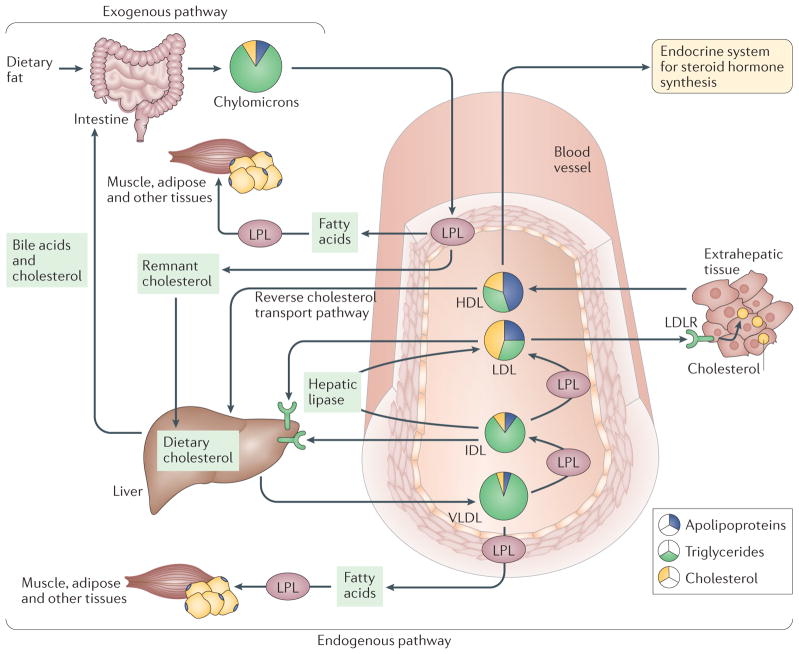
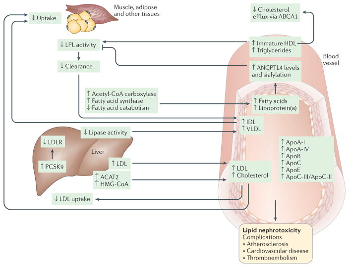
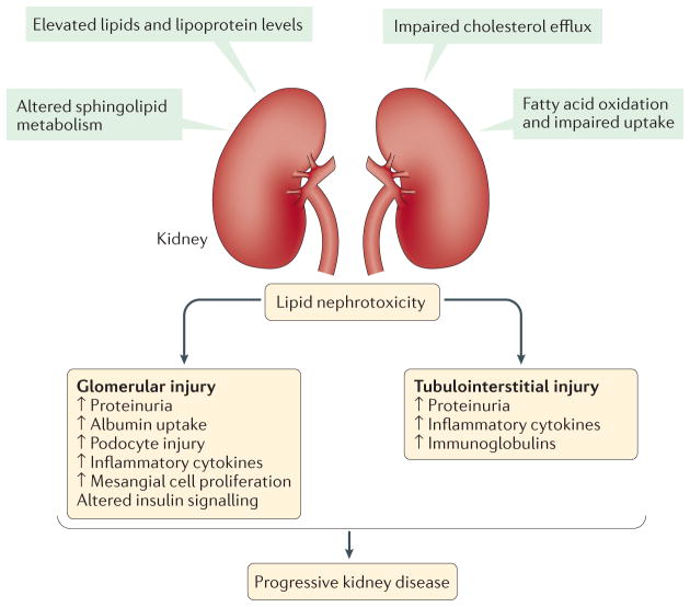
Nephrotic syndrome is a highly prevalent disease that is associated with high morbidity despite notable advances in its treatment. Many of the complications of nephrotic syndrome, including the increased risk of atherosclerosis and thromboembolism, can be linked to dysregulated lipid metabolism and dyslipidaemia. These abnormalities include elevated plasma levels of cholesterol, triglycerides and the apolipoprotein B-containing lipoproteins VLDL and IDL; decreased lipoprotein lipase activity in the endothelium, muscle and adipose tissues; decreased hepatic lipase activity; and increased levels of the enzyme PCSK9. In addition, there is an increase in the plasma levels of immature HDL particles and reduced cholesterol efflux. Studies from the past few years have markedly improved our understanding of the molecular pathogenesis of nephrotic syndrome-associated dyslipidaemia, and also heightened our awareness of the associated exacerbated risks of cardiovascular complications, progressive kidney disease and thromboembolism. Despite the absence of clear guidelines regarding treatment, various strategies are being increasingly utilized, including statins, bile acid sequestrants, fibrates, nicotinic acid and ezetimibe, as well as lipid apheresis, which seem to also induce partial or complete clinical remission of nephrotic syndrome in a substantial percentage of patients. Future potential treatments will likely also include inhibition of PCSK9 using recently-developed anti-PCSK9 monoclonal antibodies and small inhibitory RNAs, as well as targeting newly identified molecular regulators of lipid metabolism that are dysregulated in nephrotic syndrome.


Conflict of interest statement

Competing interests statement

The authors declare that they have no competing interests.

Figures

Figure 1
Figure 1. The major pathways of lipid metabolism
Lipoproteins are the major carriers of lipids in the circulation and they participate in three major pathways that are responsible for the generation and transport of lipids within the body. The two major forms of circulating lipid in the body, triglycerides and cholesterol, are packaged with apolipoproteins and phospholipids to form lipoproteins. The major forms of lipoproteins are chylomicrons, very low-density lipoprotein (VLDL), intermediate density lipoprotein (IDL), low density lipoprotein (LDL) and high density lipoprotein (HDL), and they differ in their size, density, composition and functions (detailed in TABLE 1). In the exogenous pathway, dietary lipids, which consist mainly of triglycerides (95%) and some phospholipids, free fatty acids and cholesterol, are packaged into chylomicrons by intestinal mucosal cells. These chylomicrons enter the lymphatic system and then the circulation, where triglycerides are released as free fatty acids by lipoprotein lipase (LPL) activity on the capillary endothelium. These free fatty acids are taken up by the muscle, adipose and other peripheral tissues, whereas the remnants of chylomicrons are cleared by the liver. In the endogenous pathway, the liver produces VLDL, which interacts with LPL in the circulation to form IDL, with the release of triglyceride and free fatty acids. IDL is rapidly removed by the liver via the interaction of its apolipoprotein E component with LDL receptor (LDLR). Furthermore, IDL forms LDL upon removal of triglyceride by hepatic lipase. LDL, which is very high in cholesterol content, is in turn removed from the circulation by binding to LDLR in the liver and in extrahepatic tissues. HDL is an anti-atherogenic lipoprotein or ‘good cholesterol’, as it captures the cholesterol from peripheral tissues or other lipoproteins and transports it back to liver by the third pathway, which is termed reverse cholesterol transport.
Figure 2
Figure 2. Pathophysiology of dyslipidaemia in nephrotic syndrome
Alterations of lipid and lipoprotein metabolism in nephrotic syndrome result in ‘lipid nephrotoxicity’ and other complications, such as atherosclerosis, cardiovascular disease and thromboembolism. The major lipoproteins, including intermediate density lipoprotein (IDL), very low density lipoprotein (VLDL) and low-density lipoprotein (LDL), and cholesterol are increased in the plasma of patients with nephrotic syndrome, owing mainly to impaired clearance and, to a lesser extent, increased biosynthesis. Impaired clearance is a direct result of decreased hepatic lipase activity and decreased lipoprotein lipase (LPL) activity in the endothelium and peripheral tissues, such as muscle and adipose. In addition, hepatic levels of proprotein convertase subtilisin/kexin type 9 (PCSK9) are increased in patients with nephrotic syndrome; PCSK9 degrades the LDL receptor (LDLR), and is thus a major therapeutic target for lipid lowering. Furthermore, the composition and function of the lipoproteins are also altered, with substantial increases in the plasma levels of apolipoprotein A I (ApoA I), ApoA IV, ApoB, ApoC and ApoE, and in the ApoC III/ApoC II ratio. The level of immature HDL in the plasma is also increased, resulting in reduced cholesterol efflux, which occurs mainly via ATP binding cassette subfamily A member 1 (ABCA1), in peripheral organs, including in podocytes. Another major lipid abnormality in nephrotic syndrome is hypertriglyceridaemia, as well as increased production and sialylation of ANGPTL4, which is driven primarily by increased circulating free fatty acids. ANGPTL4 in turn suppresses LPL activity by either preventing its dimerization or by inhibiting its activity noncompetitively. Hypercholesterolaemia and increased LDL levels occur in conjunction with upregulation of the expression and activity of liver acetyl CoA acetyltransferase 2 (ACAT2), which results in enhanced esterification of cholesterol and a reduction in the level of intracellular free cholesterol. Cholesterol synthesis via 3 hydroxy 3 methylglutaryl CoA (HMG CoA) reductase is also increased in experimental models of nephrotic syndrome. Evidence also exists that LDL oxidation in nephrotic syndrome may be augmented by lipoprotein(a), the level of which is also increased in patients with nephrotic syndrome. Accumulation of oxidized LDL, IDL and chylomicron remnants stimulates monocytes and macrophages to release proin ammatory cytokines and chemokines, and accelerates in ammation, which may in turn promote the progression of chronic kidney disease.
Figure 3
Figure 3. Mechanisms and consequences of lipid nephrotoxicity
The direct effects of dyslipidaemia on decreased kidney function are referred to as ‘lipid nephrotoxicity’, although the role of altered lipid metabolism in the molecular pathophysiology of nephrotic syndrome is not well understood. During dyslipidaemia, triglyceride-rich lipoproteins, such as very low density lipoprotein (VLDL) and intermediate density lipoprotein (IDL) as well as oxidized LDL, are taken up by mesangial cells, leading to the production of cytotoxic agents, cytokines and reactive oxygen species, which further damage the glomerular epithelial and endothelial cells, resulting in sclerosis. Furthermore, levels of free fatty acids are increased in patients with nephrotic syndrome, which have been reported to have toxic effects in the kidney, especially in glomeruli and podocytes, but also in the tubulointerstitium. Free fatty acids bound to albumin cause podocyte damage by enhancing macropinocytosis and activating G protein-coupled receptor (GPCR) signalling, leading to disruption of the podocyte actin cytoskeleton and podocyte morphology. In addition, these albumin bound free fatty acids cause loss of podocyte viability and increased production of several cytokines. Moreover, free cholesterol-mediated injury is another pathway of cellular injury in podocytes, and involves the ATP binding cassette sub family A member 1 (ABCA1) cholesterol transporter. Furthermore, the role of free fatty acids, and saturated fatty acids in particular, is well documented in causing damage to proximal tubule cells and tubulointerstitial injury.

References

    1. Greenbaum LA, Benndorf R, Smoyer WE. Childhood nephrotic syndrome — current and future therapies. Nat Rev Nephrol. 2012;8:445–458. - PubMed
    1. Hull RP, Goldsmith DJ. Nephrotic syndrome in adults. BMJ. 2008;336:1185–1189. - PMC - PubMed
    1. Clark AG, Barratt TM. In: Pediatric Nephrology. Barratt TM, Avner ED, Harmon WE, editors. Lippincott Williams & Wilkins; 1998. pp. 731–747.
    1. McEnery PT, Strife CF. Nephrotic syndrome in childhood. Management and treatment in patients with minimal change disease, mesangial proliferation, or focal glomerulosclerosis. Pediatr Clin North Am. 1982;89:875–894. - PubMed
    1. Nash MA, Edelmann CMJ, Bernstein J, Barnett HL. In: Pediatric Kidney Disease. Edelmann CMJ, editor. Little; 1992. pp. 1247–1266.

MeSH terms