. 2017 Nov 24;7(1):16214.
doi: 10.1038/s41598-017-15376-8.

Mesenchymal stem cells derived exosomes and microparticles protect cartilage and bone from degradation in osteoarthritis

Affiliations

Mesenchymal stem cells derived exosomes and microparticles protect cartilage and bone from degradation in osteoarthritis

Stella Cosenza et al. Sci Rep. .

Abstract

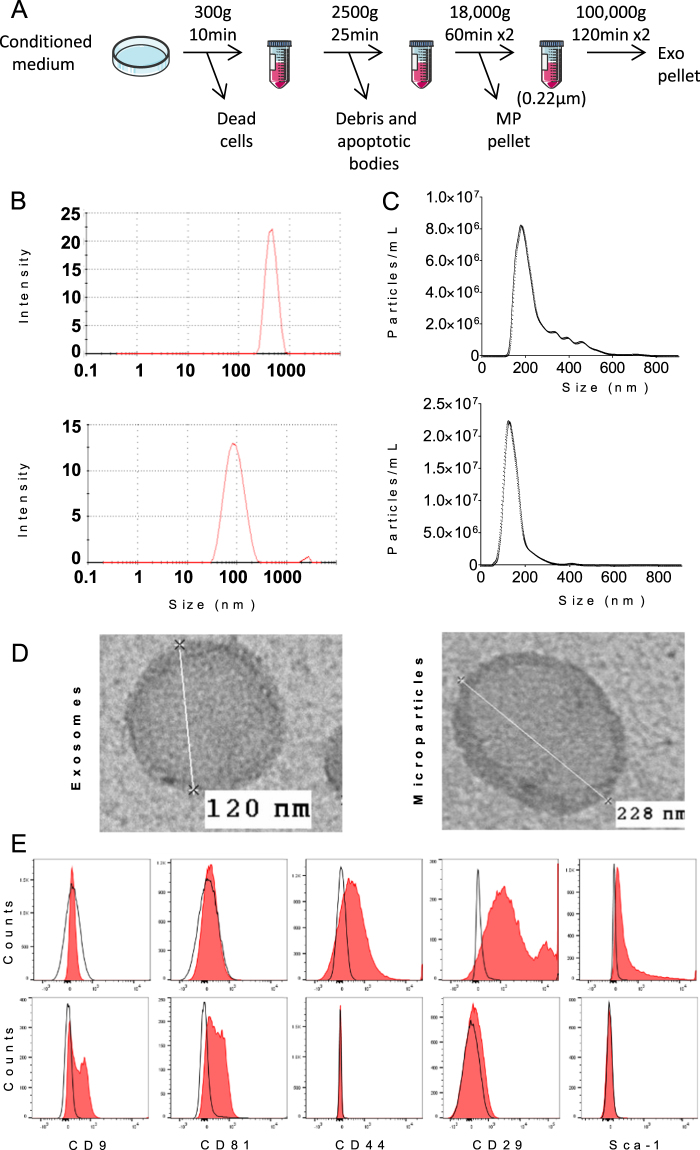
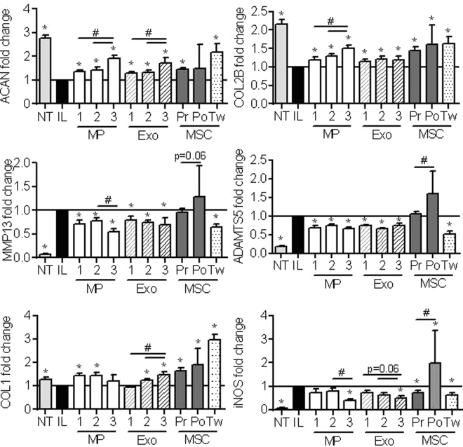
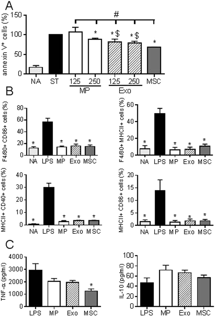
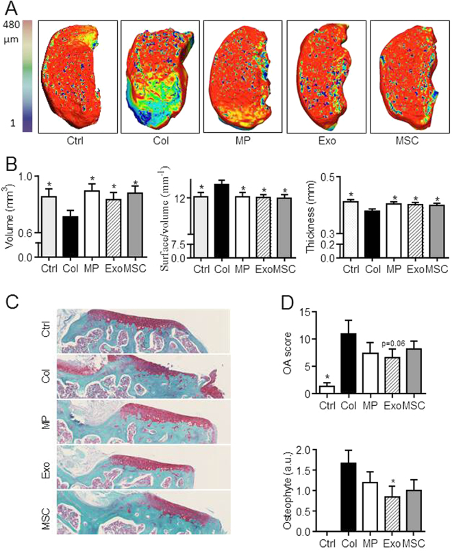
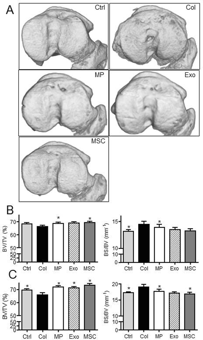
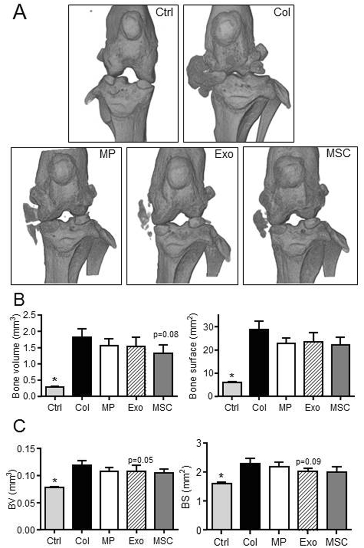
Mesenchymal stem or stromal cells (MSCs) exert chondroprotective effects in preclinical models of osteoarthritis (OA). Most of their therapeutic effects are mediated via soluble mediators, which can be conveyed within extracellular vesicles (EVs). The objective of the study was to compare the respective role of exosomes (Exos) or microvesicles/microparticles (MPs) in OA. MPs and Exos were isolated from bone marrow murine BM-MSCs through differential centrifugation. Effect of MPs or Exos was evaluated on OA-like murine chondrocytes and chondroprotection was quantified by RT-qPCR. In OA-like chondrocytes, BM-MSC-derived MPs and Exos could reinduce the expression of chondrocyte markers (type II collagen, aggrecan) while inhibiting catabolic (MMP-13, ADAMTS5) and inflammatory (iNOS) markers. Exos and MPs were also shown to protect chondrocytes from apoptosis and to inhibit macrophage activation. In vivo, Exos or MPs were injected in the collagenase-induced OA (CIOA) model and histomorphometric analyses of joints were performed by µCT and confocal laser microscopy. BM-MSCs, MPs and Exos equally protected mice from joint damage. In conclusion, MPs and Exos exerted similar chondroprotective and anti-inflammatory function in vitro and protected mice from developing OA in vivo, suggesting that either Exos or MPs reproduced the main therapeutic effect of BM-MSCs.


Conflict of interest statement

The authors declare that they have no competing interests.

Figures

Figure 1
Figure 1
Isolation and characterization of extracellular vesicles isolated from murine BM-MSCs. (A) Experimental protocol for isolation of microparticles (MPs) and exosomes (Exos) using differential ultracentrifugation. (B) Size of MPs (up) and Exos (down) detected in 200 µL by Dynamic Light Scattering analysis (C) Number and size of MPs (up) and Exos (down) detected in 1 mL (corresponding to 1 µg EV equivalent proteins) by Nano Tracking Analysis. (D) Representative images of Exos and MPs by transmission electron microscopy. (E) Expression of BM-MSC membrane markers (Sca-1, CD44, CD29) and of exosomal markers (CD9, CD81) on MPs (top) and Exos (bottom) isolated from naïve BM-MSCs as analysed by flow cytometry.
Figure 2
Figure 2
MPs and Exos isolated from TGFβ3-pretreated BM-MSCs exerted high chondroprotective effect on OA-like chondrocytes. Primary murine chondrocytes were pretreated with 1 ng/mL IL-1β (IL) or not (NT) for 24 h before addition of different amounts of MPs or Exos (1: 12.5 ng; 2: 125 ng; 3: 1.25 µg), 1 mL TGFβ3-pretreated pre-centrifugation (Pr) or post-ultracentrifugation (Po) BM-MSC-CM or TGFβ3-pretreated BM-MSCs (105 cells) on top of a transwell membrane (Tw). Expression of chondrocyte markers was quantified by RT-qPCR after 24h (n = 10). *: p < 0.05 as compared to IL1-β-treated OA-like chondrocytes; #: p < 0.05 as compared to indicated groups.
Figure 3
Figure 3
BM-MSC-derived MPs and Exos exerted anti-apoptotic effect on chondrocytes and immunosuppressive function on macrophages. (A) Percentage of annexin V+ apoptotic chondrocytes under non activated conditions (NA), staurosporine–induced apoptosis (ST) and cultured with Exos or MPs (125 ng or 250 ng) or 1 mL BM-MSC-conditioned medium (n = 5). *: p < 0.05 as compared to ST group, #: p < 0.05 as compared to BM-MSC group or $: p<0.05 s compared to MP group of same amount. (B) Expression of differentiation markers on non-activated CD11b+ macrophages (NA), on macrophages after LPS-induced activation cultured alone (LPS) or with 50 ng MPs, Exos or BM-MSCs (1 BM-MSC/5 macrophages) for 3 days (n = 4). (C) Quantification of cytokines produced by macrophages after 3 days as described in (B). *: p < 0.05 as compared to LPS.
Figure 4
Figure 4
BM-MSC-derived MPs and Exos protected mice from osteoarthritic damages in the collagenase-induced OA model. (A) Representative 3D reconstructed images of articular cartilage after confocal laser scanning microscopy analysis. Images from control mice (Ctrl), collagenase-treated mice (Col) and Col mice that received intra-articular injection of 500 ng MPs or 250 ng Exos or 2.5 × 105 BM-MSCs. (B) Histomorphometric analysis of 3D images of articular cartilages as described in (A) (n = 15). (C) Representative histological sections of tibias from mice described in (A) after Safranin O-Fast green staining. (D) OA score and osteophyte score expressed as arbitrary unit (a.u.) on histological sections of mice. *: p < 0.05 as compared to Col group.
Figure 5
Figure 5
BM-MSC-derived MPs and Exos protected mice from osteoarthritic damages in the collagenase-induced OA model. (A) Representative 3D reconstructed images of sub-chondral bone surface in tibias after µCT analysis. Images from control mice (Ctrl), collagenase-treated mice (Col) and Col mice that received intra-articular injection of 500 ng MPs or 250 ng Exos or 2.5 × 105 BM-MSCs. (B) Histomorphometric analysis of 3D images of epiphyseal bone: Bone volume/tissue volume (BV/TV) and bone surface/bone volume (BS/BV) parameters (n = 15). (C) Histomorphometric analysis of sub-chondral bone. *: p < 0.05 as compared to Col group.
Figure 6
Figure 6
BM-MSC-derived MPs and Exos protected mice from osteoarthritic damages in the collagenase-induced OA model. Images from control mice (Ctrl), collagenase-treated mice (Col) and Col mice that received intra-articular injection of 500 ng MPs or 250 ng Exos or 2.5 × 105 BM-MSCs. (A) Representative 3D reconstructed images of bone knee joints. (B) Bone volume and bone area as measured in the menisci and external ligaments. (C) Histomorphometric analysis of osteophytes at the edges of joint bone. *: p < 0.05 as compared to Col group.

References

    1. Palazzo C, Ravaud JF, Papelard A, Ravaud P, Poiraudeau S. The burden of musculoskeletal conditions. PLoS One. 2014;9:e90633. doi: 10.1371/journal.pone.0090633. - DOI - PMC - PubMed
    1. Ter Huurne M, et al. Antiinflammatory and chondroprotective effects of intraarticular injection of adipose-derived stem cells in experimental osteoarthritis. Arthritis Rheum. 2012;64:3604–3613. doi: 10.1002/art.34626. - DOI - PubMed
    1. Toupet K, et al. Survival and biodistribution of xenogenic adipose mesenchymal stem cells is not affected by the degree of inflammation in arthritis. PLoS One. 2015;10:e0114962. doi: 10.1371/journal.pone.0114962. - DOI - PMC - PubMed
    1. Murphy JM, Fink DJ, Hunziker EB, Barry FP. Stem cell therapy in a caprine model of osteoarthritis. Arthritis Rheum. 2003;48:3464–3474. doi: 10.1002/art.11365. - DOI - PubMed
    1. Desando G, et al. Intra-articular delivery of adipose derived stromal cells attenuates osteoarthritis progression in an experimental rabbit model. Arthritis Res Ther. 2013;15:R22. doi: 10.1186/ar4156. - DOI - PMC - PubMed

Publication types

MeSH terms