. 2017 Nov 27;12(11):e0188250.
doi: 10.1371/journal.pone.0188250. eCollection 2017.

Maternal-Fetal rejection reactions are unconstrained in preeclamptic women

Affiliations

Maternal-Fetal rejection reactions are unconstrained in preeclamptic women

Tina A Nguyen et al. PLoS One. .

Abstract

The risk factors for preeclampsia, extremes of maternal age, changing paternity, concomitant maternal autoimmunity, and/or birth intervals greater than 5 years, suggest an underlying immunopathology. We used peripheral blood and lymphocytes from the UteroPlacental Interface (UPI) of 3rd trimester healthy pregnant women in multicolor flow cytometry-and in vitro suppression assays. The major end-point was the characterization of activation markers, and potential effector functions of different CD4-and CD8 subsets as well as T regulatory cells (Treg). We observed a significant shift of peripheral CD4 -and CD8- T cells from naïve to memory phenotype in preeclamptic women compared to healthy pregnant women consistent with long-standing immune activation. While the proportions of the highly suppressive Cytokine and Activated Treg were increased in preeclampsia, Treg tolerance toward fetal antigens was dysfunctional. Thus, our observations indicate a long-standing inflammatory derangement driving immune activation in preeclampsia; in how far the Treg dysfunction is caused by/causes this immune activation in preeclampsia will be the object of future studies.


Conflict of interest statement

Competing Interests: The authors have declared that no competing interests exist.

Figures

Fig 1
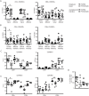
Fig 1. Naïve CD4+ T cells in the peripheral blood of preeclamptic pregnant women are significantly reduced.
Fresh peripheral blood and UPI samples from healthy 3rd trimester pregnant women and preeclamptic women were processed and analyzed as described in Materials and Methods. Gated starting with Fig 1B lymphocyte gate and subjected to Fig 1A Steps 2–5. (A, B) CD4+ (left side) or CD8+ (right side), CCR7+ or–, CD45RA+ (A) or CD45RO+ (B) sub-populations were identified as previously described[7]. (C, E) Intracellular cytokine staining of naïve (C) and memory (E) CD4+ and CD8+ T cells. PB (circles) or UPI lymphocytes (squares) were cultured for 4.5h as described in Materials and Methods including PMA/Ionomycin and Golgi Plug treatment. (E) Number of IL-2+ CD4 T cells as determined in C) (both left and right) per CD8+ T cell in the same patient in the PB (circles) and UPI (squares) of healthy (solid) or preeclamptic (open) patients. Student’s T test, unpaired *P<0.05, **P<0.009, ***P<0.0005.
Fig 2
Fig 2. Memory T cells from preeclamptic patients do not display surface markers of exhaustion.
Fresh peripheral blood and UPI samples from healthy 3rd trimester pregnant women and preeclamptic women was processed and analyzed as described in materials and methods. CD28 (A) and CD27 (B) percentage of CD4+ memory (left side) or CD8+ memory (right side), CCR7+ or—cells was determined[7]. Student’s T test, unpaired.
Fig 3
Fig 3. Regulatory T cells of preeclamptic women fail to suppress anti-fetal proliferative responses.
(A) Gating strategy for Treg identification. Gating strategy starting with Fig 1B lymphocyte gate and subjecting to S1A Fig Steps until identification of CD4+ T cells. (B) Proportion of total FoxP3+ Treg gated off the total CD4+ populations. (C) % of three Treg subtypes gated off the total CD4+ populations in the PB (left hand side) and UPI (right hand side). (D) Number of CD8+ T cells divided by Cytokine+ Activated Treg as determined in C. (E-G) Proliferative response of maternal PBL to stimulation with related- or unrelated lymphocytes. Donor PBL are exposed to lymphocytes from the indicated sources with (squares) or without (triangles) depletion of CD25+ cells. (E) Donor PBL of a healthy, non-pregnant woman are exposed to unrelated cord blood lymphocytes, (F) PBL of a healthy pregnant woman are exposed to cord blood lymphocytes from her baby and (G) PBL from a preeclamptic pregnant woman are exposed to cord blood lymphocytes from her baby. (H) Fold change in proliferation of healthy (circles) or preeclamptic (squares) pregnant women’s PBL exposed to cord blood lymphocytes of their babies after CD25-depletion. N = 8 normal pregnant women, N = 6 preeclamptic pregnant women total in two separate experiments, representative results are depicted. Statistical analysis in (B, C, D, H) used two-way Student’s T test, unpaired. (E-G) Statistical analysis used one-way ANOVA with Bonferroni post-test. In all test, *p<0.05, **p<0.01.

References

    1. Cudihy D, Lee RV. The pathophysiology of pre-eclampsia: current clinical concepts. J Obstet Gynaecol. 2009;29(7):576–82. Epub 2009/09/17. doi: 10.1080/01443610903061751 . - DOI - PubMed
    1. Siddiqui AH, Irani RA, Blackwell SC, Ramin SM, Kellems RE, Xia Y. Angiotensin receptor agonistic autoantibody is highly prevalent in preeclampsia: correlation with disease severity. Hypertension. 2010;55(2):386–93. Epub 2009/12/10. doi: 10.1161/HYPERTENSIONAHA.109.140061 . - DOI - PMC - PubMed
    1. Irani RA, Zhang Y, Blackwell SC, Zhou CC, Ramin SM, Kellems RE, et al. The detrimental role of angiotensin receptor agonistic autoantibodies in intrauterine growth restriction seen in preeclampsia. J Exp Med. 2009;206(12):2809–22. Epub 2009/11/06. doi: 10.1084/jem.20090872 . - DOI - PMC - PubMed
    1. Tal R. The role of hypoxia and hypoxia-inducible factor-1α in preeclampsia pathogenesis. Biology of reproduction. 2012;87(6):134 doi: 10.1095/biolreprod.112.102723 . - DOI - PubMed
    1. Lau SY, Guild SJ, Barrett CJ, Chen Q, McCowan L, Jordan V, et al. Tumor necrosis factor-α, interleukin-6, and interleukin-10 levels are altered in preeclampsia: a systematic review and meta-analysis. American journal of reproductive immunology. 2013;70(5):412–27. doi: 10.1111/aji.12138 . - DOI - PubMed