. 2017 Nov 27;13(11):e1006704.
doi: 10.1371/journal.ppat.1006704. eCollection 2017 Nov.

Vaccine-elicited memory CD4+ T cell expansion is impaired in the lungs during tuberculosis

Affiliations

Vaccine-elicited memory CD4+ T cell expansion is impaired in the lungs during tuberculosis

Stephen M Carpenter et al. PLoS Pathog. .

Abstract

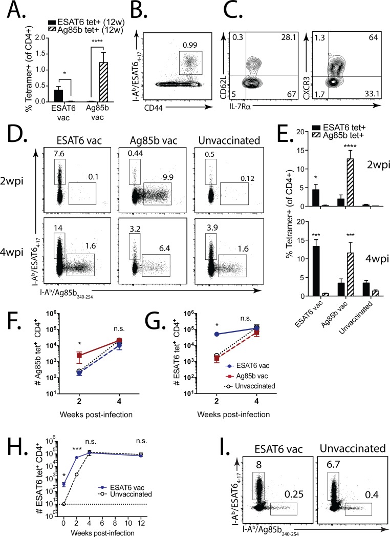
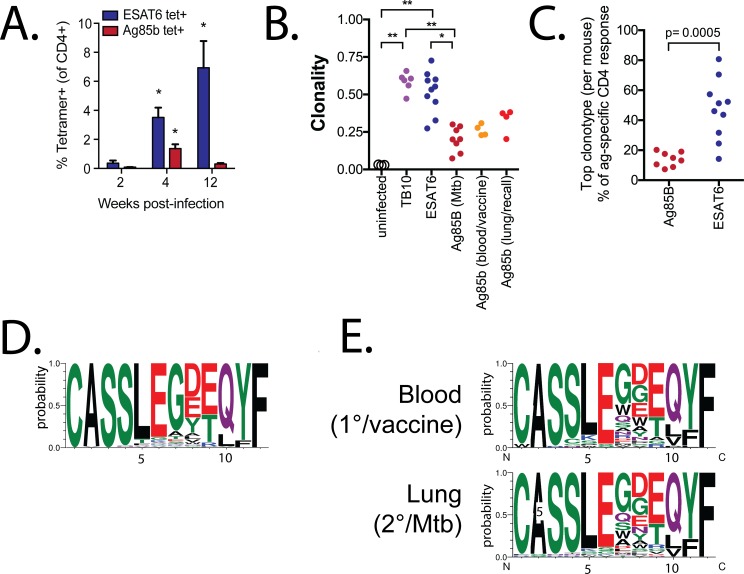
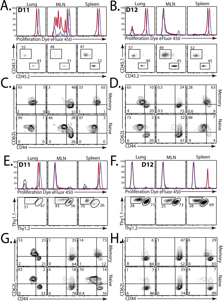
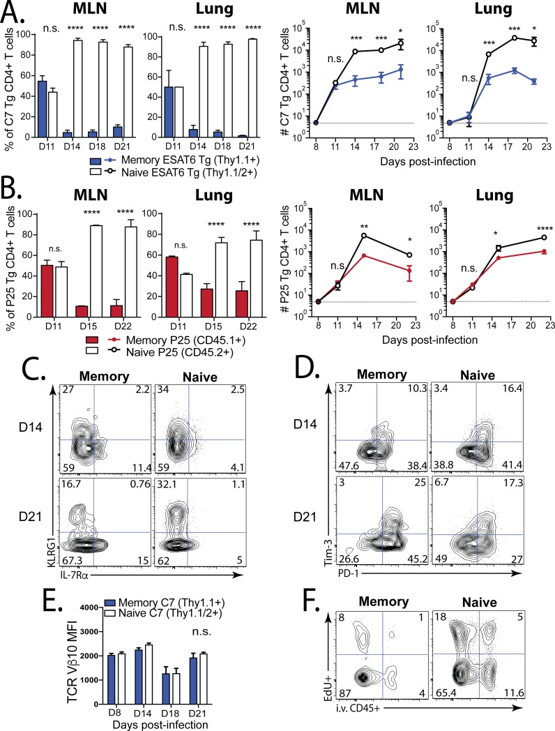
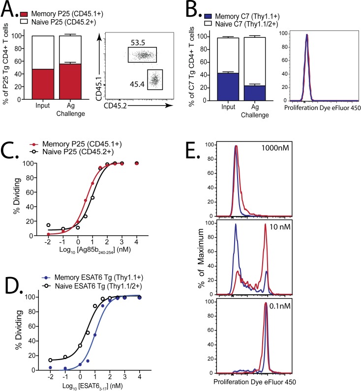
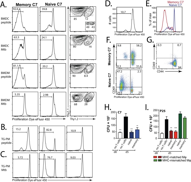
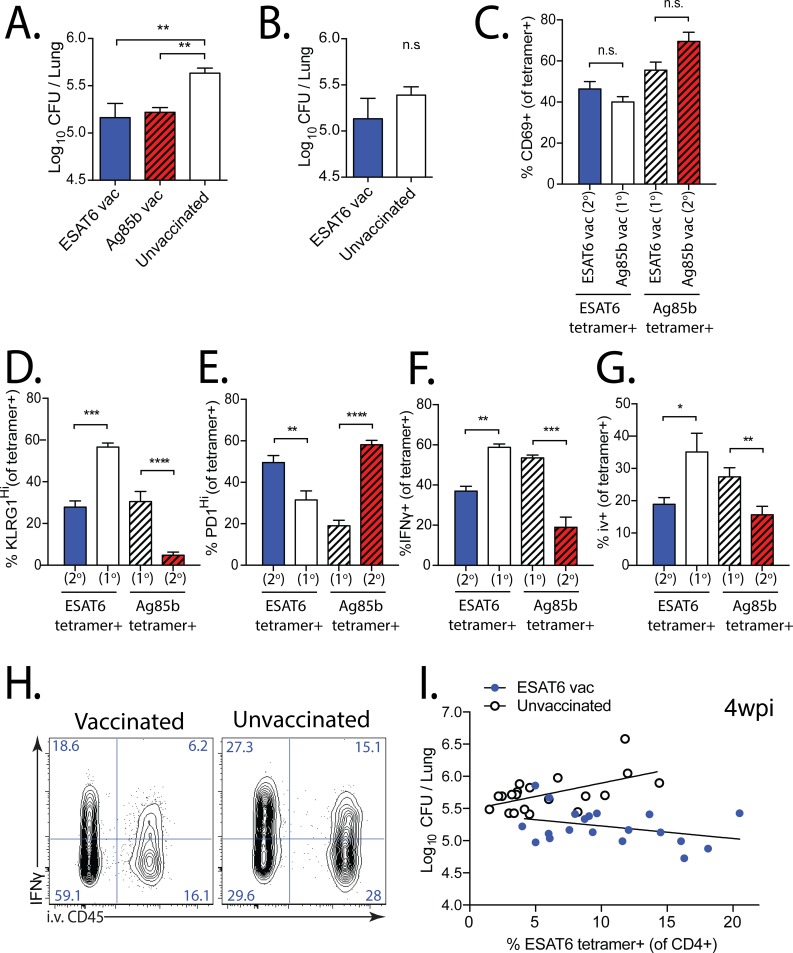
Immunological memory is the key biological process that makes vaccines possible. Although tuberculosis vaccines elicit protective immunity in animals, few provide durable protection. To understand why protection is transient, we evaluated the ability of memory CD4+ T cells to expand, differentiate, and control Mycobacterium tuberculosis. Both naïve and memory CD4+ T cells initially proliferated exponentially, and the accumulation of memory T cells in the lung correlated with early bacterial control. However, later during infection, memory CD4+ T cell proliferation was curtailed and no protection was observed. We show that memory CD4+ T cells are first activated in the LN and their recruitment to the lung attenuates bacterial growth. However, their interaction with Mtb-infected macrophages does not promote continued proliferation. We conclude that a lack of sustained expansion by memory-derived T cells in the lung limits the durability of their protection, linking their slower expansion with transient protection in vaccinated mice.


Conflict of interest statement

The authors have declared that no competing interests exist.

Figures

Fig 1
Fig 1. The augmented antigen-specific CD4+ T cell response in vaccinated mice is not durable after Mtb challenge.
(a) ESAT64-17 and Ag85b240-254 tetramer+ T cell responses in the blood 12 weeks after ESAT64-17 or Ag85b240-254 s.c. peptide vaccination. (b) Representative ESAT64-17 tetramer responses in the lung 12 weeks after ESAT6 vaccination, and (c) CD62L (left), CXCR3 (right), and IL-7Rα expression by these tetramer+CD4+ T cells. (d) Representative ESAT64-17 and Ag85b240-254 tetramer responses in the lung 2 weeks (top) and 4 weeks (bottom) after Mtb infection of ESAT6-vaccinated (left), Ag85b-vaccinated (middle), or unvaccinated mice (right). (e) Compiled ESAT6- and Ag85b-specific responses at 2 weeks (top) and 4 weeks (bottom) post-infection (wpi) from vaccinated vs. unvaccinated mice. (f) Frequency of Ag85b-specific (left) and ESAT6-specfic (right) CD4+ T cells for each vaccine group, and unvaccinated, at 2 and 4 wpi. (g) ESAT6-specific CD4+ T cells for ESAT6-vaccinated vs. unvaccinated mice at 0, 2, 4, and 12 wpi. (h) ESAT64-17 and Ag85b240-254 tetramer responses in the lungs of ESAT6-vaccinated (left) vs. unvaccinated (right) mice 12 wpi. In all figures, numbers in quadrants or gated regions of FACS plots represent percent events. * p<0.05, ** p<0.01, *** p<0.001, **** p<0.0001, n.s., not significant.
Fig 2
Fig 2. Primary and secondary Ag85b-specific CD4+ T cell responses use similar TCRs.
(a) ESAT6- and Ag85b-specific CD4+ T cell responses in infected mice at 2, 4, and 12 wpi. (b) TCR clonality of splenic T cells from normal C57BL/6 mice (n = 3), TB10.4-specific CD8+ T cells (n = 6) (both from [25]), ESAT6-specfic (n = 10) and Ag85b-specific (n = 8) CD4+ T cells from lungs 4–10 wpi; from blood 1 week after Ag85b240-254 vaccination and in the same mice 3 wpi (n = 4). (c) The top clonotypes from the Ag85b- or ESAT6-specific response from each Mtb-infected mouse. (d) CDR3β motifs for primary Ag85b-specific responses derived from TCRβs using TRBV16 with a CDR3β length of 12. (e) CDR3β motifs for Ag85b-specific CD4+ T cells that use TRBV16 with a CDR3β length of 12 in blood 10 d after vaccination (top row), or in lung 3 wpi (bottom row).
Fig 3
Fig 3. Memory CD4+ T cells are first activated in the MLN.
(a,b) eFluor450 dilution (top), and the relative proportion (bottom) of memory-derived (red, CD45.1+) and naïve-derived (blue, CD45.2+) P25 Tg Ag85b-specific CD4+ T cells isolated from the lungs, LN, and spleen on (a) d11, and (b) d12, after aerosol Mtb challenge of mice that received a 1:1 mix of memory and naive P25 cells at d7. (c, d) CD62L and CD44 expression by memory-derived (top) or naive-derived (bottom) P25 cells, isolated from lung (1st column), MLN (2nd column), or spleen (3rd column) on (c) d11, and (d) d12. (e, f) eFluor450 dilution (top), and the proportion (bottom) of memory-derived (red, Thy1.1+/+) and naive-derived (blue, Thy1.1+/1.2+) C7 Tg ESAT6-specific CD4+ T cells isolated from the lungs, MLN, or spleen on (e) d11, or (f) d12, after Mtb challenge of mice that received a 1:1 mix of memory and naive C7 cells at d7. (g, h) CD62L and CD44 expression by memory-derived (top) or naive-derived (bottom) C7 cells in these mice, isolated from lung (1st column), MLN (2nd column), and spleen (3rd column) at (g) d11, or (h) d12. Data are concatenated plots from 4 mice/group.
Fig 4
Fig 4. Memory CD4+ T cells are less fit than naive CD4+ T cells in TB.
(a) A 1:1 mix of memory and naive C7 Tg ESAT6-specific CD4+ T cells were transferred into C57BL/6 mice on d7 post-infection. The ratios (left) and numbers (right) of memory-derived (blue) and naive-derived (white) C7 cells in the LN and lung were compared at the time points indicated. (b) A 1:1 mix of memory and naive P25 Tg Ag85b-specific CD4+ T cells were transferred into C57BL/6 mice on d7. The ratios (left) and cell numbers (right) of memory-derived (red) and naive-derived (white) P25 cells in the MLN and lung were compared. Representative plots of (c) KLRG1 and IL-7Rα expression. and (d) Tim-3 and PD-1 expression by the memory-derived (1st column) and naive-derived (2nd column) C7 cells (shown in ‘a’) in the lungs at d14 (top) and d21 (bottom) after Mtb challenge. (e) TCR Vβ10 expression by memory-derived (blue) vs. naive-derived (white) C7 cells from the lungs at the indicated time points after Mtb challenge. (f) Representative plots of EdU uptake and i.v. anti-CD45 binding by memory- (left) or naive-derived (right) C7 cells in the lung at d21 post-infection. Groups contained 4 mice each. MFI, mean fluorescence intensity.
Fig 5
Fig 5. A similar antigen threshold is required to activate memory and naive CD4+ T cells.
(a) Ratios of memory (red, CD45.1+) and naive (white, CD45.2+) P25 Tg CD4+ T cells 1 d after 1:1 co-transfer into congenically-marked mice (input ratio), or 3 d after challenge with Ag85b240-254/poly(I:C)/αCD40; representative d3 plot showing ratio (right). (b) Ratios of memory (blue, Thy1.1+/+) and naive (white, Thy1.1+/1.2+) C7 Tg CD4+ T cells 1 d after 1:1 co-transfer into congenically-marked mice (input), or 3 d after challenge with ESAT63-17/poly(I:C)/αCD40; representative d3 plot showing eFluor450 dilution (right). Groups contained 4 mice each. Dose-response curves of proliferating naive or memory (c) P25, or (d) C7 cells, 3–4 d after culture with peptide-coated splenocytes. (e) Representative eFluor450 dilution by memory (red) and naive (blue) C7 cells exposed to different peptide concentrations.
Fig 6
Fig 6. Mtb-infected DC, but not macrophages, stimulate memory CD4+ T cell proliferation.
(a) eFluor450 dilution by memory (Thy1.1+/+, 1st column) or naive (Thy1.1+/1.2+, 2nd column) C7 cells, and their relative proportion (3rd column) 4 d after co-culture with ESAT63-17 peptide-coated BMDCs (1st row), Mtb-infected BMDCs (2nd row), peptide-coated BMDMs (3rd row), or Mtb-infected BMDMs (4th row). Inset (1st row, 3rd column) indicates input proportions of memory/naive C7 cells. eFluor450 dilution by memory (1st column) and naive (2nd, 3rd columns) C7 cells 5 d after co-culture with (b) peptide-loaded or (c) Mtb-infected, MHC-matched (C57BL10/J) thioglycolate-elicited peritoneal macrophages (TG-PMs) (1st, 2nd columns), or MHC-mismatched (B10.BR) TG-PMs (3rd columns). eFluor450 dilution by in vitro memory C7 cells 5d after culture with Mtb-infected (d) TG-PMs, or (e) BMDCs. (f) Representative CD69 and eFluor450 expression of in vitro memory (top) and naive (bottom) C7 cells 5 d after culture with Mtb-infected TG-PMs. (g) CD69 expression and eFluor450 dilution by vitro memory C7 cells prior to culture with TG-PMs. (h, i) Mtb-infected TG-PMs were cultured for 5 d alone or with (h) in vitro memory or naive C7 cells, or with (i) in vitro memory or naive P25 cells. The colony-forming units (CFU) were determined on the day of infection (d0) or on d5. As an additional control, in vitro memory or naive P25 cells were also cultured with MHC-mismatched (i.e., B10.BR TG-PMs).
Fig 7
Fig 7. Memory CD4+ T cells are protective early, but not late, after Mtb challenge.
(a, b) Mtb CFU quantified from the lungs of ESAT6- and Ag85b-vaccinated, or unvaccinated mice, 4 or 12 wpi. Expression of (c) CD69, (d) KLRG1, (e) PD-1, (f) IFNγ (unstimulated) by primary (1o) or secondary (2o) effector ESAT64-17 and Ag85b240-254 tetramer+CD4+ T cells in the lungs of vaccinated mice 4 wpi. (g) Proportion of tetramer+CD4+ T cells in the lung intravascular compartment (ie. those that bind CD45 mAb injected i.v. 2 min prior to sacrifice) at 4wpi. (h) Representative plots of IFNγ expression and i.v. CD45 binding in the lung 4wpi in mice vaccinated with DDA-MPL ESAT63-17 (left) or unvaccinated (right). (i) Paired CFU and ESAT6 tetramer frequency comparing ESAT6-vaccinated (blue) vs. unvaccinated (white) mice 4 wpi (from 4 experiments, each with n = 5/group). Non-linear regression with least squares fit was used to fit each group, after an extra sum of squares F-test determined that two different curves best fit the data (p<0.0001). R2 values for vaccinated and unvaccinated groups were 0.118 and 0.319, respectively.

References

    1. Mangtani P, Abubakar I, Ariti C, Beynon R, Pimpin L, Fine PEM, et al. Protection by BCG vaccine against tuberculosis: a systematic review of randomized controlled trials. CLIN INFECT DIS. 2014;58: 470–480. doi: 10.1093/cid/cit790 - DOI - PubMed
    1. Tameris MD, Hatherill M, Landry BS, Scriba TJ, Snowden MA, Lockhart S, et al. Safety and efficacy of MVA85A, a new tuberculosis vaccine, in infants previously vaccinated with BCG: a randomised, placebo-controlled phase 2b trial. Lancet. 2013;381: 1021–1028. doi: 10.1016/S0140-6736(13)60177-4 - DOI - PMC - PubMed
    1. Millet J-P, Shaw E, Orcau À, Casals M, Miro JM, Caylà JA, et al. Tuberculosis recurrence after completion treatment in a European city: reinfection or relapse? Mokrousov I, editor. PLoS ONE. 2013;8: e64898 doi: 10.1371/journal.pone.0064898 - DOI - PMC - PubMed
    1. Jameson SC, Masopust D. Diversity in T Cell Memory: An Embarrassment of Riches. Immunity. Elsevier Inc; 2009;31: 859–871. doi: 10.1016/j.immuni.2009.11.007 - DOI - PMC - PubMed
    1. Kamath AB, Behar SM. Anamnestic responses of mice following Mycobacterium tuberculosis infection. Infect Immun. 2005;73: 6110–6118. doi: 10.1128/IAI.73.9.6110-6118.2005 - DOI - PMC - PubMed

MeSH terms

Substances