. 2013 Jan 15:101:1-11.
doi: 10.1016/j.gca.2012.10.008. Epub 2012 Oct 3.

The effect of Ca2+ ions and ionic strength on Mn(II) oxidation by spores of the marine Bacillus sp. SG-1

Affiliations

The effect of Ca2+ ions and ionic strength on Mn(II) oxidation by spores of the marine Bacillus sp. SG-1

Kazuhiro Toyoda et al. Geochim Cosmochim Acta. .

Abstract

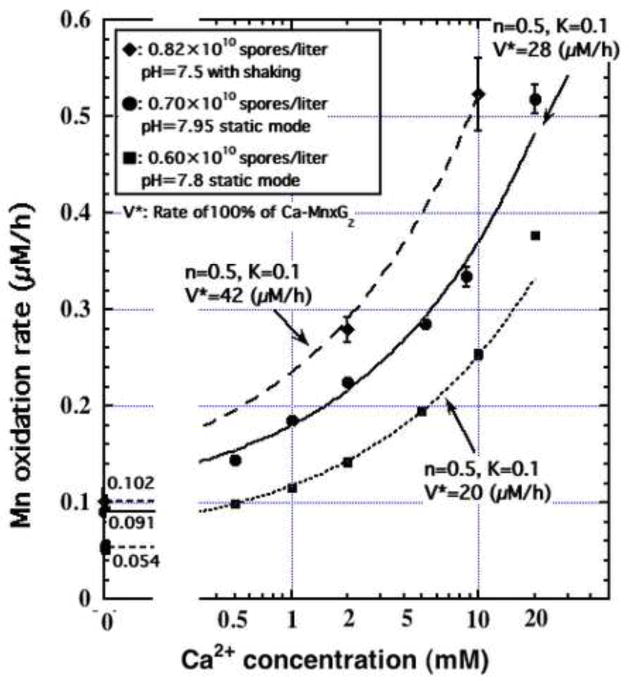
Manganese(IV) oxides, believed to form primarily through microbial activities, are extremely important mineral phases in marine environments where they scavenge a variety of trace elements and thereby control their distributions. The presence of various ions common in seawater are known to influence Mn oxide mineralogy yet little is known about the effect of these ions on the kinetics of bacterial Mn(II) oxidation and Mn oxide formation. We examined factors affecting bacterial Mn(II) oxidation by spores of the marine Bacillus sp. strain SG-1 in natural and artificial seawater of varying ionic conditions. Ca2+ concentration dramatically affected Mn(II) oxidation, while Mg2+, Sr2+, K+, Na+ and NO3- ions had no effect. The rate of Mn(II) oxidation at 10mM Ca2+ (seawater composition) was four or five times that without Ca2+. The relationship between Ca2+ content and oxidation rate demonstrates that the equilibrium constant is small (on the order of 0.1) and the binding coefficient is 0.5. The pH optimum for Mn(II) oxidation changed depending on the amount of Ca2+ present, suggesting that Ca2+ exerts a direct effect on the enzyme perhaps as a stabilizing bridge between polypeptide components. We also examined the effect of varying concentrations of NaCl or KNO3 (0 mM - 2000 mM) on the kinetics of Mn(II) oxidation in solutions containing 10 mM Ca2+. Mn(II) oxidation was unaffected by changes in ionic strength (I) below 0.2, but it was inhibited by increasing salt concentrations above this value. Our results suggest that the critical coagulation concentration is around 200 mM of salt (I = ca. 0.2), and that the ionic strength of seawater (I > 0.2) accelerates the precipitation of Mn oxides around the spores. Under these conditions, the aggregation of Mn oxides reduces the supply of dissolved O2 and/or Mn2+ and inhibits the Mn(II) -> Mn(III) step controlling the enzymatic oxidation of Mn(II). Our results suggest that the hardness and ionic strength of the aquatic environment at circumneutral pH strongly influences the rate of biologically mediated Mn(II) oxidation.


Figures

Fig. 1
Fig. 1
The effect of pH on Mn(II) oxidation. Solutions of ASW with 2 mM HEPES buffer and 8.2 × 10⁹ spores/L were adjusted to the indicated pH and incubated at 24°C under CO2-scrubbed-air bubbling.
Fig. 2
Fig. 2
Variation in the rate of Mn(II) oxidation under different pH and Ca2+ values. Solutions of ASW with the indicated Ca2+ concentrations and 2 mM HEPES were incubated as follows: Ca2+-free solutions were treated as in Figure 1; ASW with 2 mM Ca2+was maintained at 24°C with bubbling of CO2-scrubbed-air, 8.2 × 10⁹ spores/L, and initial [Mn] = ca. 47 μM; ASW with 10 mM Ca2+ was maintained at 24°C with bubbling of CO2-scrubbed-air, 12.0 × 10⁹ spores/L, and initial [Mn] = ca. 52 μM. Error bars represent one standard error (SE) of the rate measurements.
Fig. 3
Fig. 3
Mn(II) oxidation in solutions containing differing concentrations of Ca2+. Solutions of ASW containing the indicated concentrations of Ca2+ were adjusted to a total ionic strength with Mg2+. ASW also contained 2 mM HEPES adjusted to pH 7.8 and incubated with CO2-scrubbed-air and 6.0 × 10⁹ spores/L at 24°C.
Fig. 4
Fig. 4
The effect of Ca2+ content on Mn(II) oxidation rates from three experiments. The presented working curves are from the tentative estimation of the half binding site of the spores with K = 0.1 (M−1). The three closed diamonds are data from the experiment shown in Table 1; The seven closed square are data from the experiment in Figure 3; The seven closed circles are data from a third experiment. The figures of 0.054 μM/h, 0.092 μM/h and 0.113 μM/h are the calculated Mn(II) oxidation rates of SG-1 incubated without Ca2+ for each experimental set. The Mn(II) oxidation rate of the Ca-bound enzyme (V*) is higher than that of the Ca-free enzyme binding by two and half orders of magnitude. Error bars represent one standard error (SE) of the rate measurements.
Fig. 5
Fig. 5
Changes in the oxidation rate of Mn(II) under varying concentrations of NaCl. Solutions of ASW containing the indicated concentrations of NaCl, 2 mM HEPES, and 10 mM CaCl2 were maintained at pH 7.6–7.8 and incubated under CO2-scrubbed-air with 6.3 × 10⁹ spores/L at 22°C.
Fig. 6
Fig. 6
Change in the oxidation rate of Mn(II) as a function of ionic strength (I) from the data of Figure 5 and an identical experiment using a KNO3 containing solution. The closed circles represent Mn(II) removal in a NaCl containing solution (Fig. 5) and the closed squares are data obtained using a KNO3 containing solution. The logarithm of the rate was a linear function of the abscissa (√I/(√I+1)) with slopes of −3 (I>0.2) and −0.31 (I<0.2) in NaCl solution and −1.5 (I>0.2) and −0.10 (I<0.2) in KNO3 solution. Changes in the slope around 200 mM of NaCl or KNO3 with 10 mM CaCl2 cannot be explained by “activated complex theory” and it may indicate alterations in the mechanism of the rate-limiting reaction around 0.2 M ionic strength. Error bars represent one standard error (SE) of the rate measurements.

References

    1. Adams LF, Ghiorse WC. Characterization of extracellular Mn(II) oxidizing activity and isolation of and Mn(II) oxidizing protein from Leptothrix discophora SS1. J Bacteriol. 1987;169:1279–1285. - PMC - PubMed
    1. Anderson R, Johnson HA, Caputo N, Davis RE, Torpey JW, Tebo BM. Mn(II) oxidation is catalyzed by heme peroxidases in “Aurantimonas manganoxydans” strain SI85-9A1 and Erythrobacter sp. strain SD-21. Appl Environ Microbiol. 2009;75:4130–4138. - PMC - PubMed
    1. Bailey EJ, Ollis FD. Biochemical Engneering Fundamentals. McGraw-Hill; 1986.
    1. Bargar JR, Tebo BM, Villinsk JE. In situ characterization of Mn(II) oxidation by spores of the marine Bacillus sp. strain SG-1. Geochim Cosmochim Acta. 2000;64:2737–2749.
    1. Bargar JR, Tebo BM, Bergmann U, Web SM, Glatzel P, Chiu V, Villalobos M. Biotic and Abioic Products of Mn(II) Oxidation by Spores of the Marine Bacillus sp. strain SG-1. Am Miner. 2005;90:143–154.

LinkOut - more resources