

Proteomic profiling identifies markers for inflammation-related tumor-fibroblast interaction
- PMID: 29176937
- PMCID: PMC5689177
- DOI: 10.1186/s12014-017-9168-7
Proteomic profiling identifies markers for inflammation-related tumor-fibroblast interaction
Abstract
Background: Cancer associated fibroblasts are activated in the tumor microenvironment and contribute to tumor progression, angiogenesis, extracellular matrix remodeling, and inflammation.
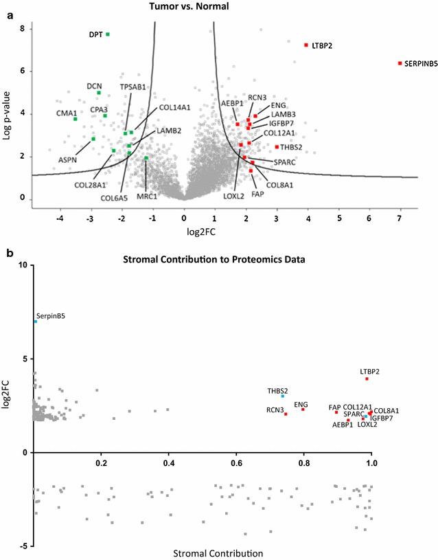
Methods: To identify proteins characteristic for fibroblasts in colorectal cancer we used liquid chromatography-tandem mass spectrometry to derive protein abundance from whole-tissue homogenates of human colorectal cancer/normal mucosa pairs. Alterations of protein levels were determined by two-sided t test with greater than threefold difference and an FDR of < 0.05. Public available datasets were used to predict proteins of stromal origin and link protein with mRNA regulation. Immunohistochemistry confirmed the localization of selected proteins.
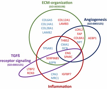
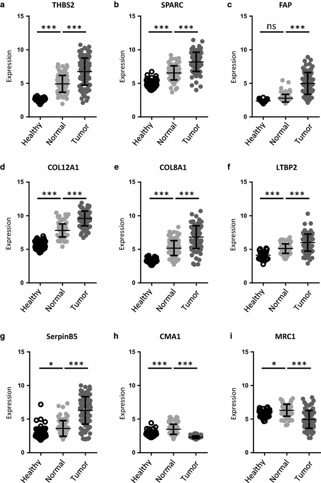
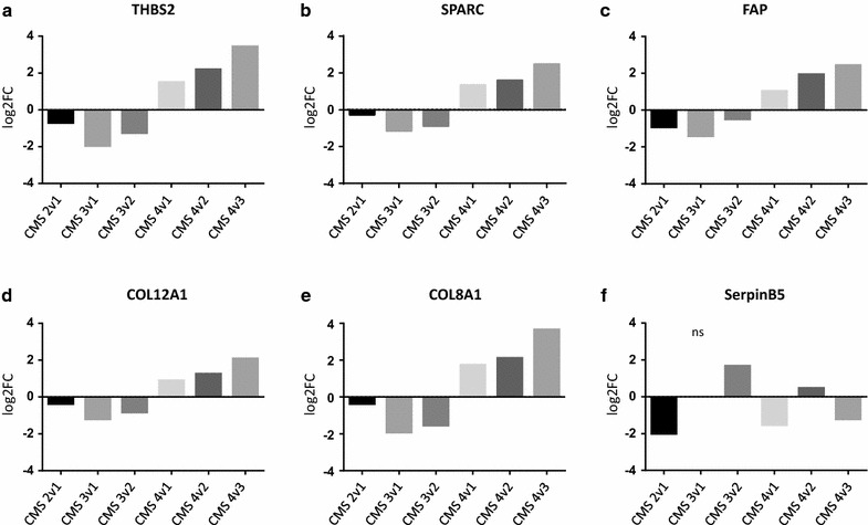
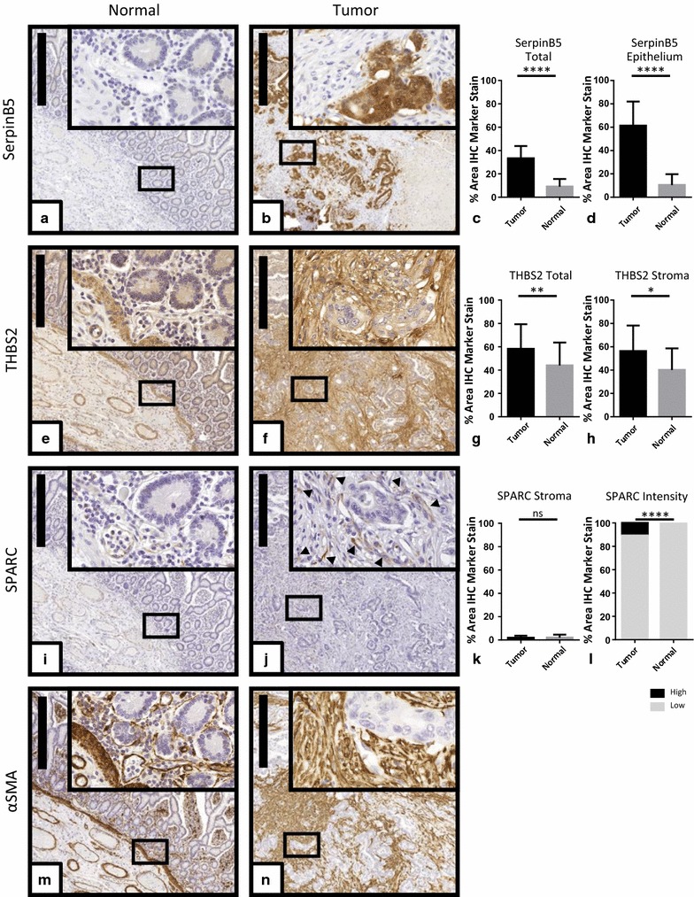
Results: We identified a set of 24 proteins associated with inflammation, matrix organization, TGFβ receptor signaling and angiogenesis mainly originating from the stroma. Most prominent were increased abundance of SerpinB5 in the parenchyme and latent transforming growth factor β-binding protein, thrombospondin-B2, and secreted protein acidic-and-cysteine-rich in the stroma. Extracellular matrix remodeling involved collagens type VIII, XII, XIV, and VI as well as lysyl-oxidase-2. In silico analysis of mRNA levels demonstrated altered expression in the tumor and the adjacent normal tissue as compared to mucosa of healthy individuals indicating that inflammatory activation affected the surrounding tissue. Immunohistochemistry of 26 tumor specimen confirmed upregulation of SerpinB5, thrombospondin B2 and secreted protein acidic-and-cysteine-rich.
Conclusions: This study demonstrates the feasibility of detecting tumor- and compartment-specific protein-signatures that are functionally meaningful by proteomic profiling of whole-tissue extracts together with mining of RNA expression datasets. The results provide the basis for further exploration of inflammation-related stromal markers in larger patient cohorts and experimental models.
Keywords: Cancer associated fibroblasts; Colorectal cancer; Extracellular matrix organization; Inflammation signature; Proteomic profiling; SPARC; THBS2.
Figures
Similar articles
-
Proteomic signatures of the desmoplastic invasion front reveal collagen type XII as a marker of myofibroblastic differentiation during colorectal cancer metastasis.Oncotarget. 2012 Mar;3(3):267-85. doi: 10.18632/oncotarget.451. Oncotarget. 2012. PMID: 22408128 Free PMC article.
-
Proteome profiling of cancer-associated fibroblasts identifies novel proinflammatory signatures and prognostic markers for colorectal cancer.Clin Cancer Res. 2013 Nov 1;19(21):6006-19. doi: 10.1158/1078-0432.CCR-13-1130. Epub 2013 Sep 11. Clin Cancer Res. 2013. PMID: 24025712
-
SPARC enhances tumor stroma formation and prevents fibroblast activation.Oncogene. 2007 Jul 5;26(31):4513-22. doi: 10.1038/sj.onc.1210247. Epub 2007 Jan 29. Oncogene. 2007. PMID: 17260013
-
Prognostic relevance of cancer-associated fibroblasts in human cancer.Semin Cancer Biol. 2014 Apr;25:61-8. doi: 10.1016/j.semcancer.2014.02.006. Epub 2014 Feb 19. Semin Cancer Biol. 2014. PMID: 24560651 Review.
-
SIBLINGs and SPARC families: their emerging roles in pancreatic cancer.World J Gastroenterol. 2014 Oct 28;20(40):14747-59. doi: 10.3748/wjg.v20.i40.14747. World J Gastroenterol. 2014. PMID: 25356037 Free PMC article. Review.
Cited by
-
An integrative network-based approach to identify novel disease genes and pathways: a case study in the context of inflammatory bowel disease.BMC Bioinformatics. 2018 Jul 13;19(1):264. doi: 10.1186/s12859-018-2251-x. BMC Bioinformatics. 2018. PMID: 30005591 Free PMC article.
-
Identification of disease-promoting stromal components by comparative proteomic and transcriptomic profiling of canine mammary tumors using laser-capture microdissected FFPE tissue.Neoplasia. 2021 Apr;23(4):400-412. doi: 10.1016/j.neo.2021.03.001. Epub 2021 Mar 29. Neoplasia. 2021. PMID: 33794398 Free PMC article.
-
Microvesicles from bone marrow-derived mesenchymal stem cells promote Helicobacter pylori-associated gastric cancer progression by transferring thrombospondin-2.Cell Commun Signal. 2023 Oct 5;21(1):274. doi: 10.1186/s12964-023-01127-y. Cell Commun Signal. 2023. PMID: 37798762 Free PMC article.
-
Deciphering the heterogeneity and plasticity of the tumor microenvironment in liver cancer provides insights for prognosis.Front Pharmacol. 2025 Jan 30;16:1495280. doi: 10.3389/fphar.2025.1495280. eCollection 2025. Front Pharmacol. 2025. PMID: 39950116 Free PMC article.
-
Impact of Fibroblast-Derived SPARC on Invasiveness of Colorectal Cancer Cells.Cancers (Basel). 2019 Sep 24;11(10):1421. doi: 10.3390/cancers11101421. Cancers (Basel). 2019. PMID: 31554208 Free PMC article.
References
-
- Alvarez MJ, Prada F, Salvatierra E, Bravo AI, Lutzky VP, Carbone C, et al. Secreted protein acidic and rich in cysteine produced by human melanoma cells modulates polymorphonuclear leukocyte recruitment and antitumor cytotoxic capacity. Can Res. 2005;65:5123–5132. doi: 10.1158/0008-5472.CAN-04-1102. - DOI - PubMed
LinkOut - more resources
Full Text Sources
Other Literature Sources
Molecular Biology Databases
Miscellaneous