Review
. 2017 Nov 10:8:1354.
doi: 10.3389/fimmu.2017.01354. eCollection 2017.

Interleukin-4 Receptor Alpha: From Innate to Adaptive Immunity in Murine Models of Cutaneous Leishmaniasis

Affiliations
Review

Interleukin-4 Receptor Alpha: From Innate to Adaptive Immunity in Murine Models of Cutaneous Leishmaniasis

Ramona Hurdayal et al. Front Immunol. .

Abstract

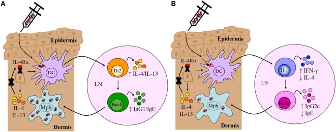
The interleukin (IL)-4 receptor alpha (IL-4Rα), ubiquitously expressed on both innate and adaptive immune cells, controls the signaling of archetypal type 2 immune regulators; IL-4 and IL-13, which elicit their signaling action by the type 1 IL-4Rα/gamma common and/or the type 2 IL-4Rα/IL-13Rα complexes. Global gene-deficient mouse models targeting IL-4, IL-13, or the IL-4Rα chain, followed by the development of conditional mice and generation of important cell-type-specific IL-4Rα-deficient mouse models, were indeed critical to gaining in-depth understanding of detrimental T helper (Th) 2 mechanisms in type 1-controlled diseases. A primary example being cutaneous leishmaniasis, which is caused by the protozoan parasite Leishmania major, among others. The disease is characterized by localized self-healing cutaneous lesions and necrosis for which, currently, not a single vaccine has made it to a stage that can be considered effective. The spectrum of human leishmaniasis belongs to the top 10 infectious diseases according to the World Health Organization. As such, 350 million humans are at risk of infection and disease, with an incidence of 1.5-2 million new cases being reported annually. A major aim of our research is to identify correlates of host protection and evasion, which may aid in vaccine design and therapeutic interventions. In this review, we focus on the immune-regulatory role of the IL-4Rα chain from innate immune responses to the development of beneficial type 1 and detrimental type 2 adaptive immune responses during cutaneous Leishmania infection. We discuss the cell-specific requirements of the IL-4Rα chain on crucial innate immune cells during L. major infection, including, IL-4Rα-responsive skin keratinocytes, macrophages, and neutrophils, as well as dendritic cells (DCs). The latter, contributing to one of the paradigm shifts with respect to the role of IL-4 instructing DCs in vivo, to promote Th1 responses against L. major. Finally, we extend these innate responses and mechanisms to control of adaptive immunity and the effect of IL-4Rα-responsiveness on T and B lymphocytes orchestrating the development of CD4+ Th1/Th2 and B effector 1/B effector 2 B cells in response to L. major infection in the murine host.

Keywords: adaptive cells; innate cells; interleukin-4 receptor alpha; interleukin-4/interleukin-13; murine cutaneous leishmaniasis.


Figures

Figure 1
Figure 1
The interleukin-4 (IL-4) and interleukin-13 (IL-13) receptor complexes. IL-4 interacts with the interleukin-4 receptor alpha (IL-4Rα) chain in combination with either the gamma common (γc) chain to form the type 1 receptor or with the IL-13-binding receptor alpha 1 (IL-13Rα1) to form the type 2 receptor complex. The IL-4Rα chain signals via the JAK1/STAT6 pathway. Definitively, JAK3 associates with the γc chain and JAK2 with the IL-13Rα1. IL-13 interacts with the type II receptor complex (through IL-13Rα1) or with the α2 chain of the IL-13 receptor (IL-13Rα2). A signaling pathway for IL-13 via IL-13Rα2 has recently been identified. TNF-α induces upregulation of IL-13Rα2 expression. IL-13 then binds to the IL-13Rα2, which activates AP-1 to induce gene expression and secretion of soluble TGF-β. Illustration adapted and redrawn from previous publications (12, 34, 42, 52, 56).
Figure 2
Figure 2
Cell-mediated immune response to Leishmania major in resistant and susceptible mice. (A) Upon interaction with L. major parasites, activated dendritic cells (DCs) process and present parasite antigen and produce IL-12 and IL-18, which interact with the IL-12 receptor β2-chain (IL-12Rβ2) in activated Th1 cells. Together, a dominant Th1 response is generated by the production of IFN-γ and TNF-α. These cytokines upregulate the expression of inducible nitric oxide synthase (iNOS) and activate infected macrophages for intracellular killing. CD8+ T cells and natural killer (NK) cells control infection by producing IFN-γ and effector killing by macrophages leading to parasite control as observed in C57BL/6 mice. (B) By contrast, parasite persistence in BALB/c mice is due to the failure of an IL-12-dependent redirection of the early Th2 response. This may be due to continued expression of early interleukin-4 (IL-4) from Vβ4+Vα8+ CD4+ T cells, mast cells, and eosinophils at the inoculation site and draining lymph nodes. This delays secretion of IL-12, or loss of responsiveness to IL-12 due to reduced expression of the IL-12Rβ2 chain on activated Th2 cells. IL-4 and IL-13 suppress Th1 responses and inhibit intracellular killing by macrophages. This leads to arginase production by macrophages that favor parasite growth. Sustained neutrophil recruitment to the site of infection and regulatory T cells may promote susceptibility and inhibit Th1 responses by the production of IL-10 and TGF-β that inhibit activation of macrophages. Solid lines represent activation, and broken lines represent inhibition. Illustration drawn from previous publications (6, 75, 76).
Figure 3
Figure 3
Disease progression in Leishmania major-infected-IL-4−/− and IL-4Rα−/− mice. BALB/c mice deficient for interleukin-4 (IL-4) and interleukin-4 receptor alpha (IL-4Rα) were infected with L. major LV39 strain and footpad swelling monitored weekly as an indication of disease progression. Illustration prepared according to published reports (, –11).
Figure 4
Figure 4
Mouse breeding strategy to create cell-specific interleukin-4 receptor alpha (IL-4Rα)-deficient mice. IL-4Rαflox/flox BALB/c mice were intercrossed with transgenic mice expressing Cre-recombinase under control of a cell-specific promoter (Zcre) and IL-4Rα−/− BALB/c mice to generate ZcreIL-4Rα−/lox mice. The “loxed” IL-4Rα allele, yellow arrows; deleted allele, red arrows. Illustration adapted and redrawn from previous publications (10, 11, 82).
Figure 5
Figure 5
Immune response in Leishmania major-infected BALB/c and C57BL/6 mice deficient for interleukin-4 receptor alpha (IL-4Rα)-responsive keratinocytes. (A) Th2/Type 2-mediated susceptibility in BALB/c mice deficient for IL-4Rα-responsive keratinocytes. KRT14creIL-4Rα−/lox BALB/c mice, generated by cre/loxP targeting, developed non-healing disease to experimental L. major LV39 and IL81 infection in the footpad, characterized by substantial interleukin-4 (IL-4)/interleukin-13 (IL-13) production and IgG1/IgE production, similar to littermate control BALB/c mice. Model prepared using information from submitted manuscript. (B) Th1/Type 1-mediated protective immunity in C57BL/6 mice deficient for IL-4Rα-responsive keratinocytes. Similar to control C57BL/6 mice, KRT14creIL-4Rα−/lox C57BL/6 mice, generated by cre/loxP targeting, controlled lesion development upon L. major LV39 and IL81 infection in the footpad or ear dermis, characterized by strong IFN-γ and IgG2c production and concomitantly reduced IL-4 and IgE (103). DC, dendritic cells; Mph, macrophage; LN, lymph node; Th1, T helper 1; Th2, T helper 2.
Figure 6
Figure 6
Immune response in BALB/c mice deficient for interleukin-4 receptor alpha (IL-4Rα)-responsive macrophages/neutrophils. Despite delayed disease progression in the footpads of L. major LV39-infected LysMcreIL-4Rα−/lox mice, unimpaired Th2/type II responses and reduced IgG2a were detected, similar to control BALB/c mice. The absence of IL-4Rα-responsive macrophages led to increased inducible nitric oxide synthase (iNOS) and reduced arginase, reflecting a shift to classically-activated macrophages likely as a consequence of increased IFN-γ compared to littermate control BALB/c mice. Illustration prepared from Hölscher et al. (13). DC, dendritic cells; Mph, macrophage; LN, lymph node; Th2, T helper 2.
Figure 7
Figure 7
Immune response in BALB/c mice deficient for interleukin-4 receptor alpha (IL-4Rα)-responsive DCs. Absence of interleukin-4 (IL-4)/interleukin-13 (IL-13) signaling on DCs led to hypersusceptibility in BALB/c mice upon Leishmania major LV39 and IL81 infection in the footpad, characterized by a shift to Th2/type II immune responses and alternatively activated macrophages stronger than that usually observed in littermate BALB/c mice. Expression of inducible nitric oxide synthase (iNOS) in DCs was also dramatically reduced with increased arginase (Arg) compared to littermate mice providing a safe haven for L. major growth, survival, and dissemination to peripheral organs. Illustration prepared from Hurdayal et al. (11). DC, dendritic cells; Mph, macrophage; LN, lymph node; Th2, T helper 2.
Figure 8
Figure 8
CD4+ T cell-specific interleukin-4 receptor alpha (IL-4Rα)-deficient mice develop a polarized Th1/type I immune response. In the absence of interleukin-4 (IL-4)-responsive T cells, normally susceptible BALB/c mice developed a healing phenotype upon experimental infection in the footpad, characterized by increased IFN-γ and IgG2a and concomitantly reduced IL-10 and IgG1 compared to littermate BALB/c mice. Interestingly, CD4+ T cell-specific IL-4Rα-deficient mice maintained equivalent IL-4 production to control littermate BALB/c mice. Illustration prepared from Radwanska et al. (10). DC, dendritic cells; Mph, macrophage; LN, lymph node; Th0, naive T cell; Th1, T helper 1.
Figure 9
Figure 9
Absence of interleukin-4 (IL-4)-responsive B cells transforms non-healer BALB/c mice to a healer phenotype. Mb1creIL-4Rα−/lox mice developed an IL-12-induced protective Th1/type I immune response compared to littermate control BALB/c mice following Leishmania major LV39 and IL81 infection in the footpad. IL-4Rα-deficient B cells secreted increased ifn-γ and reduced il-4 transcripts, as early as day 1 postinfection, resembling a B effector 1 phenotype in comparison to IL-4Rα-sufficient B cells. This in turn led to improved macrophage leishmanicidal functions and control of parasite replication at the site of infection. Illustration prepared from Hurdayal et al. (135). DC, dendritic cells; Mph, macrophage; LN, lymph node; B, B cells; Be1, B effector 1 B cells; Th1, T helper 1.

Similar articles

Cited by

References

    1. Kaye P, Scott P. Leishmaniasis: complexity at the host-pathogen interface. Nat Rev Microbiol (2011) 9(8):604–15.10.1038/nrmicro2608 - DOI - PubMed
    1. Savoia D. Recent updates and perspectives on leishmaniasis. J Infect Dev Ctries (2015) 9(6):588–96.10.3855/jidc.6833 - DOI - PubMed
    1. Gupta G, Oghumu S, Satoskar AR. Mechanisms of immune evasion in leishmaniasis. Adv Appl Microbiol (2013) 82:155–84.10.1016/B978-0-12-407679-2.00005-3 - DOI - PMC - PubMed
    1. Alexander J, Brombacher F. T helper1/T helper2 cells and resistance/susceptibility to Leishmania infection: is this paradigm still relevant? Front Immunol (2012) 3:80.10.3389/fimmu.2012.00080 - DOI - PMC - PubMed
    1. Hurdayal R, Brombacher F. The role of IL-4 and IL-13 in cutaneous leishmaniasis. Immunol Lett (2014) 161(2):179–83.10.1016/j.imlet.2013.12.022 - DOI - PubMed