. 2017 Nov 10:8:1523.
doi: 10.3389/fimmu.2017.01523. eCollection 2017.

US6 Gene Deletion in Herpes Simplex Virus Type 2 Enhances Dendritic Cell Function and T Cell Activation

Affiliations

US6 Gene Deletion in Herpes Simplex Virus Type 2 Enhances Dendritic Cell Function and T Cell Activation

Angello Retamal-Díaz et al. Front Immunol. .

Abstract

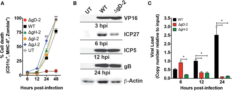
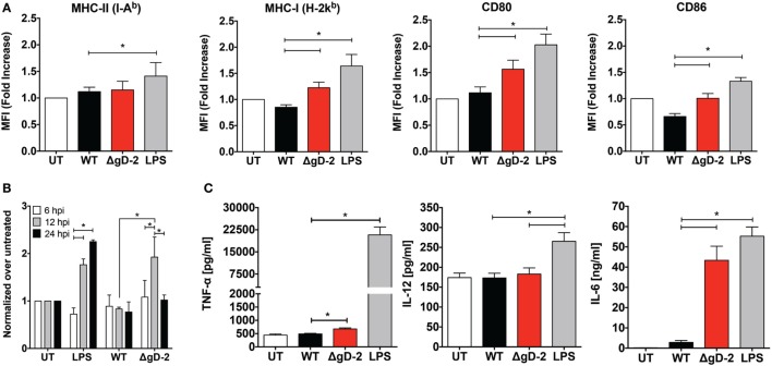
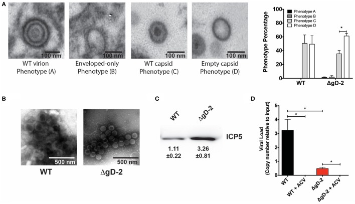
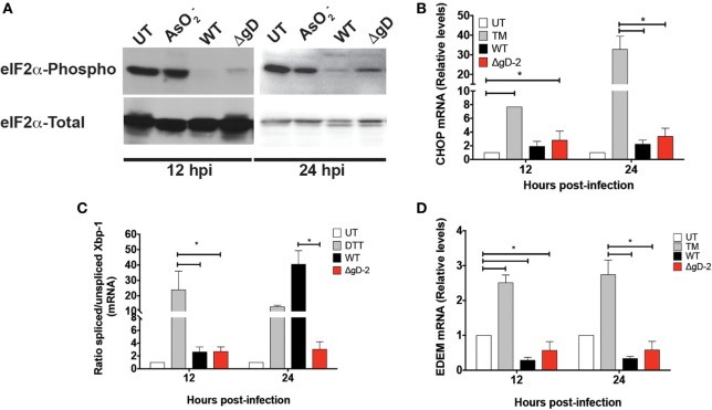
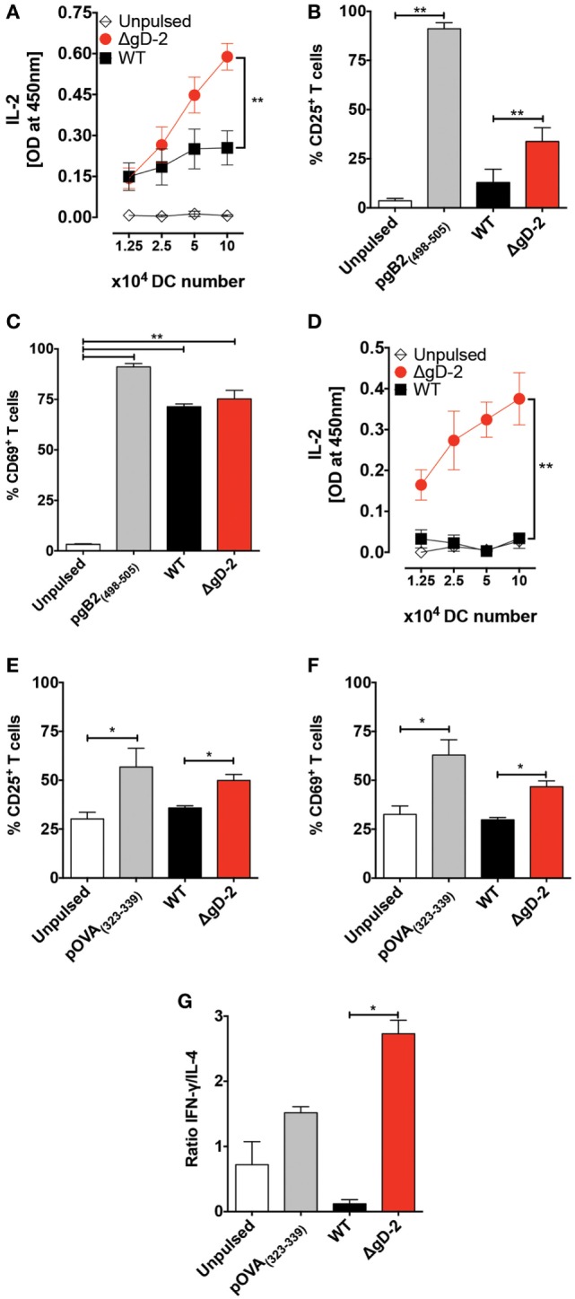
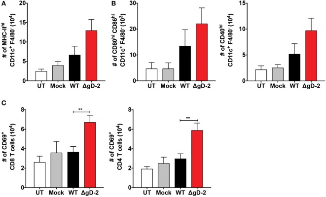
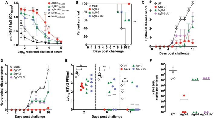
Herpes simplex virus (HSV) type 1 (HSV-1) and type 2 (HSV-2) produce lifelong infections that are associated with frequent asymptomatic or clinically apparent reactivation. Importantly, HSV express multiple virulence factors that negatively modulate innate and adaptive immune components. Notably, HSV interfere with dendritic cell (DC) viability and function, likely hindering the capacity of the host to mount effective immunity against these viruses. Recently, an HSV-2 virus that was deleted in glycoprotein D was engineered (designated ΔgD-2). The virus is propagated on a complementing cell line that expresses HSV-1 gD, which permits a single round of viral replication. ΔgD-2 is safe, immunogenic, and provided complete protection against vaginal or skin challenges with HSV-1 and HSV-2 in murine models. Here, we sought to assess the interaction of ΔgD-2 with DCs and found that, in contrast to wild-type (WT) virus which induces DC apoptosis, ΔgD-2 promoted their migration and capacity to activate naïve CD8+ and CD4+ T cells in vitro and in vivo. Furthermore, DCs exposed to the WT and ΔgD-2 virus experienced different unfolded protein responses. Mice primed with DCs infected with ΔgD-2 in vitro displayed significantly reduced infection and pathology after genital challenge with virulent HSV-2 compared to non-primed mice, suggesting that DCs play a role in the immune response to the vaccine strain.

Keywords: HSV type 2; adaptive immunity; apoptosis; dendritic cells; glycoprotein D; migration; unfolded protein response.


Figures

Figure 1
Figure 1
ΔgD-2 HSV type 2 (HSV-2) is attenuated in dendritic cells (DCs). (A) DC viability, as determined by flow cytometry (gated on CD11c+, I-Ab+, Zombie+ cells) at different time points after virus inoculation with an multiplicity of infection (MOI) of 1. UT, untreated. (B) Western blot analyses of structural (VP16), early (ICP5, ICP27), and late (gB) viral proteins post-inoculation of DCs. (C) Viral genome loads in inoculated DCs determined by qPCR for the UL30 gene. Data are means ± SEM of three independent experiments. Representative images are shown for western blots. Two-way analysis of variance and Tukey’s multiple comparison test were used for statistical analyses (**p < 0.01).
Figure 2
Figure 2
HSV type 2 (HSV-2) ΔgD-2 induces dendritic cell (DC) maturation and cytokine secretion. (A) Expression of surface maturation markers CD80, CD86, MHC-I (H-2Kb), and MHC-II (I-Ab), determined by flow cytometry on DCs (gated on CD11c+, Zombie- cells) at 24 h post-virus inoculation. (B) Flow cytometry analysis of reactive oxygen species (ROS) production in DCs treated as indicated. (C) Supernatants from virus-inoculated DCs were assessed by ELISA for the presence TNF-α, IL-12, and IL-6. Data shown are means ± SEM from eight (A) and three (B,C) independent experiments. Two-way analysis of variance and Tukey’s multiple comparison test were used for statistical analyses (*p < 0.05).
Figure 3
Figure 3
Dendritic cells (DCs) inoculated with ΔgD-2 or wild-type (WT) virus mainly release defective herpes simplex virus particles. (A) Four predominant viral particle phenotypes, indicated in the four panels, were observed in the extracellular space of virus-inoculated DCs (extracellular space data shown). Transmission electron microscopy. Extreme left: WT-like particles displaying an envelope, an electron-dense tegument, and an electron-dense capsid; middle left: envelope-only phenotype displaying an envelope-like structure, harboring spikes in the outer region, yet no apparent capsid or tegument; middle right: WT-like capsids, yet no envelope; extreme right: empty capsids with no envelope and no electron-dense content in capsids. Right panel: quantification of viral particle phenotypes observed. (B) Transmission electron microscopy (negative staining) of viral particles recovered from ultracentrifugated supernatant of virus-inoculated DCs. (C) Western blot analysis of the major capsid protein ICP5 in virus particles recovered from ultracentrifugated supernatants of DCs 18 h post-virus inoculation. Values indicate relative expression of ICP5. (D) Quantification of viral genome copies (determined by qPCR) in viral particles obtained from ultracentrifugated supernatants derived from virus-inoculated DCs with or without acyclovir treatment. One-way analysis of variance and Tukey’s multiple comparison test were used for statistical analyses (*p < 0.05).
Figure 4
Figure 4
Differential Unfolded protein response in WT- and ΔgD-2 virus-inoculated dendritic cells (DCs). (A) Western blot analyses of the phosphorylation of eIF2-α at 12 and 24 h post-inoculation with the indicated treatments. AsO2: sodium arsenite was used as a positive control for PERK activation and eIF2-α phosphorylation. (B) Relative expression of CHOP mRNA in virus-inoculated DCs at 12 and 24 h post-inoculation with the indicated viruses. Here, tunicamycin (TM) was used as positive control for ATF-6 activation. (C) Ratio of spliced to non-spliced XBP-1 mRNA in DCs at 12 and 24 h post-inoculation. Dithiothreitol (DTT) was used as a positive control for activating the IRE-1-α pathway. (D) Relative expression of EDEM in DCs at 12 and 24 h post-inoculation. The Western blots in (A) are representative of three independent experiments. Data for (B) and (D) are means ± SEM of five independent experiments. One-way analysis of variance and Tukey’s multiple comparison test were used for statistical analyses (*p < 0.05).
Figure 5
Figure 5
Dendritic cells (DCs) pulsed with ΔgD-2 activate CD4+ and CD8+ T cells in vitro. (A) IL-2 secretion in the supernatants of cocultures of virus-pulsed DCs with virus-specific gBT-I CD8+ T cells. (B) Surface expression of CD25 in T cells from cocultures of DCs and gBT-I cells. (C) Surface expression of CD69 in T cells from cocultures of DCs and gBT-I cells. (D) IL-2 secretion in the supernatants of cocultures of virus-pulsed DCs with OT-II CD4+ T cells. (E) Surface expression of CD25 in OT-II T cells from DC and T cell cocultures. (F) Surface expression of CD69 in OT-II T cells from DC and T cell cocultures. (G) Secretion of IFN-γ and IL-4 from OT-II CD4+ T cells in DC and T cell cocultures was measured by ELISA and is presented as a ratio. One-way analysis of variance and Tukey’s multiple comparison test were used for statistical analyses (*p < 0.05, **p < 0.01, ***p < 0.001).
Figure 6
Figure 6
ΔgD-2 promotes dendritic cell (DC) migration and CD4+ and CD8+ T cell activation in vivo. (A) DC migration from the hind limb to inguinal lymph nodes (LNs) 48 h after subcutaneous injection of 106 PFU of virus. (B) Phenotype of DCs in inguinal LNs after virus inoculation in the hind limb. The following surface markers were used: CD11c, F4/80, MHC-II, CD80, CD86, and CD40. (C) T cell numbers and phenotypes in inguinal LNs 48 after subcutaneous injection of 106 PFU. One-way analysis of variance with Dunnett’s post-test was used. Data are means ± SEM of three independent experiments (n = 7–12 mice/group, **p < 0.01).
Figure 7
Figure 7
ΔgD-2 increases the relocation of CD103+ migrating dendritic cells (DCs) to draining lymph nodes (LNs). (A) Evaluation of DC migration from the footpads to popliteal LNs 24 h after injection of 106 PFU of virus and CFSE tracking dye (CFSE+ gated, then CD11c+-MHC-II+ gated). (B) Quantification of CD207+-CD103+ migrating dermal DCs (CFSE+ gated, then CD11c+-MHC-II+ gated) relocating to the draining LNs after injection of 106 PFU of virus and CFSE tracking dye. (C) Quantification of CD207+-CD103 Langerhans cells (CFSE+ gated, then CD11c+-MHC-II+ gated) relocating to the draining LNs after injection of 106 PFU of virus and CFSE tracking dye. One-way analysis of variance with Tukey’s post-test was used. Data are means ± SEM (n = 4 mice/group, *p < 0.05).
Figure 8
Figure 8
Transfer of dendritic cells (DCs) infected with ΔgD-2 confer protection against intravaginal lethal challenge with HSV type 2 (HSV-2). Mice were immunized subcutaneously with DCs infected in vitro with ΔgD-2, ΔgH-2, or UV-inactivated ΔgD-2 and administered three times over 7-day intervals and 2 weeks after the last dose challenged intravaginally with virulent HSV-2 (10× LD90). (A) Anti-HSV-2 antibodies in the serum of mice detected by ELISA at day 7 post-challenge. (B) Survival curves, (C) epithelial pathology score, 0: no disease; 1: slight erythema, edema; 2: moderate erythema, edema; 3: small lesion, hair loss; severe erythema, edema; 4: large lesion or multiple lesions, hair loss; severe erythema, edema, and (D) neurological pathology score, 0: no disease; 1: constipation; 2: hind limb paresis; 3: urinary retention; 4: hind limb paralysis; urinary retention; constipation after lethal challenge with HSV-2. (E) HSV-2 titers in vaginal lavages (VALs) at days 2, 6, and 11 (or at the time of euthanasia). (F) Quantification of viral genome copies in the dorsal root ganglia of mice injected with virus-treated DCs and challenged with a lethal dose of HSV-2 as determined by qPCR (UL30 gene). Statistics was performed using two-way analysis of variance and Sidak’s post-test at 95% of confidence interval, data are means ± SEM (n = 5 mice/group **p < 0.05).

Similar articles

Cited by

References

    1. Paz-Bailey G, Ramaswamy M, Hawkes SJ, Geretti AM. Herpes simplex virus type 2: epidemiology and management options in developing countries. Sex Transm Infect (2007) 83(1):16–22.10.1136/sti.2006.020966 - DOI - PMC - PubMed
    1. Smith JS, Robinson NJ. Age-specific prevalence of infection with herpes simplex virus types 2 and 1: a global review. J Infect Dis (2002) 186(Suppl 1):S3–28.10.1086/343739 - DOI - PubMed
    1. Looker KJ, Magaret AS, May MT, Turner KM, Vickerman P, Gottlieb SL, et al. Global and regional estimates of prevalent and incident herpes simplex virus type 1 infections in 2012. PLoS One (2015) 10(10):e0140765.10.1371/journal.pone.0140765 - DOI - PMC - PubMed
    1. Suazo PA, Tognarelli EI, Kalergis AM, Gonzalez PA. Herpes simplex virus 2 infection: molecular association with HIV and novel microbicides to prevent disease. Med Microbiol Immunol (2015) 204(2):161–76.10.1007/s00430-014-0358-x - DOI - PMC - PubMed
    1. Benedetti J, Corey L, Ashley R. Recurrence rates in genital herpes after symptomatic first-episode infection. Ann Intern Med (1994) 121(11):847–54.10.7326/0003-4819-121-11-199412010-00004 - DOI - PubMed