. 2017 Dec 1;140(12):3204-3214.
doi: 10.1093/brain/awx275.

Synaptic phosphorylated α-synuclein in dementia with Lewy bodies

Affiliations

Synaptic phosphorylated α-synuclein in dementia with Lewy bodies

Martí Colom-Cadena et al. Brain. .

Abstract

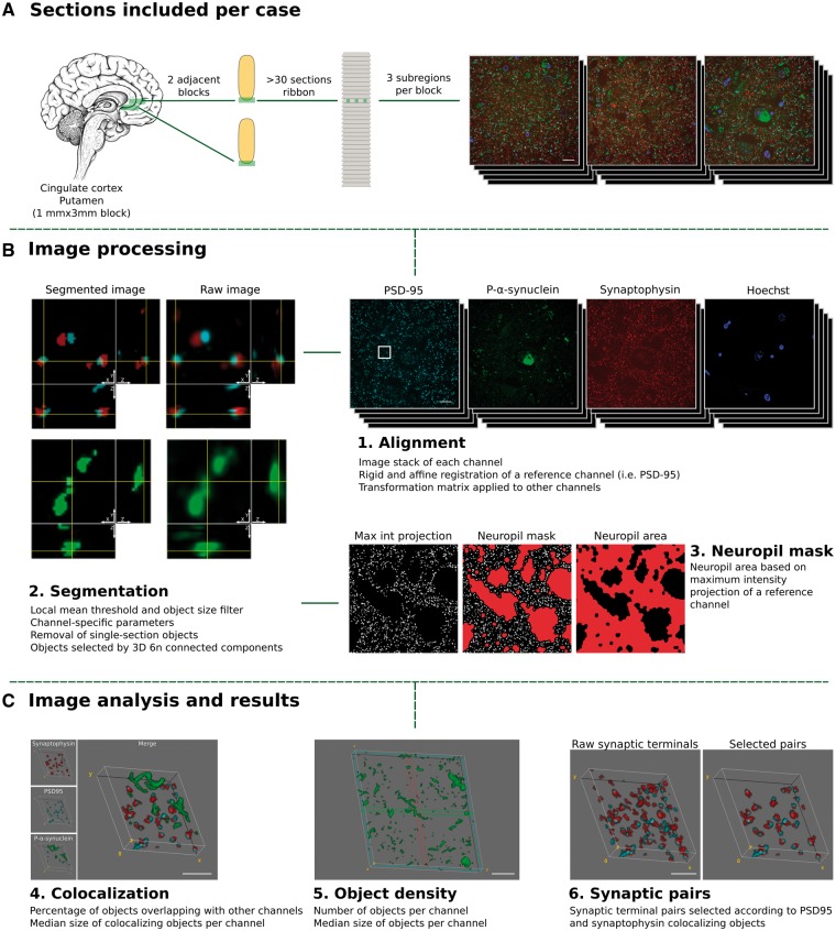
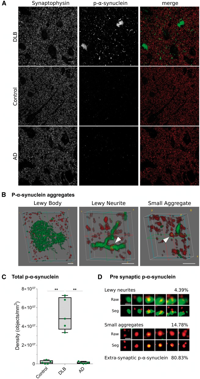
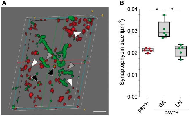
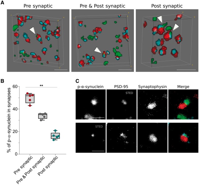
Dementia with Lewy bodies is characterized by the accumulation of Lewy bodies and Lewy neurites in the CNS, both of which are composed mainly of aggregated α-synuclein phosphorylated at Ser129. Although phosphorylated α-synuclein is believed to exert toxic effects at the synapse in dementia with Lewy bodies and other α-synucleinopathies, direct evidence for the precise synaptic localization has been difficult to achieve due to the lack of adequate optical microscopic resolution to study human synapses. In the present study we applied array tomography, a microscopy technique that combines ultrathin sectioning of tissue with immunofluorescence allowing precise identification of small structures, to quantitatively investigate the synaptic phosphorylated α-synuclein pathology in dementia with Lewy bodies. We performed array tomography on human brain samples from five patients with dementia with Lewy bodies, five patients with Alzheimer's disease and five healthy control subjects to analyse the presence of phosphorylated α-synuclein immunoreactivity at the synapse and their relationship with synapse size. Main analyses were performed in blocks from cingulate cortex and confirmed in blocks from the striatum of cases with dementia with Lewy bodies. A total of 1 318 700 single pre- or postsynaptic terminals were analysed. We found that phosphorylated α-synuclein is present exclusively in dementia with Lewy bodies cases, where it can be identified in the form of Lewy bodies, Lewy neurites and small aggregates (<0.16 µm3). Between 19% and 25% of phosphorylated α-synuclein deposits were found in presynaptic terminals mainly in the form of small aggregates. Synaptic terminals that co-localized with small aggregates of phosphorylated α-synuclein were significantly larger than those that did not. Finally, a gradient of phosphorylated α-synuclein aggregation in synapses (pre > pre + post > postsynaptic) was observed. These results indicate that phosphorylated α-synuclein is found at the presynaptic terminals of dementia with Lewy bodies cases mainly in the form of small phosphorylated α-synuclein aggregates that are associated with changes in synaptic morphology. Overall, our data support the notion that pathological phosphorylated α-synuclein may disrupt the structure and function of the synapse in dementia with Lewy bodies.

Keywords: array tomography; dementia with Lewy bodies; human tissue; p-α-synuclein; synapses.


Figures

Figure 1
Figure 1
Study design. Tissue collection and processing of each case is shown in A. For each case, two adjacent tissue sections of cingulate cortex or the striatum (putamen nucleus) were processed and embedded in LR white. For each block a ribbon of >40 consecutive sections of 70 nm was produced. Each ribbon was immunostained for synaptophysin (red), p-α-synuclein (green) and PSD-95 (cyan), synapsin I or α-synuclein, and nuclei were visualized with Hoechst 33258 (blue). Three subregions were imaged through the entire ribbon. (B and C) Processing and analysis of the images. 1. First, individual channel stacks were produced, with all consecutive sections imaged. Consecutive sections of a reference channel (i.e. synaptophysin) were registered using a rigid and an affine transformation. Transformation matrices were applied to other channels. 2. Second, images were segmented using an in-house algorithm based on local mean threshold segmentation, removing single section objects, filtering by size, and detecting 3D objects as six neighbour connected components. Raw images (right) and segmented (left) representative images are shown. Each image corresponds to a single 70 nm section with its corresponding orthogonal views. 3. Neuropil area was calculated based on a maximum intensity projection of synaptophysin channel. 4. Co-localization between the channels of interest and the sizes of co-localizing objects was calculated. 5. The object density and object size were quantified and (6) for entire synaptic studies, segmented images of synaptophysin (presynaptic) and PSD-95 (postsynaptic) channels combined to remove all those objects without pre- and postsynaptic pairs. Max Int = maximum intensity; 6 n = six neighbour. Scale bars in A and B = 10 µm; C = 2 µm.
Figure 2
Figure 2
P-α-synuclein immunoreactivity patterns and synaptic localization. (A) Representative images of DLB, control and Alzheimer’s disease cases stained with antibodies against synaptophysin (red) and p-α-synuclein (green). Each image is a maximum intensity projection of 31 consecutive sections. (B) Representative 3D representations of types of p-α-synuclein aggregates (green) found in DLB cases and synaptophysin terminals (red). Lewy bodies did not co-localize with synaptophysin terminals, while Lewy neurites and small aggregates did (arrowheads). (C) The quantification of total p-α-synuclein objects revealed that pathology was found almost exclusively in DLB cases (**P < 0.01). (D) Synaptograms representing six 70 nm consecutive sections (from left to right) of the Lewy neurite (LN) or small aggregate (SA) indicated by the arrowheads in B. Both the raw and segmented images are shown. The percentages of presynaptic p-α-synuclein found of Lewy neurites or small aggregate and extra-synaptic p-α-synuclein are indicated. AD = Alzheimer disease. Scale bar in A = 10 µm; B = 2 µm; D = 1 µm.
Figure 3
Figure 3
Median volume of presynaptic terminals co-localizing with p-α-synuclein. (A) 3D reconstruction of 20 consecutive sections from a DLB case. P-α-synuclein (green) and synaptophysin (red) are shown. Black arrowheads indicate synaptophysin objects that co-localize with small aggregates of p-α-synuclein, grey arrowheads point to synaptic terminals that co-localize with Lewy neurites, and white arrowheads those synaptophysin objects that do not co-localize with p-α-synuclein. (B) Median volumes of synaptic terminals according to co-localization with p-α-synuclein (p-α-synuclein +, or −) and their characteristic pattern (small aggregate or Lewy neurite, *P < 0.05). LN = Lewy neurite; Psyn = p-α-synuclein; SA = small aggregate. Scale bar in A = 2 µm.
Figure 4
Figure 4
Trans-synaptic localization of p-α-synuclein. (A) 3D reconstruction of 20 consecutive sections from a DLB case. P-α-synuclein (green), synaptophysin (red) and PSD-95 (blue) are shown. White arrowheads point to zones where p-α-synuclein co-localizes with synaptophysin (presynaptic), PSD-95 (postsynaptic) or both (pre- and postsynaptic). In B, p-α-synuclein objects that co-localized with synaptic pairs (synaptophysin and PSD-95 objects) are classified depending on its presynaptic, pre- and postsynaptic or postsynaptic localization. The relative co-localization of p-α-synuclein with the synaptophysin presynaptic marker was significantly higher than the PSD-95 postsynaptic co-localization. (C) Representative images of the co-localization between between p-α-synuclein, synaptophysin and PSD-95 using array tomography combined with STED microscopy. A single 70 nm-thick section stained for p-α-synuclein (green), synaptophysin (red) and PSD-95 (blue) is shown. STED was applied to image PSD-95 (top row) or p-α-synuclein (bottom row). Scale bar in A = 2 µm; C = 1 µm.

Similar articles

Cited by

References

    1. Abeliovich A, Gitler AD. Defects in trafficking bridge Parkinson’s disease pathology and genetics. Nature 2016; 539: 207–16. - PubMed
    1. Anderson JP, Walker DE, Goldstein JM, De Laat R, Banducci K, Caccavello RJ, et al.Phosphorylation of Ser-129 is the dominant pathological modification of α-synuclein in familial and sporadic lewy body disease. J Biol Chem 2006; 281: 29739–52. - PubMed
    1. Bereczki E, Francis PT, Howlett D, Pereira JB, Höglund K, Bogstedt A, et al.Synaptic proteins predict cognitive decline in Alzheimer’s disease and Lewy body dementia. Alzheimers Dement 2016; 12: 1149–58. - PubMed
    1. Braak H, Del Tredici K, Rüb U, De Vos RAI, Jansen Steur ENH, Braak E. Staging of brain pathology related to sporadic Parkinson’s disease. Neurobiol Aging 2003; 24: 197–211. - PubMed
    1. Brown DF, Risser RC, Bigio EH, Tripp P, Stiegler A, Welch E, et al.Neocortical synapse density and Braak stage in the Lewy body variant of Alzheimer disease: a comparison with classic Alzheimer disease and normal aging. J Neuropathol Exp Neurol 1998; 57: 955–60. - PubMed