Review
. 2018 Apr 1;59(4):651-660.
doi: 10.1093/pcp/pcx182.

Manipulation of Bryophyte Hosts by Pathogenic and Symbiotic Microbes

Affiliations
Review

Manipulation of Bryophyte Hosts by Pathogenic and Symbiotic Microbes

Philip Carella et al. Plant Cell Physiol. .

Erratum in

  • Erratum.
    [No authors listed] [No authors listed] Plant Cell Physiol. 2018 Apr 1;59(4):876. doi: 10.1093/pcp/pcy075. Plant Cell Physiol. 2018. PMID: 29718476 Free PMC article. No abstract available.

Abstract

The colonization of plant tissues by pathogenic and symbiotic microbes is associated with a strong and directed effort to reprogram host cells in order to permit, promote and sustain microbial growth. In response to colonization, hosts accommodate or sequester invading microbes by activating a set of complex regulatory programs that initiate symbioses or bolster defenses. Extensive research has elucidated a suite of molecular and physiological responses occurring in plant hosts and their microbial partners; however, this information is mostly limited to model systems representing evolutionarily young plant lineages such as angiosperms. The extent to which these processes are conserved across land plants is therefore poorly understood. In this review, we outline key aspects of host reprogramming that occur during plant-microbe interactions in early diverging land plants belonging to the bryophytes (liverworts, hornworts and mosses). We discuss how further knowledge of bryophyte-microbe interactions will advance our understanding of how plants and microbes co-operated and clashed during the conquest of land.


Figures

Fig. 1
Fig. 1
Differential colonization of the liverwort thallus by pathogenic and symbiotic microbes. (A) Light micrograph of a sectioned liverwort thallus (Lunularia cruciata) demonstrating air chambers (AC) on the dorsal photosynthetic layer, the central storage region (CSR) and rhizoids (Rz) emanating from the ventral epidermis. (B) Colonization of L. cruciata thalli by the hemi-biotrophic oomycete pathogen Phytophthora palmivora (Accession P3914) constitutively expressing tdTomato. Confocal fluorescence microscopy of sectioned thalli of 2-month-old L. cruciata plants 7 d after infection. Merged z-stack images display red fluorescence and bright field images. Plants were grown under a short-day photoperiod (50 μE m−2 s−1 light levels) on mycorrhization (M) media. (C) Colonization of L. cruciata thalli by the symbiotic fungus Rhizophagus irregularis. Confocal fluorescence microscopy of sectioned thalli stained with WGA (wheat germ agglutinin)–Alexa488 to detect fungal chitin. Plants were grown on vermiculite supplemented with crude R. irregularis inoculum for 2 months under a long-day photoperiod (16 h light) with attenuated light (approximately 50–70 μE m−2 s−1). Merged z-stack images display green fluorescence and bright field images. Scale bars = 100 μm.
Fig. 2
Fig. 2
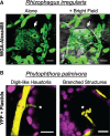
Intracellular biotrophic structures of symbiotic and pathogenic microbes in liverwort cells. (A) Arbusculated liverwort cells. Confocal fluorescence microscopy of sectioned Lunularia cruciata thalli stained with WGA (wheat germ agglutinin)–Alexa488 to detect fungal chitin. Plants were grown on vermiculite supplemented with crude Rhizophagus irregularis inoculum for 2 months under a long-day photoperiod (16 h light) with attenuated light (approximately 50–70 μE m−2 s−1). Z-stack images display green fluorescence alone or merged with bright field images. Arrows indicate an arbuscule-like structure occupying a cell within the non-photosynthetic storage layer. Scale bars = 10 μm. (B) Digit-like haustoria and branched intracellular structures of the hemi-biotrophic oomycete pathogen Phytophthora palmivora deployed in photosynthetic cells of the liverwort Marchantia polymorpha (TAK1). Three-week-old TAK1 plants grown on 1/2 MS-B5 (Musharige and Skoog + B5 vitamins) medium(pH 6.5) were inoculated with zoospores of P. palmivora (Accession P16830) constitutively expressing endoplasmic reticulum-retained yellow fluorescent protein (YFP). Confocal fluorescence microscopy of TAK1 thalli was performed 3 d after infection. Merged z-stack images display YFP fluorescence and plastid autofluorescence (magenta). Scale bars = 10 μm.

Similar articles

Cited by

References

    1. Adams D.G., Duggan P.S. (2008) Cyanobacteria–bryophyte symbioses. J. Exp. Bot. 59: 1047–1058. - PubMed
    1. Alcaraz L.D., Peimbert M., Dorantes-Acosta A.E., Bowman J.L., Arteaga-Vazquez M.A. (2017) Marchantia liverworts as a proxy to plants basal microbiomes. bioRxiv. doi: 10.1101/103861. - DOI - PMC - PubMed
    1. Alvarez A., Montesano M., Schmelz E., Ponce de Leon I. (2016) Activation of shikimate, phenylpropanoid, oxylipins, and auxin pathways in Pectobacterium carotovorum elicitors-treated moss. Front. Plant Sci. 7: 328. - PMC - PubMed
    1. Balestrini R., Bonfante P. (2014) Cell wall remodeling in mycorrhizal symbiosis: a way towards biotrophism. Front. Plant Sci. 5: 237. - PMC - PubMed
    1. Balestrini R., Cosgrove D.J., Bonfante P. (2005) Differential location of alpha-expansin proteins during the accommodation of root cells to an arbuscular mycorrhizal fungus. Planta 220: 889–899. - PubMed

LinkOut - more resources