

Disagreement between splenic switch-off and myocardial T1-mapping after caffeine intake
- PMID: 29177579
- PMCID: PMC5859139
- DOI: 10.1007/s10554-017-1274-0
Disagreement between splenic switch-off and myocardial T1-mapping after caffeine intake
Abstract
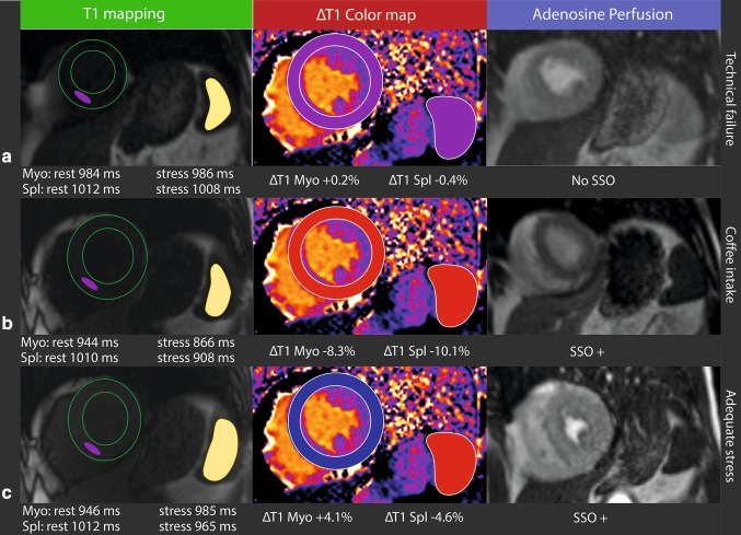
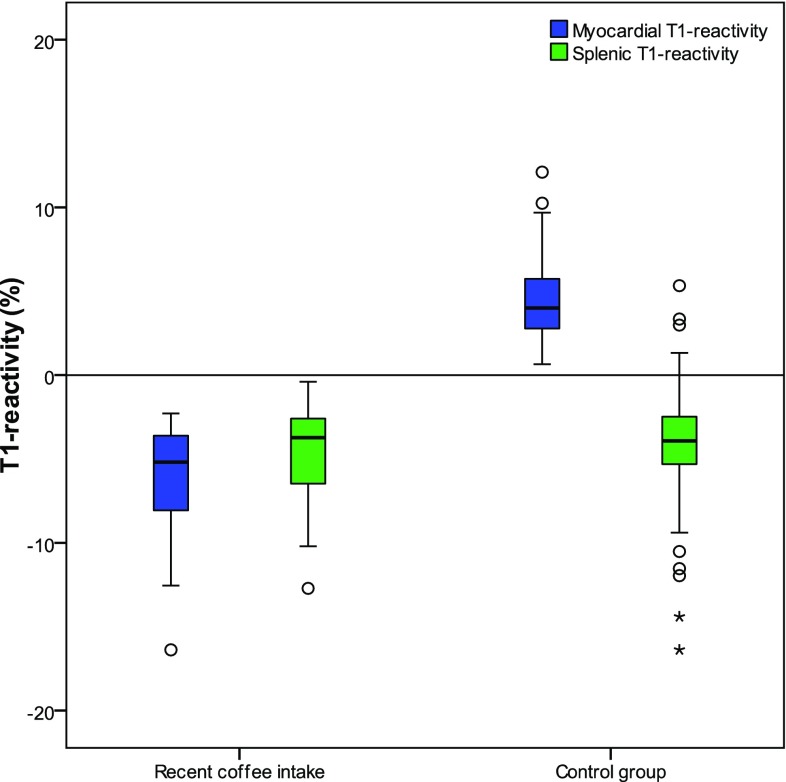
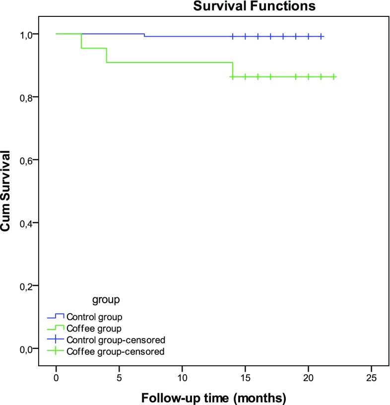
Caffeine is an adenosine receptor antagonist and a possible cause of inadequate stress perfusion. Splenic switch-off (SSO) and splenic rest-stress T1-mapping have been proposed as indicators of stress adequacy during perfusion cardiac magnetic resonance (CMR). We compared myocardial rest-stress T1-mapping with SSO and splenic rest-stress T1-mapping in patients with and without recent coffee intake. We analyzed 344 consecutive patients suspected of myocardial ischemia with adenosine perfusion CMR. All 146 normal CMR studies with a normal T1-rest of the myocardium, used as standard of reference, were included and divided in two groups. 22 patients accidentally ingested coffee < 4 h before CMR, compared to control group of 124 patients without self-reported coffee intake. Two independent readers graded SSO visually. T1-reactivity (ΔT1) was defined as percentual difference in T1-rest and T1-stress. Follow-up data were extracted from electronic patients records. In patients with recent coffee intake SSO was identified in 96%, which showed no significant difference with SSO in controls (94%, p = 0.835), however event rates were significantly different (13.6 and 0.8%, respectively (p < 0.001), median FU 17 months). Myocardial ΔT1 in the coffee group (- 5.2%) was significantly lower compared to control (+ 4.0%, p < 0.001), in contrast to the splenic ΔT1 (- 3.7 and - 4.0%, p = 0.789). The splenic T1-mapping results failed to predict false negative results. SSO and splenic rest-stress T1-mapping are not reliable indicators of stress adequacy in patients with recent coffee intake. Therefore, the dark spleen sign does not indicate adequate myocardial stress in patients with recent caffeine intake. Myocardial rest-stress T1-mapping is an excellent indicator of stress adequacy during adenosine perfusion CMR.
Keywords: Adenosine; Cardiac magnetic resonance; Splenic switch-off; Stress CMR; T1-mapping.
Conflict of interest statement
Conflict of interest
The authors declare that they have no conflict of interest.
Ethical approval
The institutional review board approved this study and all subjects gave written informed consent.
Figures
Similar articles
-
Impact of caffeine on myocardial perfusion reserve assessed by semiquantitative adenosine stress perfusion cardiovascular magnetic resonance.J Cardiovasc Magn Reson. 2019 Jun 24;21(1):33. doi: 10.1186/s12968-019-0542-7. J Cardiovasc Magn Reson. 2019. PMID: 31230593 Free PMC article.
-
Caffeine intake inverts the effect of adenosine on myocardial perfusion during stress as measured by T1 mapping.Int J Cardiovasc Imaging. 2016 Oct;32(10):1545-53. doi: 10.1007/s10554-016-0949-2. Epub 2016 Jul 29. Int J Cardiovasc Imaging. 2016. PMID: 27473274 Free PMC article.
-
Effects of caffeine intake prior to stress cardiac magnetic resonance perfusion imaging on regadenoson- versus adenosine-induced hyperemia as measured by T1 mapping.Int J Cardiovasc Imaging. 2017 Nov;33(11):1753-1759. doi: 10.1007/s10554-017-1157-4. Epub 2017 May 25. Int J Cardiovasc Imaging. 2017. PMID: 28547666 Free PMC article.
-
Effects of Caffeine on Myocardial Blood Flow: A Systematic Review.Nutrients. 2018 Aug 13;10(8):1083. doi: 10.3390/nu10081083. Nutrients. 2018. PMID: 30104545 Free PMC article.
-
Cardiovascular magnetic resonance in patients with magnetic resonance conditional pacemaker systems at 1.5 T: influence of pacemaker related artifacts on image quality including first pass perfusion, aortic and mitral valve assessment, flow measurement, short tau inversion recovery and T1-weighted imaging.Int J Cardiovasc Imaging. 2017 Mar;33(3):383-394. doi: 10.1007/s10554-016-1012-z. Epub 2016 Nov 4. Int J Cardiovasc Imaging. 2017. PMID: 27815793 Review.
Cited by
-
Splenic switch-off as a novel marker for adenosine response in nitrogen-13 ammonia PET myocardial perfusion imaging: Cross-validation against CMR using a hybrid PET/MR device.J Nucl Cardiol. 2022 Jun;29(3):1205-1214. doi: 10.1007/s12350-020-02448-y. Epub 2020 Dec 22. J Nucl Cardiol. 2022. PMID: 33354759 Free PMC article.
-
Impact of caffeine on myocardial perfusion reserve assessed by semiquantitative adenosine stress perfusion cardiovascular magnetic resonance.J Cardiovasc Magn Reson. 2019 Jun 24;21(1):33. doi: 10.1186/s12968-019-0542-7. J Cardiovasc Magn Reson. 2019. PMID: 31230593 Free PMC article.
-
MRI perfusion in patients with stable chest-pain.Br J Radiol. 2020 Sep 1;93(1113):20190881. doi: 10.1259/bjr.20190881. Epub 2020 Jan 6. Br J Radiol. 2020. PMID: 31834813 Free PMC article. Review.
-
Cardiovascular imaging 2018 in the International Journal of Cardiovascular Imaging.Int J Cardiovasc Imaging. 2019 Jul;35(7):1175-1188. doi: 10.1007/s10554-019-01579-9. Int J Cardiovasc Imaging. 2019. PMID: 30868339 No abstract available.
-
Splenic switch-off as a predictor for coronary adenosine response: validation against 13N-ammonia during co-injection myocardial perfusion imaging on a hybrid PET/CMR scanner.J Cardiovasc Magn Reson. 2021 Jan 7;23(1):3. doi: 10.1186/s12968-020-00696-y. J Cardiovasc Magn Reson. 2021. PMID: 33407586 Free PMC article.
References
-
- Windecker S, Kolh P, Alfonso F, Collet JP, Cremer J, Falk V, Filippatos G, Hamm C, Head SJ, Jüni P, Kappetein AP, Kastrati A, Knuuti J, Landmesser U, Laufer G, Neumann FJ, Richter DJ, Schauerte P, Uva MS, Stefanini GG, Taggart DP, Torracca L, Valgimigli M, Wijns W, Witkowski A. 2014 ESC/EACTS guidelines on myocardial revascularization. Rev Esp Cardiol (Engl Ed) 2015;68(2):144. - PubMed
-
- Schwitter J, Wacker CM, Wilke N, Al-Saadi N, Sauer E, Huettle K, Schönberg SO, Debl K, Strohm O, Ahlstrom H, Dill T, Hoebel N, Simor T. Superior diagnostic performance of perfusion-cardiovascular magnetic resonance versus SPECT to detect coronary artery disease: the secondary endpoints of the multicenter multivendor MR-IMPACT II (magnetic resonance imaging for myocardial perfusion assessment in coronary artery disease trial) J Cardiovasc Magn Res. 2012;14:61–71. doi: 10.1186/1532-429X-14-61. - DOI - PMC - PubMed
-
- Greenwood JP, Maredia N, Younger JF, Brown JM, Nixon J, Everett CC, Bijsterveld P, Ridgway JP, Radjenovic A, Dickinson CJ, Ball SG, Plein S. Cardiovascular magnetic resonance and single-photon emission computed tomography for diagnosis of coronary heart disease (CE-MARC): a prospective trial. Lancet. 2012;379(9814):453–460. doi: 10.1016/S0140-6736(11)61335-4. - DOI - PMC - PubMed
-
- Greenwood JP, Herzog BA, Brown JM, Everett CC, Nixon J, Bijsterveld P, Maredia N, Motwani M, Dickinson CJ, Ball SG, Plein S. Prognostic value of cardiovascular magnetic resonance and single-photon emission computed tomography in suspected coronary heart disease: long-term follow-up of a prospective, diagnostic accuracy cohort study. Ann Intern Med. 2016;165:1–9. doi: 10.7326/M15-1801. - DOI - PubMed
-
- Mishra RK, Dorbala S, Logsetty G, Hassan A, Heinonen T, Schelbert HR, et al. Quantitative relation between hemodynamic changes during intravenous adenosine infusion and the magnitude of coronary hyperemia: implications for myocardial perfusion imaging. J Am Coll Cardiol. 2005;45(4):553–558. doi: 10.1016/j.jacc.2004.10.064. - DOI - PubMed
MeSH terms
Substances
LinkOut - more resources
Full Text Sources
Other Literature Sources
Medical
