

Parallel in vivo and in vitro transcriptomics analysis reveals calcium and zinc signalling in the brain as sensitive targets of HBCD neurotoxicity
- PMID: 29177809
- PMCID: PMC5866835
- DOI: 10.1007/s00204-017-2119-2
Parallel in vivo and in vitro transcriptomics analysis reveals calcium and zinc signalling in the brain as sensitive targets of HBCD neurotoxicity
Abstract
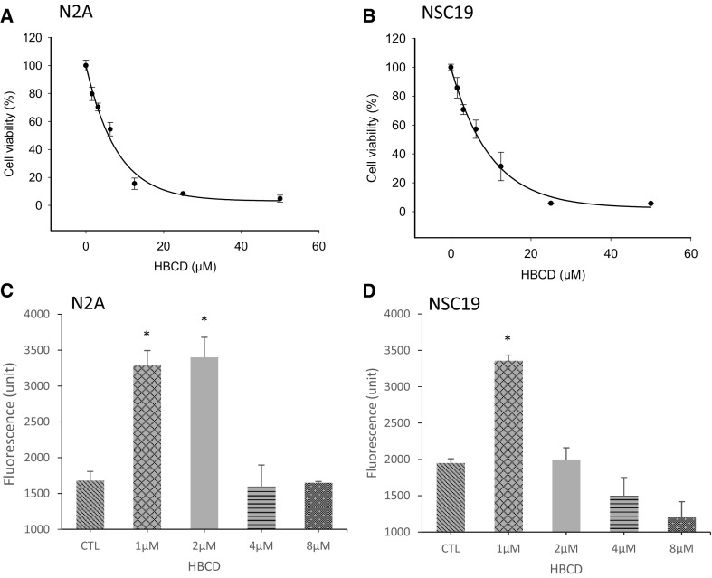
Hexabromocyclododecane (HBCD) is a brominated flame retardant (BFR) that accumulates in humans and affects the nervous system. To elucidate the mechanisms of HBCD neurotoxicity, we used transcriptomic profiling in brains of female mice exposed through their diet to HBCD (199 mg/kg body weight per day) for 28 days and compared with those of neuronal N2A and NSC-19 cell lines exposed to 1 or 2 µM HBCD. Similar pathways and functions were affected both in vivo and in vitro, including Ca2+ and Zn2+ signalling, glutamatergic neuron activity, apoptosis, and oxidative stress. Release of cytosolic free Zn2+ by HBCD was confirmed in N2A cells. This Zn2+ release was partially quenched by the antioxidant N-acetyl cysteine indicating that, in accordance with transcriptomic analysis, free radical formation is involved in HBCD toxicity. To investigate the effects of HBCD in excitable cells, we isolated mouse hippocampal neurons and monitored Ca2+ signalling triggered by extracellular glutamate or zinc, which are co-released pre-synaptically to trigger postsynaptic signalling. In control cells application of zinc or glutamate triggered a rapid rise of intracellular [Ca2+]. Treatment of the cultures with 1 µM of HBCD was sufficient to reduce the glutamate-dependent Ca2+ signal by 50%. The effect of HBCD on zinc-dependent Ca2+ signalling was even more pronounced, resulting in the reduction of the Ca2+ signal with 86% inhibition at 1 µM HBCD. Our results show that low concentrations of HBCD affect neural signalling in mouse brain acting through dysregulation of Ca2+ and Zn2+ homeostasis.
Keywords: Androgen; BFR; Dihydrotestosterone; Glutamate; GnRH; Neurotoxicity; Oestrogen; Prolactin; Transcriptomics.
Figures


Similar articles
-
Insights into brominated flame retardant neurotoxicity: mechanisms of hippocampal neural cell death and brain region-specific transcriptomic shifts in mice.Toxicol Sci. 2024 Oct 1;201(2):282-299. doi: 10.1093/toxsci/kfae090. Toxicol Sci. 2024. PMID: 38995820
-
Diastereoisomer-specific neurotoxicity of hexabromocyclododecane in human SH-SY5Y neuroblastoma cells.Sci Total Environ. 2019 Oct 10;686:893-902. doi: 10.1016/j.scitotenv.2019.06.008. Epub 2019 Jun 3. Sci Total Environ. 2019. PMID: 31200309
-
Cross-omics gene and protein expression profiling in juvenile female mice highlights disruption of calcium and zinc signalling in the brain following dietary exposure to CB-153, BDE-47, HBCD or TCDD.Toxicology. 2014 Jul 3;321:1-12. doi: 10.1016/j.tox.2014.03.006. Epub 2014 Mar 27. Toxicology. 2014. PMID: 24680724
-
Toxic effects of brominated flame retardants in man and in wildlife.Environ Int. 2003 Sep;29(6):841-53. doi: 10.1016/S0160-4120(03)00107-7. Environ Int. 2003. PMID: 12850100 Review.
-
The potential of selected brominated flame retardants to affect neurological development.J Toxicol Environ Health B Crit Rev. 2010;13(5):411-48. doi: 10.1080/10937401003751630. J Toxicol Environ Health B Crit Rev. 2010. PMID: 20582854 Review.
Cited by
-
Unveiling the potential of proteomics in addressing food and feed safety challenges.EFSA J. 2023 Nov 30;21(Suppl 1):e211013. doi: 10.2903/j.efsa.2023.e211013. eCollection 2023 Nov. EFSA J. 2023. PMID: 38047126 Free PMC article.
-
Polymer microchamber arrays for geometry-controlled drug release: a functional study in human cells of neuronal phenotype.Biomater Sci. 2019 May 28;7(6):2358-2371. doi: 10.1039/c8bm01499j. Biomater Sci. 2019. PMID: 30916673 Free PMC article.
-
A PBPK model describing the pharmacokinetics of γ-HBCD exposure in mice.Toxicol Appl Pharmacol. 2021 Oct 1;428:115678. doi: 10.1016/j.taap.2021.115678. Epub 2021 Aug 11. Toxicol Appl Pharmacol. 2021. PMID: 34390738 Free PMC article.
-
Roadmap for the development of alternative test methods.Arch Toxicol. 2020 Oct;94(10):3597-3598. doi: 10.1007/s00204-020-02888-y. Epub 2020 Aug 28. Arch Toxicol. 2020. PMID: 32857209 Free PMC article. No abstract available.
-
Update of the risk assessment of hexabromocyclododecanes (HBCDDs) in food.EFSA J. 2021 Mar 8;19(3):e06421. doi: 10.2903/j.efsa.2021.6421. eCollection 2021 Mar. EFSA J. 2021. PMID: 33732387 Free PMC article.
References
-
- American Chemistry Council (2001) Draft Screening Assessment of Hexabromocyclododecane (HBCD). http://www.ec.gc.ca/lcpe-cepa/default.asp?lang=En&n=A27E7A60-1&offset=13... (consulted on May 2011)
MeSH terms
Substances
Grants and funding
LinkOut - more resources
Full Text Sources
Other Literature Sources
Miscellaneous
