

Educational and practice gaps in the management of volar melanocytic lesions
- PMID: 29178552
- PMCID: PMC5967984
- DOI: 10.1111/jdv.14712
Educational and practice gaps in the management of volar melanocytic lesions
Abstract
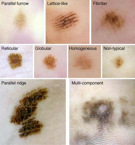
Background: The benign and malignant patterns of acral melanocytic naevi (AMN) and acral melanomas (AM) have been defined in a series of retrospective studies. A three-step algorithm was developed to determine when to biopsy acral melanocytic lesions. This algorithm has only been applied to a Japanese population.
Objectives: Our study aimed to review the current management strategy of acral melanocytic lesions and to investigate the utility of the three-step algorithm in a predominately Caucasian cohort.
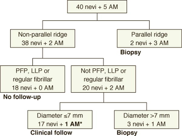
Methods: A retrospective search of the pathology and image databases at Mayo Clinic was performed between the years 2006 and 2016. Only cases located on a volar surface with dermoscopic images were included. Two dermatologists reviewed all dermoscopic images and assigned a global dermoscopic pattern. Clinical and follow-up data were gathered by chart review. All lesions with known diameter and pathological diagnosis were used for the three-step algorithm.
Results: Regular fibrillar and ridge patterns were more likely to be biopsied (P = 0.01). The majority of AMN (58.1%) and AM (60%) biopsied were due to physician-deemed concerning dermoscopic patterns. 39.2% of these cases were parallel furrow, lattice-like or regular fibrillar. When patients were asked to follow-up within a 3- to 6-month period, only 16.7% of the patients returned within that interval. The three-step algorithm would have correctly identified four of five AM for biopsy, missing a 6 mm, multicomponent, invasive melanoma.
Conclusion: We found one major educational gap in the recognition of low-risk lesions with high rates of biopsy of the fibrillary pattern. Recognizing low-risk dermoscopic patterns could reduce the rate of biopsy of AMN by 23.3%. We identified two major practice gaps, poor patient compliance with follow-up and the potential insensitivity of the three-step algorithm to small multicomponent acral melanocytic lesions.
© 2017 European Academy of Dermatology and Venereology.
Conflict of interest statement
None of the authors have any conflicts of interest to disclose.
Figures
References
-
- Altamura D, Altobelli E, Micantonio T, Piccolo D, Fargnoli MC, Peris K. Dermoscopic patterns of acral melanocytic nevi and melanomas in a white population in central Italy. Archives of dermatology. 2006;142(9):1123–8. - PubMed
-
- Miyazaki A, Saida T, Koga H, Oguchi S, Suzuki T, Tsuchida T. Anatomical and histopathological correlates of the dermoscopic patterns seen in melanocytic nevi on the sole: a retrospective study. J Am Acad Dermatol. 2005;53(2):230–6. - PubMed
-
- Saida T, Miyazaki A, Oguchi S, Ishihara Y, Yamazaki Y, Murase S, et al. Significance of dermoscopic patterns in detecting malignant melanoma on acral volar skin: results of a multicenter study in Japan. Arch Dermatol. 2004;140(10):1233–8. - PubMed
-
- Saida T, Oguchi S, Miyazaki A. Dermoscopy for acral pigmented skin lesions. Clin Dermatol. 2002;20(3):279–85. - PubMed
MeSH terms
Grants and funding
LinkOut - more resources
Full Text Sources
Other Literature Sources
Medical
Miscellaneous
