. 2017 Nov 27;19(1):94.
doi: 10.1186/s12968-017-0405-z.

3D whole-heart phase sensitive inversion recovery CMR for simultaneous black-blood late gadolinium enhancement and bright-blood coronary CMR angiography

Affiliations

3D whole-heart phase sensitive inversion recovery CMR for simultaneous black-blood late gadolinium enhancement and bright-blood coronary CMR angiography

Giulia Ginami et al. J Cardiovasc Magn Reson. .

Abstract

Background: Phase sensitive inversion recovery (PSIR) applied to late gadolinium enhancement (LGE) imaging is widely used in clinical practice. However, conventional 2D PSIR LGE sequences provide sub-optimal contrast between scar tissue and blood pool, rendering the detection of subendocardial infarcts and scar segmentation challenging. Furthermore, the acquisition of a low flip angle reference image doubles the acquisition time without providing any additional diagnostic information. The purpose of this study was to develop and test a novel 3D whole-heart PSIR-like framework, named BOOST, enabling simultaneous black-blood LGE assessment and bright-blood visualization of cardiac anatomy.

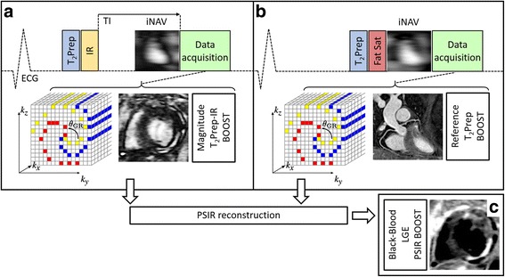
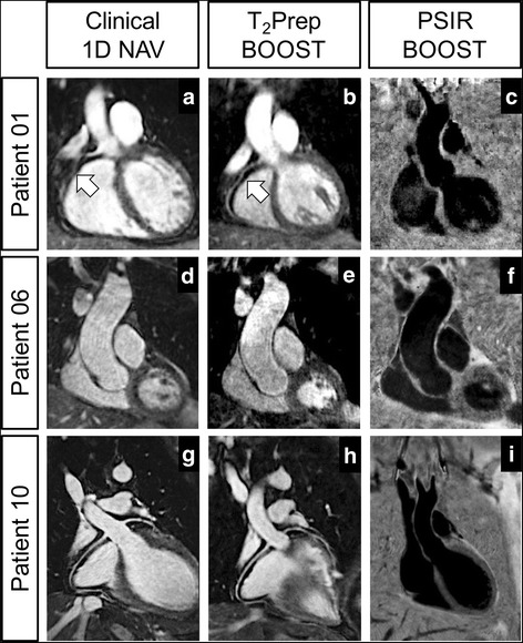
Methods: The proposed approach alternates the acquisition of a 3D volume preceded by a T2-prepared Inversion Recovery (T2Prep-IR) module (magnitude image) with the acquisition of a T2-prepared 3D volume (reference image). The two volumes (T2Prep-IR BOOST and bright-blood T2Prep BOOST) are combined in a PSIR-like reconstruction to obtain a complementary 3D black-blood volume for LGE assessment (PSIR BOOST). The black-blood PSIR BOOST and the bright-blood T2Prep BOOST datasets were compared to conventional clinical sequences for scar detection and coronary CMR angiography (CMRA) in 18 patients with a spectrum of cardiovascular disease (CVD).

Results: Datasets from 12 patients were quantitatively analysed. The black-blood PSIR BOOST dataset provided statistically improved contrast to noise ratio (CNR) between blood and scar when compared to a clinical 2D PSIR sequence (15.8 ± 3.3 and 4.1 ± 5.6, respectively). Overall agreement in LGE depiction was found between 3D black-blood PSIR BOOST and clinical 2D PSIR acquisitions, with 11/12 PSIR BOOST datasets considered diagnostic. The bright-blood T2Prep BOOST dataset provided high quality depiction of the proximal coronary segments, with improvement of visual score when compared to a clinical CMRA sequence. Acquisition time of BOOST (~10 min), providing information on both LGE uptake and heart anatomy, was comparable to that of a clinical single CMRA sequence.

Conclusions: The feasibility of BOOST for simultaneous black-blood LGE assessment and bright-blood coronary angiography was successfully tested in patients with cardiovascular disease. The framework enables free-breathing multi-contrast whole-heart acquisitions with 100% scan efficiency and predictable scan time. Complementary information on 3D LGE and heart anatomy are obtained reducing examination time.

Keywords: Black-blood; Bright-blood; Coronary MR angiography; Late gadolinium enhancement (LGE); Whole-heart.


Conflict of interest statement

Ethics approval and consent to participate

The study was approved by the National Research Ethics Service and written informed consent was obtained for all the subjects. Anonymized data were analyzed at the School of Biomedical Engineering and Imaging Sciences (King’s College London) at the St. Thomas’ Hospital.

Consent for publication

All the subjects provided written informed consent for the publication of accompanying images in this manuscript. The consent forms are held in the patients’ clinical notes and are available to the Editor-in-Chief upon request.

Competing interests

R.N. is employed by Siemens Healthcare Limited. All the other Authors declare that they do not have competing interests.

Publisher’s Note

Springer Nature remains neutral with regard to jurisdictional claims in published maps and institutional affiliations.

Figures

Fig. 1
Fig. 1
Proposed post-contrast BOOST framework for simultaneous 3D whole-heart bright-blood coronary angiography and black-blood late gadolinium enhancement (LGE) assessment. A T2-prepared inversion recovery (T2Prep-IR) module is applied at odd heartbeats (T2Prep-IR BOOST, magnitude image) (a), whereas data acquisition is T2 prepared and performed with a high flip angle at even heartbeats (bright-blood T2Prep-BOOST, reference image) (b). A 3D Cartesian trajectory with spiral profile order [28] is used for data acquisition; data collection is segmented over multiple heartbeats (yellow, red, blue) to minimize the effects of cardiac motion. Even heartbeat acquisitions include a SPIR pulse for fat saturation, while a STIR-like fat suppression is employed in odd heartbeats. 3D data acquisition at each heartbeat is preceded by a low-resolution 2D image-based navigator (iNAV) that is used to estimate translational respiratory motion along the superior-inferior and right-left directions. The two motion corrected datasets (T2Prep-IR BOOST and T2Prep BOOST) are combined in a PSIR-like reconstruction to generate a third, complementary, black-blood dataset (PSIR BOOST) for LGE visualization (c). The motion corrected bright-blood T2Prep BOOST dataset (reference image, b) provides adequate contrast for heart anatomy, great vessel, and coronary lumen visualization
Fig. 2
Fig. 2
Sequence simulations and phantom images comparing BOOST and conventional sequences for LGE assessment and CMRA. Simulated magnetization of the post-contrast BOOST sequence (a, b), of a conventional PSIR sequence (f, g), and of a dedicated post-contrast CMRA sequence (k, l) are displayed. The expected longitudinal magnetization (Mz/M0) is reported for blood (red lines), healthy viable myocardium (blue lines), and scar tissue (orange lines). Furthermore, results from the phantom experiments are displayed (BOOST: c, d, e; PSIR: h, i, j; CMRA: m) and the vial of interest are highlighted (blood – red circle, healthy viable myocardium – blue circle, and scar tissue – orange circle). Comparable contrast between the scar tissue and healthy viable myocardium can be observed in the PSIR reconstructions obtained with the BOOST sequence (PSIR BOOST) and the conventional PSIR sequence. Differently, improved contrast between the scar tissue and the healthy viable myocardium can be observed in the PSIR BOOST dataset when compared to the conventional PSIR sequence (phantom images in e and j). Data acquired with BOOST at even heartbeats (T2Prep BOOST, d) exhibit higher signal when compared to the reference image of the conventional PSIR sequence, acquired at a low flip-angle (i). In particular, T2Prep BOOST shows comparable signal and tissue contrast to that of a dedicated T2 prepared CMRA sequence (m)
Fig. 3
Fig. 3
Phantom images obtained with the BOOST and the conventional PSIR sequence. Imaging data were acquired by nulling the signal from the healthy viable myocardium (blue vial) in the magnitude images (a, e). Differently from f, the T2Prep BOOST dataset, acquired at a high flip-angle, exhibits both high signal from the blood (red vial) and pronounced contrast between blood and healthy viable myocardium (b). The PSIR reconstruction obtained with BOOST and using intensity normalization (d) shows reduced tissue contrast, which is restored once intensity normalization is not applied (c). Furthermore, such restored contrast between the scar tissue (orange vial) and the healthy viable myocardium is comparable to that of the PSIR reconstruction in (g), while improved contrast between scar and blood can be appreciated
Fig. 4
Fig. 4
Improvement in BOOST image quality after translational motion correction in two representative patients. The use of translational motion correction along the SI and RL directions reduces blurring artefacts and improves coronary vessel sharpness in the bright-blood T2Prep BOOST datasets (arrows in a, c, e, and g). Motion compensation recovers also small details as showed in the zoomed images. Furthermore, improved image sharpness can be observed on the black-blood PSIR-like reconstructions (arrows in b, d, f, and h), where a sharper delineation of the LGE uptake can be appreciated following motion correction (f versus h)
Fig. 5
Fig. 5
Comparison between the proposed 3D whole-heart BOOST framework and the clinical 2D PSIR acquisition. Images in a, e, and i show the LGE uptake as depicted in the T2Prep-IR BOOST datasets (white arrows), where signal from the blood pool is present and the viable myocardium is suppressed. Reformats in b, f, and j show the coronary reformats obtained from the 3D whole-heart bright-blood T2Prep BOOST dataset. Complementary 3D black-blood LGE images obtained with BOOST are shown in c, g, and k. All the images from the T2Prep-IR BOOST and the PSIR BOOST datasets were reformatted to match the orientation of the clinical 2D PSIR acquisitions (d, h, and l). The LGE uptake identified in both the T2Prep-IR BOOST and PSIR BOOST datasets matches that of the clinical 2D PSIR acquisition. Furthermore, improved contrast between the scar tissue and the blood pool can be appreciated in the 3D PSIR BOOST datasets when compared to the 2D PSIR acquisition (g, k versus h, l, orange arrows). LGE uptake appears more shallow and blood pool signal is not entirely suppressed in Patient 04 with myocarditis (cd, orange arrows), due to a longer TI
Fig. 6
Fig. 6
Comparison between conventional 3D whole-heart acquisition with diaphragmatic navigator and the proposed bright-blood T2Prep BOOST. Improved delineation of the RCA can be appreciated in Patient 01 with T2Prep BOOST when compared to the conventional CMRA acquisition (arrows in a, b). Dilated aorta can be observed in Patient 06 due to the presence of hypertensive heart disease (d, e). Excellent coronary delineation was obtained with both sequences in Patient 10 (g, h). Furthermore, the complementary black-blood PSIR BOOST datasets for LGE assessment are shown in c, f, and i
Fig. 7
Fig. 7
Fusion of the bright-blood T2Prep BOOST and the black blood PSIR BOOST datasets. Images correspond to two representative patients with positive LGE findings. Bright-blood images for visualization of the heart anatomy are shown in a, d (T 2 Prep-BOOST). Complementary visualization of scar tissue (PSIR BOOST) is shown in b, e; these datasets could potentially be used for an easy scar segmentation, as unclear border between the surrounding tissues and the scar itself have disappeared. Fusion images, where the anatomical localization of the scar can be retrieved, are shown in c, f

References

    1. Kim RJ, Fieno DS, Parrish TB, Harris K, Chen EL, Simonetti O, Bundy J, Finn JP, Klocke FJ, Judd RM. Relationship of MRI delayed contrast enhancement to irreversible injury, infarct age, and contractile function. Circulation. 1999;100:1992–2002. doi: 10.1161/01.CIR.100.19.1992. - DOI - PubMed
    1. Fieno DS, Kim RJ, Chen EL, Lomasney JW, Klocke FJ, Judd RM. Contrast-enhanced magnetic resonance imaging of myocardium at risk: distinction between reversible and irreversible injury throughout infarct healing. J Am Coll Cardiol. 2000;36:1985–1991. doi: 10.1016/S0735-1097(00)00958-X. - DOI - PubMed
    1. Friedrich MG, Strohm O, Schulz-Menger J, Marciniak H, Luft FC, Dietz R. Contrast media-enhanced magnetic resonance imaging visualizes myocardial changes in the course of viral myocarditis. Circulation. 1998;97:1802–1809. doi: 10.1161/01.CIR.97.18.1802. - DOI - PubMed
    1. Abdel-Aty H, Boye P, Zagrosek A, Wassmuth R, Kumar A, Messroghli D, Bock P, Dietz R, Friedrich MG, Schulz-Menger J. Diagnostic performance of cardiovascular magnetic resonance in patients with suspected acute myocarditis: comparison of different approaches. J Am Coll Cardiol. 2005;45:1815–1822. doi: 10.1016/j.jacc.2004.11.069. - DOI - PubMed
    1. Friedrich MG, Sechtem U, Schulz-Menger J, Holmvang G, Alakija P, Cooper LT, White JA, Abdel-Aty H, Gutberlet M, Prasad S, et al. Cardiovascular magnetic resonance in myocarditis: a JACC white paper. J Am Coll Cardiol. 2009;53:1475–1487. doi: 10.1016/j.jacc.2009.02.007. - DOI - PMC - PubMed

MeSH terms