Genome-wide 5-hydroxymethylcytosine patterns in human spermatogenesis are associated with semen quality
- PMID: 29179435
- PMCID: PMC5687605
- DOI: 10.18632/oncotarget.18331
Genome-wide 5-hydroxymethylcytosine patterns in human spermatogenesis are associated with semen quality
Abstract
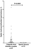
We performed immunofluorescent analysis of DNA hydroxymethylation and methylation in human testicular spermatogenic cells from azoospermic patients and ejaculated spermatozoa from sperm donors and patients from infertile couples. In contrast to methylation which was present throughout spermatogenesis, hydroxymethylation was either high or almost undetectable in both spermatogenic cells and ejaculated spermatozoa. On testicular cytogenetic preparations, 5-hydroxymethylcytosine was undetectable in mitotic and meiotic chromosomes, and was present exclusively in interphase spermatogonia Ad and in a minor spermatid population. The proportions of hydroxymethylated and non-hydroxymethylated diploid and haploid nuclei were similar among samples, suggesting that the observed alterations of 5-hydroxymethylcytosine patterns in differentiating spermatogenic cells are programmed. In ejaculates, a few spermatozoa had high 5-hydroxymethylcytosine level, while in the other ones hydroxymethylation was almost undetectable. The percentage of highly hydroxymethylated (5-hydroxymethylcytosine-positive) spermatozoa varied strongly among individuals. In patients from infertile couples, it was higher than in sperm donors (P<0.0001) and varied in a wider range: 0.12-21.24% versus 0.02-0.46%. The percentage of highly hydroxymethylated spermatozoa correlated strongly negatively with the indicators of good semen quality - normal morphology (r=-0.567, P<0.0001) and normal head morphology (r=-0.609, P<0.0001) - and strongly positively with the indicator of poor semen quality: sperm DNA fragmentation (r=0.46, P=0.001). Thus, the immunocytochemically detected increase of 5hmC in individual spermatozoa is associated with infertility in a couple and with deterioration of sperm parameters. We hypothesize that this increase is not programmed, but represents an induced abnormality and, therefore, it can be potentially used as a novel indicator of semen quality.
Keywords: 5-hydroxymethylcytosine; Pathology Section; human spermatogenesis; semen quality; sperm DNA fragmentation; testicular spermatogenic cells.
Conflict of interest statement
CONFLICTS OF INTEREST The authors declare that there is no conflict of interest.
Figures


Similar articles
-
Aberrant DNA methylation patterns of spermatozoa in men with unexplained infertility.Hum Reprod. 2015 May;30(5):1014-28. doi: 10.1093/humrep/dev053. Epub 2015 Mar 9. Hum Reprod. 2015. PMID: 25753583
-
Testicular function in a birth cohort of young men.Hum Reprod. 2015 Dec;30(12):2713-24. doi: 10.1093/humrep/dev244. Epub 2015 Sep 25. Hum Reprod. 2015. PMID: 26409015
-
A comparison of ejaculated and testicular spermatozoa aneuploidy rates in patients with high sperm DNA damage.Syst Biol Reprod Med. 2012 Jun;58(3):142-8. doi: 10.3109/19396368.2012.667504. Epub 2012 Mar 20. Syst Biol Reprod Med. 2012. PMID: 22432504
-
Genome-wide alteration in DNA hydroxymethylation in the sperm from bisphenol A-exposed men.PLoS One. 2017 Jun 5;12(6):e0178535. doi: 10.1371/journal.pone.0178535. eCollection 2017. PLoS One. 2017. PMID: 28582417 Free PMC article.
-
[Apoptosis during spermatogenesis and in ejaculated spermatozoa: importance for fertilization].Ann Biol Clin (Paris). 2001 Sep-Oct;59(5):531-45. Ann Biol Clin (Paris). 2001. PMID: 11602383 Review. French.
Cited by
-
Telomere Length in Chromosomally Normal and Abnormal Miscarriages and Ongoing Pregnancies and Its Association with 5-hydroxymethylcytosine Patterns.Int J Mol Sci. 2021 Jun 21;22(12):6622. doi: 10.3390/ijms22126622. Int J Mol Sci. 2021. PMID: 34205622 Free PMC article.
-
A View on Uterine Leiomyoma Genesis through the Prism of Genetic, Epigenetic and Cellular Heterogeneity.Int J Mol Sci. 2023 Mar 17;24(6):5752. doi: 10.3390/ijms24065752. Int J Mol Sci. 2023. PMID: 36982825 Free PMC article. Review.
-
A Decade of Exploring the Mammalian Sperm Epigenome: Paternal Epigenetic and Transgenerational Inheritance.Front Cell Dev Biol. 2018 May 15;6:50. doi: 10.3389/fcell.2018.00050. eCollection 2018. Front Cell Dev Biol. 2018. PMID: 29868581 Free PMC article. Review.
-
Environmental Impact on Male (In)Fertility via Epigenetic Route.J Clin Med. 2020 Aug 5;9(8):2520. doi: 10.3390/jcm9082520. J Clin Med. 2020. PMID: 32764255 Free PMC article. Review.
-
Telomere Length in Human Spermatogenic Cells as a New Potential Predictor of Clinical Outcomes in ART Treatment with Intracytoplasmic Injection of Testicular Spermatozoa.Int J Mol Sci. 2023 Jun 21;24(13):10427. doi: 10.3390/ijms241310427. Int J Mol Sci. 2023. PMID: 37445605 Free PMC article.
References
-
- White CR, MacDonald WA, Mann MR. Conservation of DNA Methylation Programming Between Mouse and Human Gametes and Preimplantation Embryos. Biol Reprod. 2016;95:61. - PubMed
LinkOut - more resources
Full Text Sources
Other Literature Sources

