

Osteoblast-oriented differentiation of BMSCs by co-culturing with composite scaffolds constructed using silicon-substituted calcium phosphate, autogenous fine particulate bone powder and alginate in vitro
- PMID: 29179436
- PMCID: PMC5687606
- DOI: 10.18632/oncotarget.19015
Osteoblast-oriented differentiation of BMSCs by co-culturing with composite scaffolds constructed using silicon-substituted calcium phosphate, autogenous fine particulate bone powder and alginate in vitro
Abstract
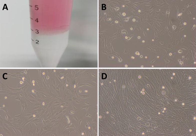
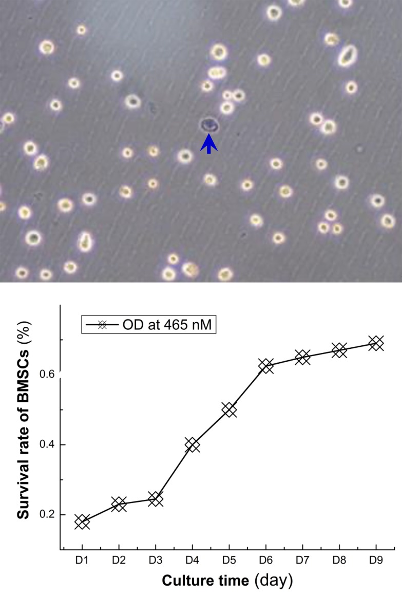
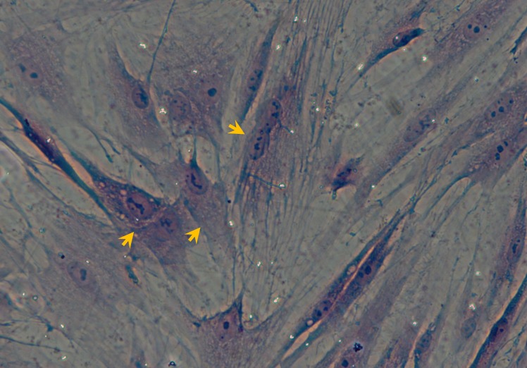
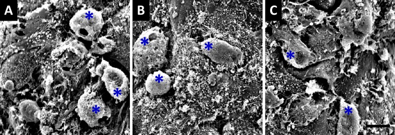
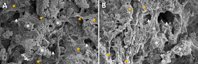
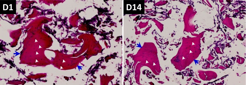
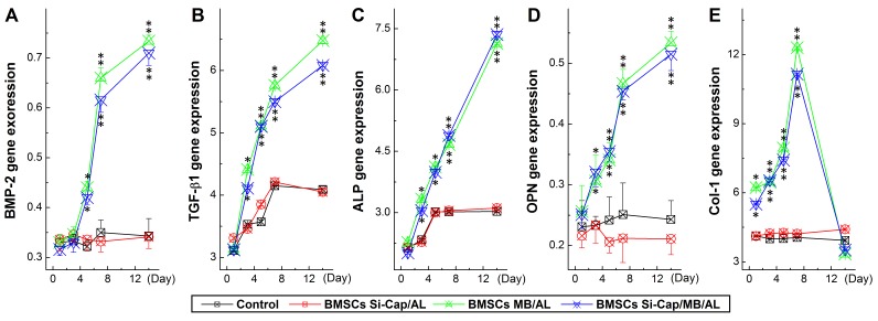
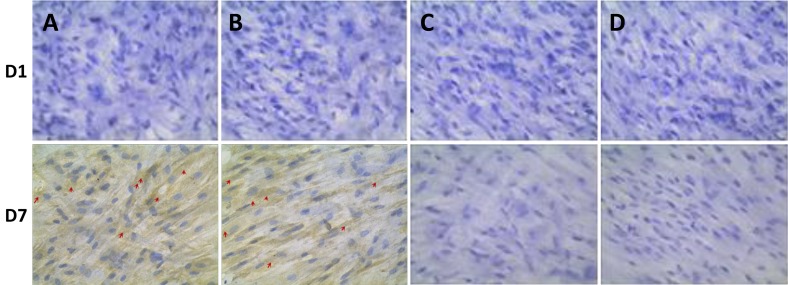
Autogenous bone graft is the best for spinal fusion in clinics, however, lacking sources, bleeding and infection are limited its practice. Seeking alternative materials are urgent for orthopaedic surgeon. Here, we evaluated osteoblast-oriented differentiation of rabbit BMSCs by co-culturing with composite scaffolds constructed using silicon-substituted-CaP-fine particulate bone powder-alginate. Using CCk8-kit, biocompatibility was evaluated by testing BMSCs proliferation; morphology and survival of osteoblasts within scaffolds were observed using EM and HE staining; growth factors and related genes were detected using RT-PCR. HE staining showed spindle-shaped BMSCs after the 3rd passage; EM data showed that uneven surface and longitudinal section were observed with scattered distribution of 5-100 mm interspaces, which leave enough space for BMSCs adhesion and growth. Interestingly, at 14-day culture with HE staining, osteocytes within the scaffolds grew well with regular shape and integrate structure. RT-PCR results showed that expression levels of BMP2, TGF-b and COL-I, ALP, OPN were increased significantly and time-dependently. Collectively, all mentioned effects were more obvious in co-culture BMSCs with scaffolds than those with other components. Immunohistochemistry showed that positive OPN expression was detected at 7-day co-culturing BMSCs with scaffold, rather than other situations. These results suggest that composite scaffolds constructed with Si-CaP-fine particulate bone powder-alginate have a certain degree of biocompatibility and bioactivity to promote osteoblast-oriented BMSCs differentiation.
Keywords: Pathology Section; autogenous fine particulate bone powder; bone marrow stromal cells; silicon-substituted calcium phosphate; spinal fusion; tissue-engineering.
Conflict of interest statement
CONFLICTS OF INTERESTS No conflict of interests.
Figures
Similar articles
-
A novel tissue-engineered bone graft composed of silicon-substituted calcium phosphate, autogenous fine particulate bone powder and BMSCs promotes posterolateral spinal fusion in rabbits.J Orthop Translat. 2020 Sep 14;26:151-161. doi: 10.1016/j.jot.2020.06.003. eCollection 2021 Jan. J Orthop Translat. 2020. PMID: 33437634 Free PMC article.
-
[Effect of osteoblast conditioned culture medium on differentiation of BMSCs].Zhongguo Xiu Fu Chong Jian Wai Ke Za Zhi. 2009 Feb;23(2):145-50. Zhongguo Xiu Fu Chong Jian Wai Ke Za Zhi. 2009. PMID: 19275092 Chinese.
-
Osteoinduction and proliferation of bone-marrow stromal cells in three-dimensional poly (ε-caprolactone)/ hydroxyapatite/collagen scaffolds.J Transl Med. 2015 May 8;13:152. doi: 10.1186/s12967-015-0499-8. J Transl Med. 2015. PMID: 25952675 Free PMC article.
-
In vitro and in vivo evaluation of differentially demineralized cancellous bone scaffolds combined with human bone marrow stromal cells for tissue engineering.Biomaterials. 2005 Jun;26(16):3173-85. doi: 10.1016/j.biomaterials.2004.08.020. Biomaterials. 2005. PMID: 15603812
-
Synthesis of and in vitro and in vivo evaluation of a novel TGF-β1-SF-CS three-dimensional scaffold for bone tissue engineering.Int J Mol Med. 2016 Aug;38(2):367-80. doi: 10.3892/ijmm.2016.2651. Epub 2016 Jun 21. Int J Mol Med. 2016. PMID: 27352815 Free PMC article.
Cited by
-
A novel tissue-engineered bone graft composed of silicon-substituted calcium phosphate, autogenous fine particulate bone powder and BMSCs promotes posterolateral spinal fusion in rabbits.J Orthop Translat. 2020 Sep 14;26:151-161. doi: 10.1016/j.jot.2020.06.003. eCollection 2021 Jan. J Orthop Translat. 2020. PMID: 33437634 Free PMC article.
-
Corrosion Resistance and Biocompatibility Assessment of a Biodegradable Hydrothermal-Coated Mg-Zn-Ca Alloy: An in Vitro and in Vivo Study.ACS Omega. 2020 Feb 25;5(9):4548-4557. doi: 10.1021/acsomega.9b03889. eCollection 2020 Mar 10. ACS Omega. 2020. PMID: 32175501 Free PMC article.
-
Expression of microRNA-21 in osteoporotic patients and its involvement in the regulation of osteogenic differentiation.Exp Ther Med. 2019 Jan;17(1):709-714. doi: 10.3892/etm.2018.6998. Epub 2018 Nov 21. Exp Ther Med. 2019. PMID: 30651854 Free PMC article.
-
Let-7i-5p functions as a putative osteogenic differentiation promoter by targeting CKIP-1.Cytotechnology. 2021 Feb;73(1):79-90. doi: 10.1007/s10616-020-00444-1. Epub 2021 Jan 4. Cytotechnology. 2021. PMID: 33505116 Free PMC article.
-
Autogenous bone particles combined with platelet-rich plasma can stimulate bone regeneration in rabbits.Exp Ther Med. 2020 Dec;20(6):279. doi: 10.3892/etm.2020.9409. Epub 2020 Oct 27. Exp Ther Med. 2020. PMID: 33200004 Free PMC article.
References
-
- Younger EM, Chapman MW. Morbidity at bone graft donor sites. J Orthop Trauma. 1989;3:192–95. https://doi.org/10.1097/00005131-198909000-00002. - DOI - PubMed
-
- Banwart JC, Asher MA, Hassanein RS. Iliac crest bone graft harvest donor site morbidity. A statistical evaluation. Spine. 1995;20:1055–60. https://doi.org/10.1097/00007632-199505000-00012. - DOI - PubMed
-
- Kruppke B, Farack J, Wagner AS, Beckmann S, Heinemann C, Glenske K, Rößler S, Wiesmann HP, Wenisch S, Hanke T. Gelatine modified monetite as a bone substitute material: an in vitro assessment of bone biocompatibility. Acta Biomater. 2016;32:275–85. https://doi.org/10.1016/j.actbio.2015.12.035. - DOI - PubMed
-
- Zan X, Sitasuwan P, Feng S, Wang Q. Effect of Roughness on in Situ Biomineralized CaP-Collagen Coating on the Osteogenesis of Mesenchymal Stem Cells. Langmuir. 2016;32:1808–17. https://doi.org/10.1021/acs.langmuir.5b04245. - DOI - PubMed
-
- Sun C, Tian Y, Xu W, Zhou C, Xie H, Wang X. Development and performance analysis of Si-CaP/fine particulate bone powder combined grafts for bone regeneration. Biomed Eng Online. 2015;14:47. https://doi.org/10.1186/s12938-015-0042-4. - DOI - PMC - PubMed
LinkOut - more resources
Full Text Sources
Other Literature Sources
Research Materials
Miscellaneous
