. 2016 Aug 19;8(51):88386-88400.
doi: 10.18632/oncotarget.11393. eCollection 2017 Oct 24.

SH003 suppresses breast cancer growth by accumulating p62 in autolysosomes

Affiliations

SH003 suppresses breast cancer growth by accumulating p62 in autolysosomes

Youn Kyung Choi et al. Oncotarget. .

Abstract

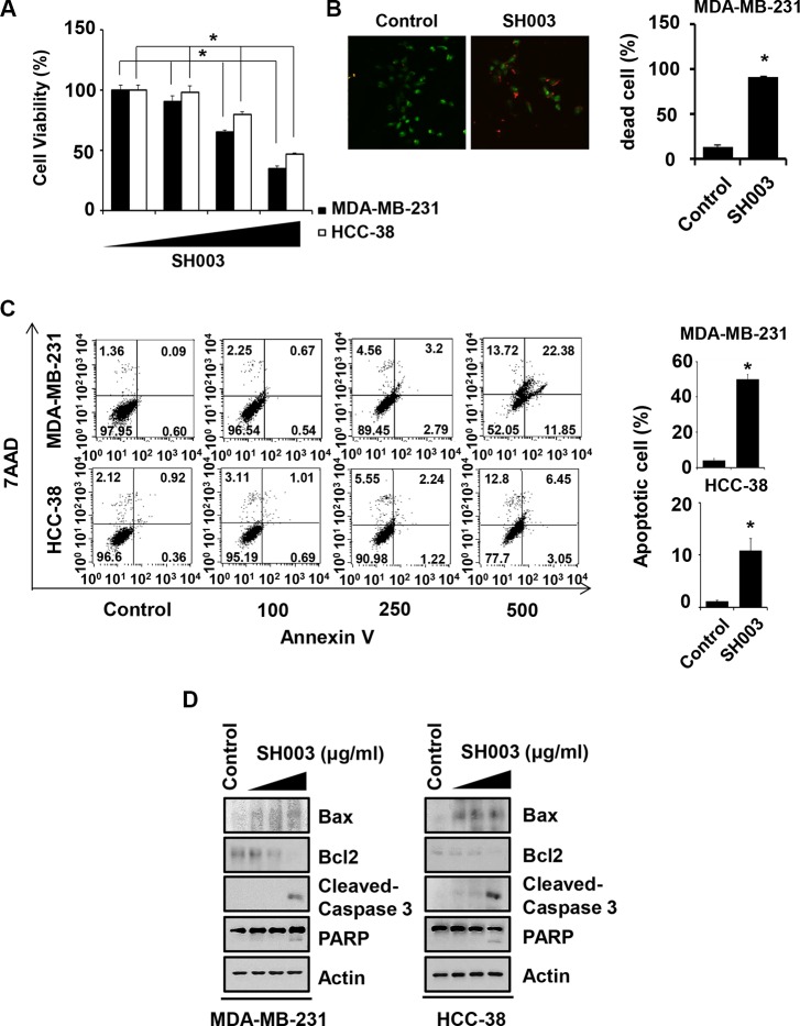
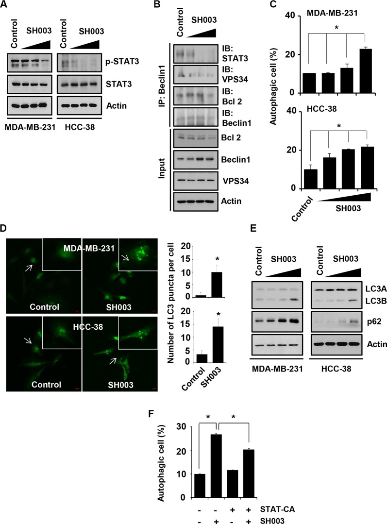
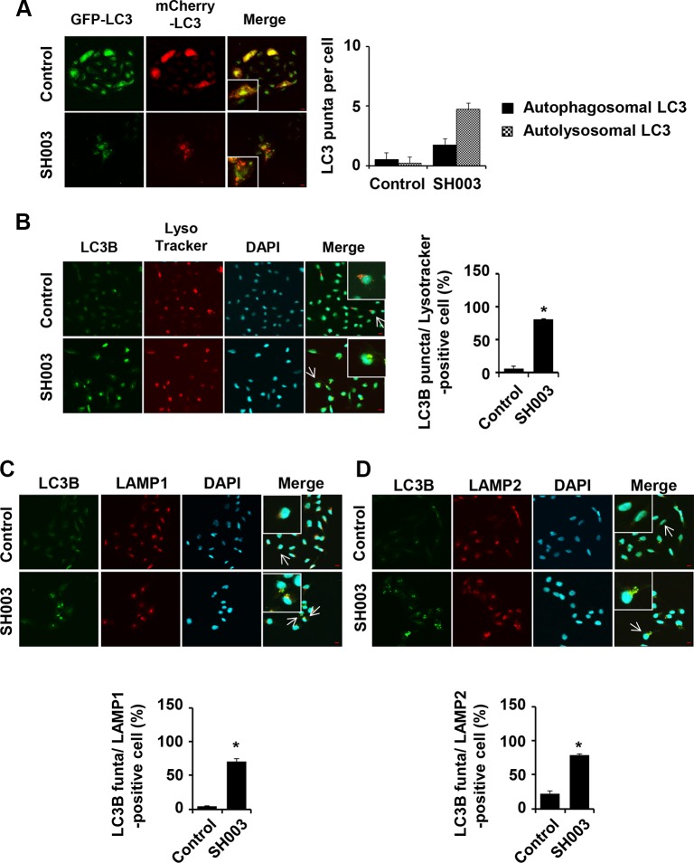
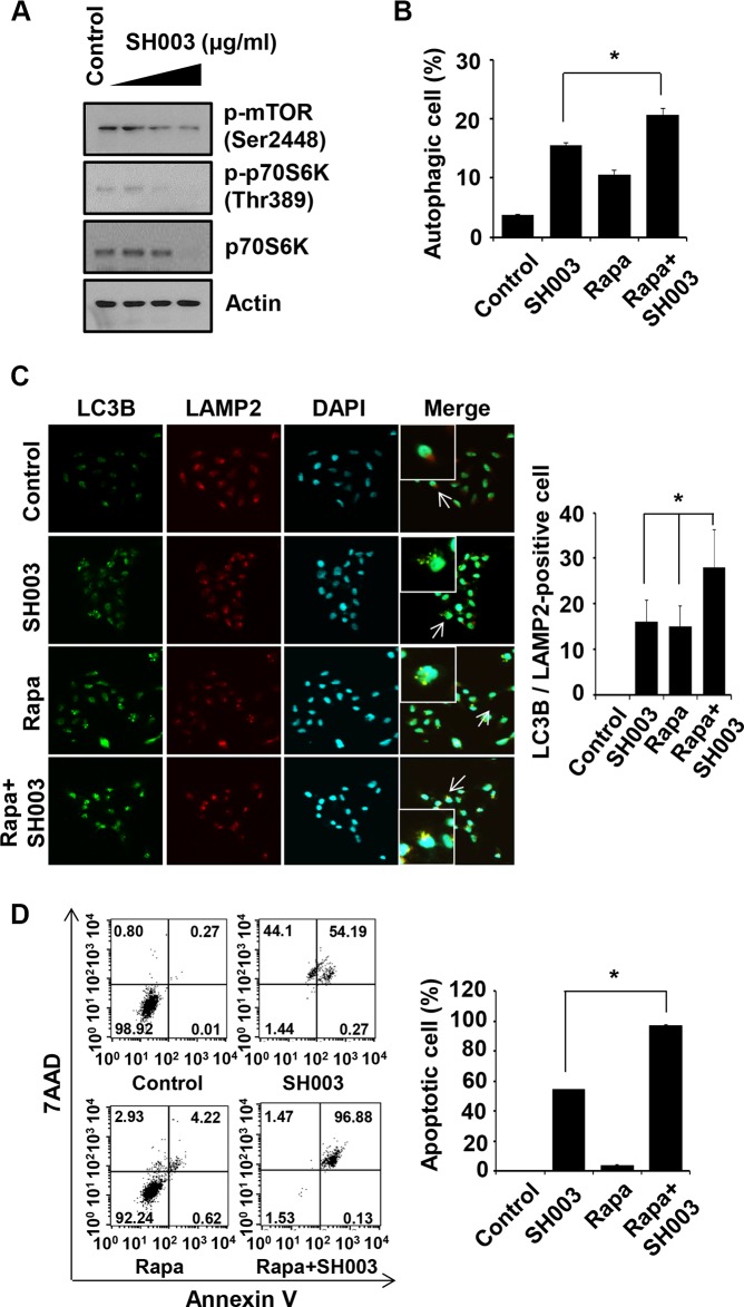
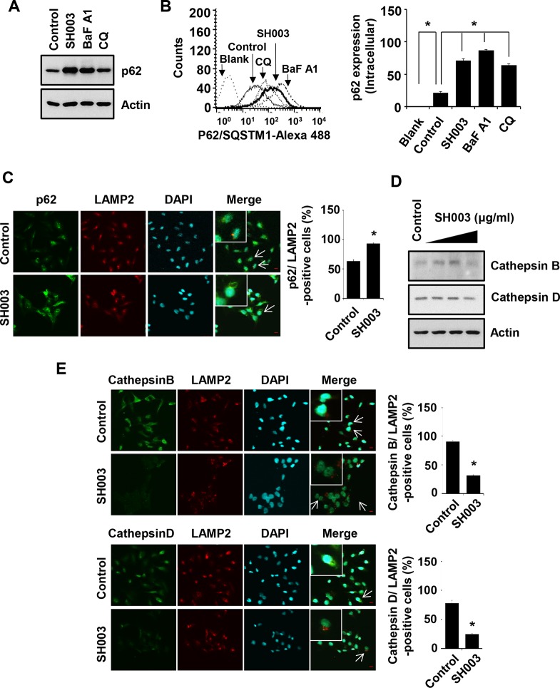
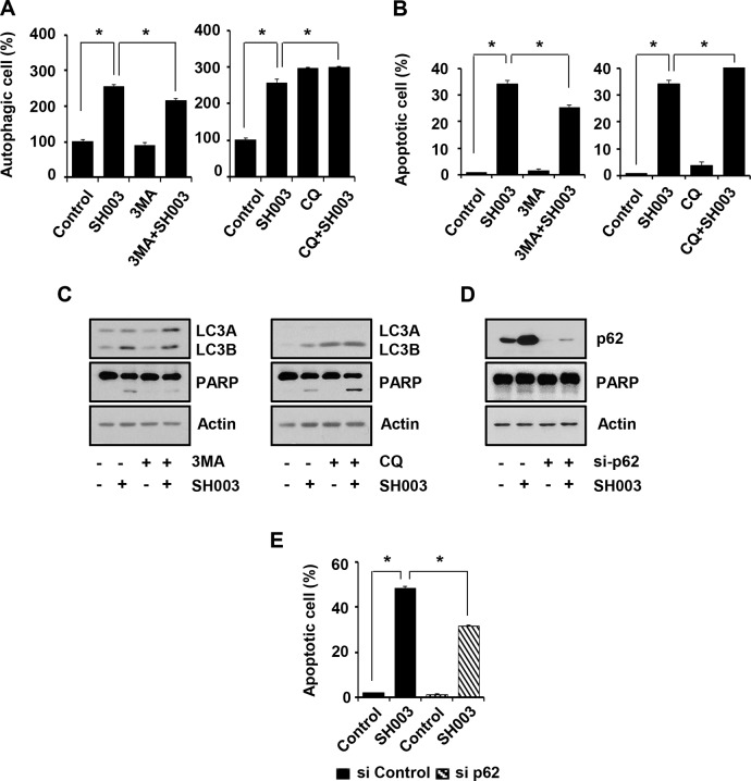
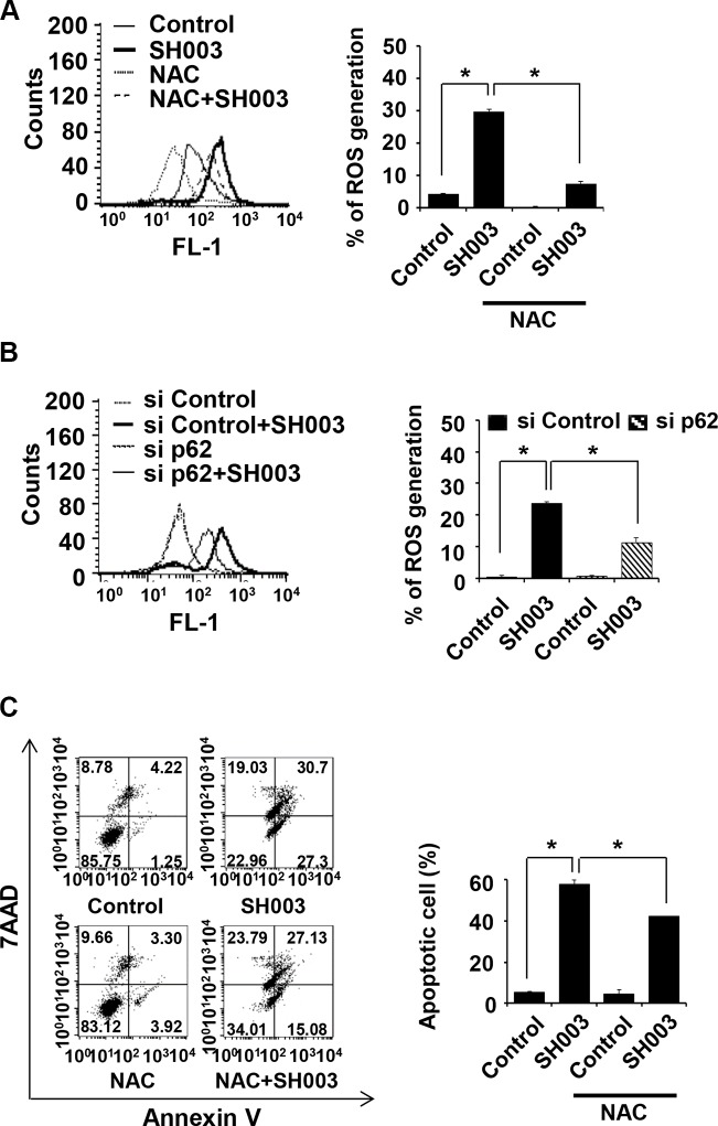
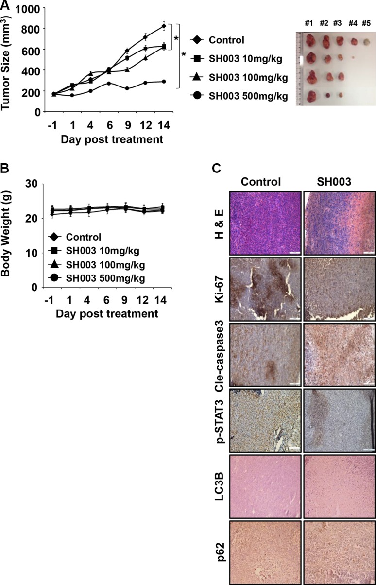
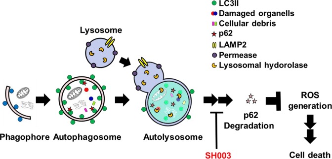
Drug markets revisits herbal medicines, as historical usages address their therapeutic efficacies with less adverse effects. Moreover, herbal medicines save both cost and time in development. SH003, a modified version of traditional herbal medicine extracted from Astragalus membranaceus (Am), Angelica gigas (Ag), and Trichosanthes Kirilowii Maximowicz (Tk) with 1:1:1 ratio (w/w) has been revealed to inhibit tumor growth and metastasis on highly metastatic breast cancer cells, both in vivo and in vitro with no toxicity. Meanwhile, autophagy is imperative for maintenance cellular homeostasis, thereby playing critical roles in cancer progression. Inhibition of autophagy by pharmacological agents induces apoptotic cell death in cancer cells, resulting in cancer treatment. In this study, we demonstrate that SH003-induced autophagy via inhibiting STAT3 and mTOR results in an induction of lysosomal p62/SQSTM1 accumulation-mediated reactive oxygen species (ROS) generation and attenuates tumor growth. SH003 induced autophagosome and autolysosome formation by inhibiting activation of STAT3- and mTOR-mediated signaling pathways. However, SH003 blocked autophagy-mediated p62/SQSTM1 degradation through reducing of lysosomal proteases, Cathepsins, resulting in accumulation of p62/SQSTM1 in the lysosome. The accumulation of p62/SQSTM1 caused the increase of ROS, which resulted in the induction of apoptotic cell death. Therefore, we conclude that SH003 suppresses breast cancer growth by inducing autophagy. In addition, SH003-induced p62/SQSTM1 could function as an important mediator for ROS generation-dependent cell death suggesting that SH003 may be useful for treating breast cancer.

Keywords: SH003; apoptosis; autophagy; breast cancer; p62.


Conflict of interest statement

CONFLICTS OF INTEREST The authors declare that they no conflicts interests.

Figures

Figure 1
Figure 1. SH003 induces apoptotic cell death
(A) Cell viability was measured by MTT assay. MDA-MB-231 and HCC-38 cells were seed in 96-well plates and treated with various concentration of SH003 (0, 100, 250 and 500 μg/ml) for 48 hours. Data were analyzed by ANOVA with P < 0.05. (B) After MDA-MB-231 cells were treated with 500 μg/ml of SH003 for 48 hours, live and dead assay was done by using live and dead cell assay kit. Dead cells (Red fluorescence-positive cells) were counted. *P < 0.05. (C) MDA-MB-231 and HCC-38 cells were treated with different doses of SH003 for 48 hours. Cells were stained with Annexin V and 7AAD at room temperature in the dark. Annexin V-positive apoptotic cells were detected using FACSCalibur. *P < 0.05. Graph shows annexin V-positive apoptotic cells (%) calculated from the total amount of right-upper and -lower portion. (D) Cells were treated with 500 μg/ml of SH003 for 24 hours and then performed western blots with anti-Bax, -Bcl2, -Cleaved caspase 3 and PARP. Actin was used for the internal control. Experiments were performed in triplicate. Bars indicate means that standard deviations (SD).
Figure 2
Figure 2. SH003 induces autophagy by suppressing STAT3 phosphorylation
(A) MDA-MB-231 and HCC-38 cells were treated with different doses of SH003 for 15 minutes and then performed western blots with anti-p-STAT3 and STAT3. Actin was used for the internal control. (B) Cells were treated with SH003 for 24 hours and whole-cell lysates were immunoprecipitated with anti-Beclin1 antibody. The immunoprecipitants and input proteins were then blotted with the antibodies for STAT3, VPS34, BCl-2, Beclin1 and actin. (C) MDA-MB-231 and HCC-38 cells were treated with SH003 (0, 100, 250 and 500 μg/ml) for 24 hours and then stained with Cyto-ID fluorescence dye for 30 minutes at room temperature in the dark. Data analyzed using a FACSCalibur. Data were analyzed by ANOVA with P < 0.05. (D) MDA-MB-231 and HCC-38 cells were treated with 500 μg/ml of SH003 for 24 hours and then stained with anti-LC3B antibody (1 μg/ml) and anti-Alexa Fluor-488 (1:250) antibody. LC3 punctate in the cells were analyzed using Olympus FV10i Self Contained Confocal Laser System. The object was 20× and scale bar indicates 10 μm. *P < 0.05. (E) Analysis of autophagy-related molecules. Cells were treated with SH003 for 24 hours and whole-cell lysates were analyzed by western blots with anti-LC3A/B and p62/SQSTM1. Actin was used for the loading control. (F) Cells were transfected with STAT3-CA and treated with SH003 for 24 hours. Autophagosome formation was stained with Cyto-ID fluorescence. *P < 0.05. Experiments were performed in triplicate. Bars indicate means that standard deviations (SD).
Figure 3
Figure 3. SH003 induces autolysosome formation
(A) Stable expression of mCherry-GFP-LC3 MDA-MB-231 cells were treated with 500 μg/ml of SH003 for 24 hours and images were obtained with using Olympus FV10i Self Contained Confocal Laser System. Yellow (double staining with GFP and RFP) and red (staining with only RFP) florescence were stained for autophagosome and autolysosome, respectively. The object was 20× and scale bar indicates 10 μm. *P < 0.05. (B) MDA-MB-231 cells were treated with SH003 for 24 hours and stained with DND-99 lysotracker dye (75 nM) for 1 hour at 37°C. After fixation, permeabilization and blocking, cells were stained with anti-LC3B and Alexa-488 antibodies. DAPI was used for nucleus staining. (C) Cells were treated with SH003 for 24 hours and stained with anti-LC3B and LAMP1 (1:100) antibodies. (D) MDA-MB-231 cells were stained with anti-LC3B and LAMP2 (1:100) antibody. Colocalization with LC3B and LAMP2 was analyzed using Olympus FV10i Self Contained Confocal Laser System. The object was 20× and scale bar indicates 10 μm. *P < 0.05.
Figure 4
Figure 4. Rapamycin enhances SH003-induced autophagy-mediated apoptosis
(A) MDA-MB-231 cells were treated with different concentrations of SH003 (0, 100, 250 and 500 μg/ml) for 24 hours and then subjected to western blots with the antibodies indicated (anti-p-mTOR, -p-p70S6K and -p70S6K). Actin was used as internal control. (B) Cells were treated with 10 μM of rapamycin (Rapa) and 500 μg/ml of SH003 and then autophagosome vacuoles were measured by Cyto-ID fluorescence. Data analyzed using a FACSCalibur. *P < 0.05. (C) MDA-MB-231 cells were treated with rapamycin and SH003 and then stained with anti-LC3B and LAMP2 antibodies. Colocalization with LC3B and LAMP2 were analyzed using Olympus FV10i Self Contained Confocal Laser System. The object was 20× and scale bar indicates 10 μm. *P < 0.05. (D) Cells were treated with rapamycin and SH003 for 48 hours and then stained with Annexin V and 7AAD at room temperature in the dark. Annexin V-positive apoptotic cells were detected using FACSCalibur. *P < 0.05. Representative data were presented as the means and standard deviations (SD).
Figure 5
Figure 5. SH003 induces p62 accumulation via reduction of Cathepsin expression
(A) MDA-MB-231 cells were treated with SH003 (500 μg/ml), BaFA1 (400 nM) and CQ (10 μM) for 24 hours. p62 protein expression was objected with western blots. Actin was used for the internal control. (B) Cells were treated with SH003 and autophagy inhibitors (BaFA1 and CQ) for 24 hours and stained with p62 -Alexa 488-conjugated p62 antibody for 30 minutes. p62 accumulation in the cells were detected using FACSCalibur. *P < 0.05. (C) MDA-MB-231 cells were treated with SH003 for 24 hours and stained with p62 (1 μg/ml) and LAMP2. DAPI was used as nucleus staining. The object was 20× and scale bar indicates 10 μm. *P < 0.05. (D) Cells were treated with SH003 for 24 hours and then performed western blots with anti-Cathepsin B and -Cathepsin D. Actin was used for loading control. (E) MDA-MB-231 cells were treated with 500 μg/ml of SH003 for 24 hours and stained with Cathepsin B (1:50)/LAMP2 and Cathepsin D (1:50)/LAMP2. Images were obtained with using Olympys FV10i Self Contained Confocal Laser System. The object was 20× and scale bar indicates 10 μm. *P < 0.05. Experiments were performed in triplicate. Bars indicate means that standard deviations (SD).
Figure 6
Figure 6. SH003-induced p62 accumulation causes autophagy-mediated apoptosis
(A) MDA-MB-231 cells were pretreated with 1 mM of 3MA and 10 μM of CQ for 30 minutes and then treated with 500 μg/ml of SH003. After 24 hours, cells were stained with Cyto-ID fluorescence dye for 30 minutes and analyzed using a FACSCalibur. *P < 0.05. (B) Cells were treated with SH003 for 24 hours and then stained with annexin V and 7AAD at room temperature in the dark. Autophagy inhibitors (3MA and CQ) were treated 30 minutes before SH003 treatment. Annexin V-positive apoptotic cells were detected using FACSCalibur. *P < 0.05. (C) MDA-MB-231 cells were pretreated with 3MA and CQ for 30 minutes and then treated with SH003. 24 hours after treatment, LC3A/B, PARP and actin levels were examined. (D) MDA-MB-231 cells were transfected with p62 siRNA, treated with SH003 and performed western blots with anti-p62 and -PARP antibodies. Actin was used as internal controls. (E) Cells were transfected with control siRNA and p62 siRNA and then treated with 500 μg/ml of SH003. After 48 hours, cells were stained with Annexin V and 7AAD at room temperature in the dark. Annexin V-positive apoptotic cells were detected using FACSCalibur. *P < 0.05. Experiments were performed in triplicate. Bars indicate means that standard deviations (SD).
Figure 7
Figure 7. SH003-induced p62 accumulation causes ROS-mediated apoptotic cell death
(A) MDA-MB-231 cells were pretreated with or without NAC (2.5 mM) for 1 hour, followed by exposure to SH003. After 24 hours, cells were stained with H2DCFDA for 1 hour at 37°C. ROS generation was detected with using FACSCalibur by the FL1 channel. (B) Cells were transfected with control siRNA and p62 siRNA and then treated with SH003 for 24 hours. ROS generation was measured by FACSCalibur. *P < 0.05. (C) Cells were pretreated with NAC for 1 hour and then treated with 500 μg/ml of SH003 for 48 hours. Cells were stained with annexin V and 7AAD. *P < 0.05. Experiments were performed in triplicate. Bars indicate means that standard deviations (SD).
Figure 8
Figure 8. SH003 suppresses in vivo tumor growth
(A) 5 × 10⁵ MDA-MB-231 cells were s.c. injected and mice (n = 5/group) were p.o. administrated daily with different concentrations of SH003 (10, 100 and 500 mg/kg) for 15 days. Xenograft tumor volumes and body weight of mice were measured three times a week. Tumor volumes were calculated using the following: Tumor volume (cubic millimeters) = width2 × length/2. Data were analyzed by ANOVA with P < 0.05. (B) Body weight (C) Tumor tissues were stained with hematoxylin, eosin and antibodies indicated (anti-Ki-67, -Cleaved caspase 3, -p-STAT3, -LC3B and -p62). Images were obtained at 20× magnification. The scale bar indicates 100 μm. Bars indicate means that standard deviations (SD).
Figure 9
Figure 9. A schematic representation of the mechanisms for SH003 suppression of breast cancer growth

References

    1. Puissant A, Fenouille N, Auberger P. When autophagy meets cancer through p62/SQSTM1. Am J Cancer Res. 2012;2:397–413. - PMC - PubMed
    1. Codogno P, Meijer AJ. Autophagy and signaling: their role in cell survival and cell death. Cell Death Differ. 2005;12:1509–1518. - PubMed
    1. Mijaljica D, Prescott M, Klionsky DJ, Devenish RJ. Autophagy and vacuole homeostasis: a case for self-degradation? Autophagy. 2007;3:417–421. - PubMed
    1. Amaravadi RK, Lippincott-Schwartz J, Yin XM, Weiss WA, Takebe N, Timmer W, DiPaola RS, Lotze MT, White E. Principles and current strategies for targeting autophagy for cancer treatment. Clin Cancer Res. 2011;17:654–666. - PMC - PubMed
    1. Ryter SW, Lee SJ, Smith A, Choi AM. Autophagy in vascular disease. Proc Am Thorac Soc. 2010;7:40–47. - PMC - PubMed