. 2017 Aug 3;8(51):88563-88574.
doi: 10.18632/oncotarget.19902. eCollection 2017 Oct 24.

Traditional Chinese medicine Danggui Buxue Tang inhibits colorectal cancer growth through induction of autophagic cell death

Affiliations

Traditional Chinese medicine Danggui Buxue Tang inhibits colorectal cancer growth through induction of autophagic cell death

Shun-Ting Chen et al. Oncotarget. .

Abstract

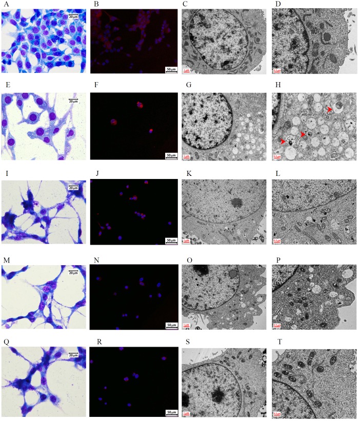
Purpose: The induction of autophagic cell death is an important process in the development of anticancer therapeutics. We aimed to evaluate the activity of the ancient Chinese decoction Danggui Buxue Tang (DBT) against colorectal cancer (CRC) and the associated autophagy-related mechanism.

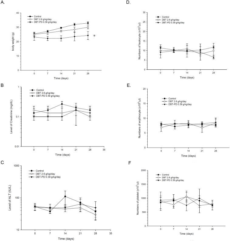
Materials and methods: CT26 CRC cells were implanted into syngeneic BALB/c mice for the tumor growth assay. DBT extracts and DBT-PD (polysaccharide-depleted) fractions were orally administered. The toxicity profiles of the extracts were analyzed using measurements of body weight, hemogram, and biochemical parameters. The morphology of tissue sections was observed using light and transmission electron microscopy. Western blotting and small interference RNA assays were used to determine the mechanism.

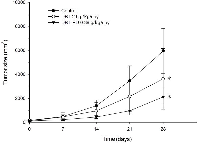
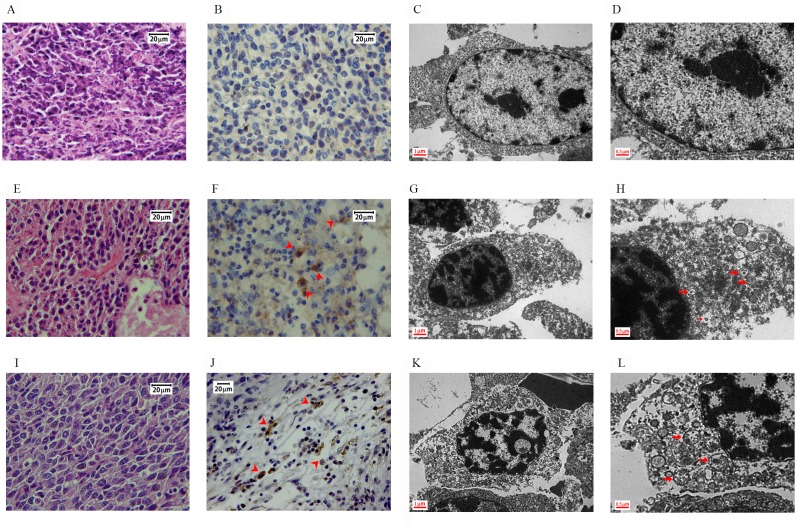
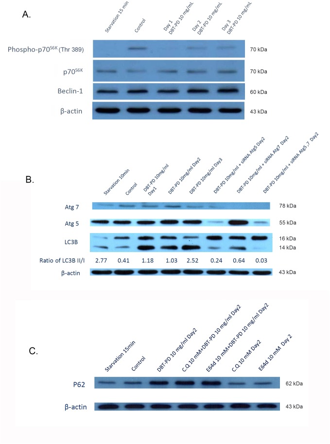
Results: DBT-PD and DBT, which contained an equal amount of DBT-PD, inhibited CT26 syngeneic tumor growth. In the tumor specimen, the expression of microtubule-associated proteins 1A/1B light chain 3B (LC3B) was upregulated by DBT-PD and DBT. The development of autophagosomes was observed via transmission electron microscopy in tumors treated with DBT-PD and DBT. In vitro experiments for mechanism clarification demonstrated that DBT-PD could induce autophagic death in CT26 cells accompanied by LC3B lipidation, downregulation of phospho-p70s6k, and upregulation of Atg7. RNA interference of Atg7, but not Atg5, partially reversed the effect of DBT-PD on LC3B lipidation and expression of phospho-p70s6k and Atg7. The changes in ultrastructural morphology and LC3B expression induced by DBT-PD were also partially blocked by the knockdown of Atg7 mRNA.

Conclusion: DBT induced autophagic death of colorectal cancer cells through the upregulation of Atg7 and modulation of the mTOR/p70s6k signaling pathway.

Keywords: Danggui Buxue Tang; autophagy; colorectal cancer; mammalian target of rapamycin (mTOR); traditional Chinese medicine.


Conflict of interest statement

CONFLICTS OF INTEREST The authors declare no conflicts of interest.

Figures

Figure 1
Figure 1. Tumor size in BALB/c mice implanted with CT26 colorectal adenocarcinoma cells
Both DBT 2.6 g/kg/day (○) and DBT-PD 0.39 g/kg/day (▼) produced better inhibition of tumor growth than that observed in the control group (●) (p<0.05 *). DBT-PD demonstrated the most dramatic efficacy of these three groups, although the components of polysaccharide-depleted fraction in 2.6 g DBT were equal to that in 0.39 g DBT-PD.
Figure 2
Figure 2. Changes in body weight, serum chemistry, and blood cells of BALB/c mice implanted with CT26 colorectal adenocarcinoma cells
Although a mild decrease of body weight in the DBT-PD group was observed (p<0.05 *), no significant differences in plasma ALT, creatinine levels, and blood cell counts were observed (p>0.05).
Figure 3
Figure 3. Morphology of BALB/c mice implanted with CT26 colorectal adenocarcinoma cells
Images (A to D): control group; (E to H): 2.6 g/kg DBT-treated group; (I to L): 0.39 g/kg DBT-PD-treated group. Hematoxylin and eosin staining showed a more condensed nucleus in the control group (Figure 3A) than in the treatment groups (Figure 3E and 3I). After the immunohistochemistry staining for LC3 protein, both treatment groups (Figure 3F and 3J) showed more brown color (arrowhead) than the control group (Figure 3B), which was and indicator of the activity of the LC3 protein. When examined under a transmission electron microscope (TEM), both the DBT- and DBT-PD-treatment groups showed the presence of autophagosomes (arrow), including the appearance of multiple cytoplasmic vacuoles with double-layered membranes (Figure 3G, 3H, 3K, and 3L), which were autophagosomes that contained degraded organelles, such as markedly swollen mitochondria. Some fusion vacuoles were autolysosomes (asterisk).
Figure 4
Figure 4. Western blot of CT26 colorectal adenocarcinoma cells after treatment of 10 mg/mL DBT-PD
In (A), after the treatment with DBT-PD, phospho-p70S6K decreased dramatically and LC3-I was increase of amount with sequentially transformed to LC3-II. Atg5, Atg7 (B) and p62 (C) were activated after treated of DBT-PD but not in chloroquine (CQ) and E64D groups to p62 proteins.
Figure 5
Figure 5. Morphology of CT26 colorectal adenocarcinoma cells after treatment with 10 mg/mL DBT-PD for 48 h
Images (A to D): control group; (E to H): 10 mg/mL DBT-PD-treatment, (I to L): Atg5 siRNA plus DBT-PD treatment; (M to P): Atg7 siRNA plus DBT-PD treatment; (Q to T): Atg5 and Atg7 siRNA plus DBT-PD treatment. Following Liu's staining, abundant cytoplasmic vacuoles were observed in CT26 cells treated with 10 mg/mL DBP-PD relative to control cells (Figure 5A and 5E), but not in cells transfected with siRNA to Atg5 and Atg7 (Figure 5I, 5M and 5Q). The LC3 protein was expressed diffusely in untreated cells and transformed to a punctate distribution pattern of intense acridine orange in DBT-PD-treated cells; however, this phenomenon disappeared after the transfection of Atg5 and Atg7 siRNA (Figure 5B, 5F, 5J, 5N and 5R). When compared to the treatment group alone, the siRNA-treated groups filed to form a complete autophagosome (arrowhead), although the phagophore (arrow) formation had already commenced (Figure 5D, 5H, 5L, 5P and 5T).

References

    1. Torre LA, Bray F, Siegel RL, Ferlay J, Lortet-Tieulent J, Jemal A. Global cancer statistics, 2012. CA Cancer J Clin. 2015;65:87–108. - PubMed
    1. Eddy DM. Screening for colorectal cancer. Ann Intern Med. 1990;113:373–384. - PubMed
    1. Siegel RL, Miller KD, Jemal A. Cancer statistics, 2016. CA Cancer J Clin. 2016;66:7–30. - PubMed
    1. Center MM, Jemal A, Smith RA, Ward E. Worldwide variations in colorectal cancer. CA Cancer J Clin. 2009;59:366–378. - PubMed
    1. O'Connell JB, Maggard MA, Ko CY. Colon cancer survival rates with the new American Joint Committee on Cancer sixth edition staging. J Natl Cancer Inst. 2004;96:1420–1425. - PubMed

LinkOut - more resources