. 2018 Feb 15;35(4):671-680.
doi: 10.1089/neu.2017.5015. Epub 2018 Jan 11.

Erythropoietin Attenuates the Brain Edema Response after Experimental Traumatic Brain Injury

Affiliations

Erythropoietin Attenuates the Brain Edema Response after Experimental Traumatic Brain Injury

Jonas Blixt et al. J Neurotrauma. .

Abstract

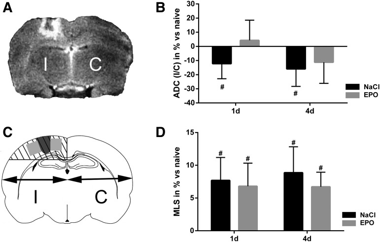
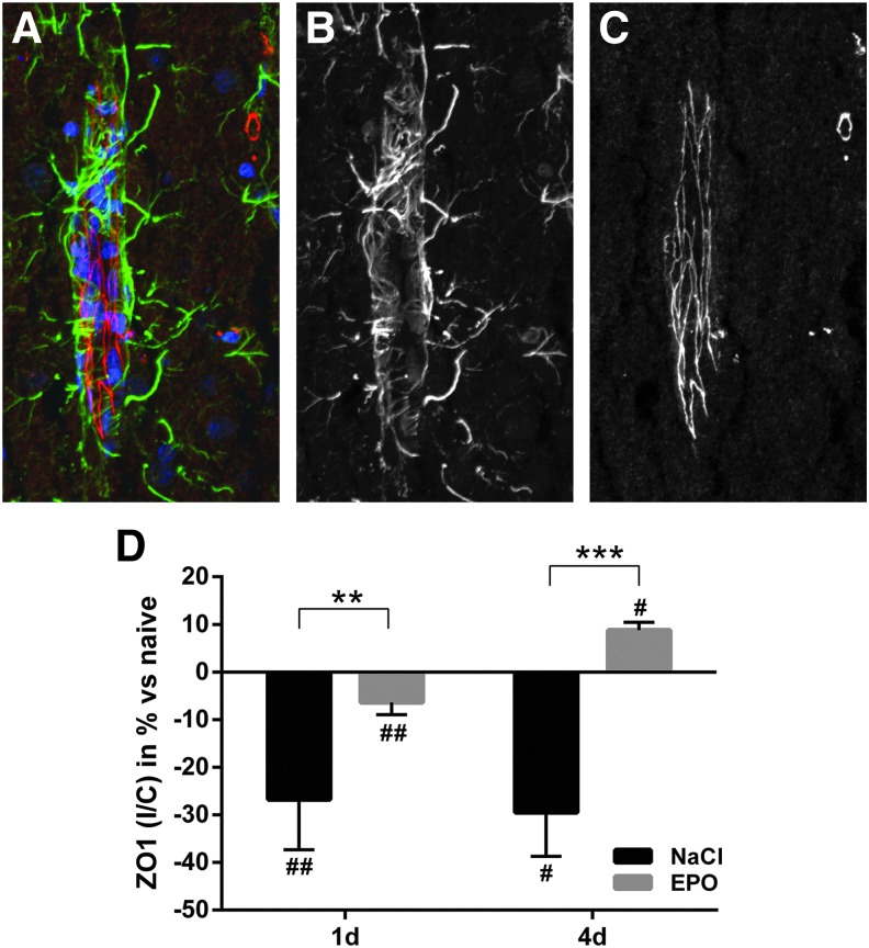
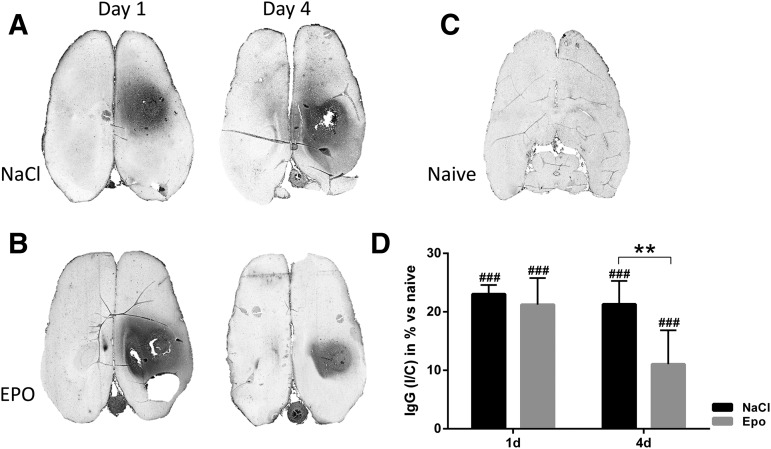
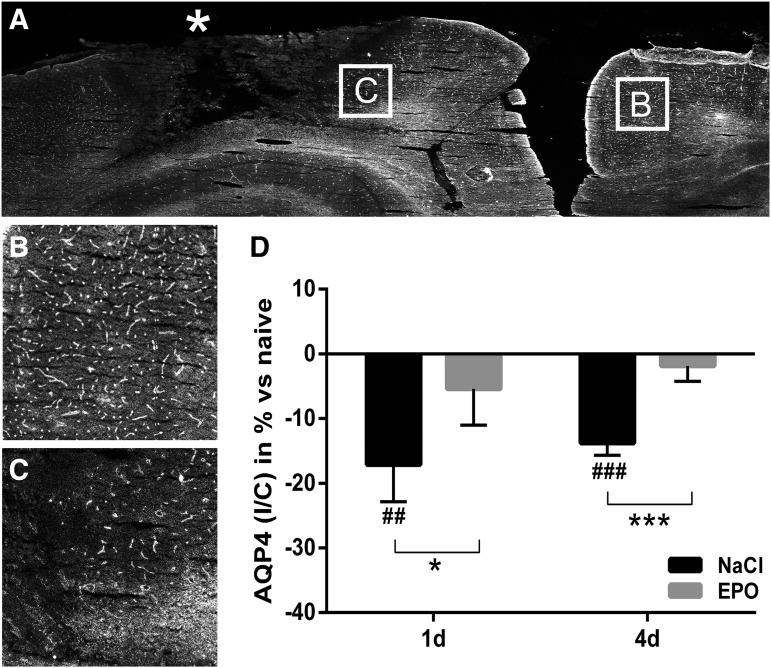
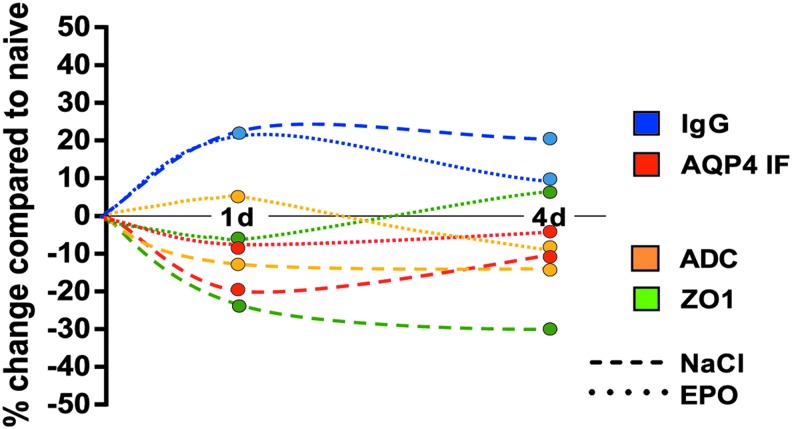
Erythropoietin (EPO) has neuroprotective effects in multiple central nervous system (CNS) injury models; however EPO's effects on traumatic brain edema are elusive. To explore EPO as an intervention in traumatic brain edema, male Sprague-Dawley (SD) rats were subjected to blunt, controlled traumatic brain injury (TBI). Animals were randomized to EPO 5000 IU/kg or saline (control group) intraperitoneally within 30 min after trauma and once daily for 4 consecutive days. Brain MRI, immunohistofluorescence, immunohistochemistry, and quantitative protein analysis were performed at days 1 and 4 post- trauma. EPO significantly prevented the loss of the tight junction protein zona occludens 1 (ZO-1) observed in control animals after trauma. The decrease of ZO-1 in the control group was associated with an immunoglobulin (Ig)G increase in the perilesional parenchyma, indicating blood-brain barrier (BBB) dysfunction and increased permeability. EPO treatment attenuated decrease in apparent diffusion coefficient (ADC) after trauma, suggesting a reduction of cytotoxic edema, and reduced the IgG leakage, indicating that EPO contributed to preserve BBB integrity and attenuated vasogenic edema. Animals treated with EPO demonstrated conserved levels of aquaporin 4 (AQP4) protein expression in the perilesional area, whereas control animals showed a reduction of AQP4. We show that post TBI administration of EPO decreases early cytotoxic brain edema and preserves structural and functional properties of the BBB, leading to attenuation of the vasogenic edema response. The data support that the mechanisms involve preservation of the tight junction protein ZO-1 and the water channel AQP4, and indicate that treatment with EPO may have beneficial effects on the brain edema response following TBI.

Keywords: BBB; EPO; TBI; aquaporin; brain edema.


Conflict of interest statement

No competing financial interests exist.

Figures

<b>FIG. 1.</b>
FIG. 1.
(A) Representative T2-weighted coronal MRI section of adult rat brain at day 4 after traumatic brain injury (contusion) in the right hemisphere. The focal injury in the ipsilateral hemisphere (I) is surrounded by a hyperdense area, indicating edema. No hyperdense MRI changes are observed in the contralateral hemisphere (C). (B) Relative changes of apparent diffusion coefficient (ADC) in the perilesional region at day 1 and 4 in control animals and erythropoietin (EPO)-treated animals (EPO); ipsilateral/contralateral (I/C) values in percent. ADC I/C was significantly decreased at both day 1 and day 4 after trauma in controls. EPO treatment attenuated the ADC decrease at both time points. Resultant ADC values were similar to ADC in naïve animals without trauma. (C) Schematic illustration of a coronal section of a rat brain. Midline shift (MLS) was estimated in percent using the distance between the outer border of the cortex and the middle of the third ventricle in the ipsilateral and contralateral hemispheres, respectively. The dark gray area illustrates the contusion, and the light gray quadrants depict the perilesional regions analyzed in all other experiments. (D) MLS at days 1 and 4 after trauma in control animals and EPO-treated animals. A significant MLS toward the uninjured side was present at both time points in both groups; i.e., EPO treatment did not affect MLS. Values presented as mean ± SD. d, days after trauma; #p < 0.05)
<b>FIG. 2.</b>
FIG. 2.
(A) High magnification confocal immunofluorescence image of zona occludens 1 (ZO-1) (red), the glial cell marker glial fibrillary acidic protein (GFAP) (green), and the nucleus marker DAPI (blue) from brain area with blood vessels (longitudinal view of blood vessel to the left), to illustrate the specificity and localization of the antibody binding. (B) Monochromatic presentation of GFAP immunofluorescence. (C) Monochromatic presentation of ZO-1 immunofluorescence. (D) Summary data on ZO-1 immunofluorescence in the traumatized, ipsilateral, hemisphere normalized against the contralateral side at days 1 and 4 after trauma in control and erythropoietin (EPO)-treated animals. ZO-1 was significantly reduced after trauma in the control group. EPO prevented the loss of ZO-1 at both time points after trauma. Values presented as mean ± SD. DAPI, 4',6-diamidino-2-phenylindole; #p < 0.05, ##p < 0.01 compared with naïve animals; **p < 0.01, ***p < 0.001 between groups.
<b>FIG. 3.</b>
FIG. 3.
Immunoglobulin (Ig)G immunohistochemistry on representative axial brain sections. (A) Control animals day 1 and 4 post-trauma, (B) erythropoietin (EPO)-treated animal day 1 and day 4 post-trauma, (C) naïve animal without trauma. IgG-positive immunoreactivity (dark area) was observed around the site of the trauma in the right hemisphere. (D) Relative changes (in percent) of IgG immunoreactivity at days 1 and 4 after trauma and compared with naïve animals (baseline). The perilesional area is normalized against the contralateral side. IgG was significantly increased in control animals. EPO treatment significantly decreased IgG in the perilesional area at day 4 after trauma. Values presented as mean ± SD. d, days post trauma; I/C, ipsilateral/contralateral; ###p < 0.001 compared with naïve animals; **p < 0.01 between groups.
<b>FIG. 4.</b>
FIG. 4.
(A) Representative monochromatic immunofluorescence image with aquaporin 4 (AQP4) (white) on part of the coronal brain section after blunt controlled parietal trauma (*) in the right hemisphere in a control animal. The core of the injury shows disrupted microscopic structure. Measurements of AQP4 protein abundance were performed in the perilesional area (as shown in Fig. 1C) with a visually intact structure. (B) High magnification of representative contralateral and (C) perilesional and areas. (D) Summary data on relative changes of AQP4 immunofluorescence intensity in the perilesional area at days 1 and 4 after trauma in control and erythropoietin (EPO)-treated animals. AQP4 was significantly reduced at both time points in control animals. EPO treatment normalized AQP4 protein levels in the perilesional area at both days 1 and 4 after trauma. Values presented as mean ± SD. I/C, ipsilateral/contralateral; d, days post trauma; ##p < 0.01, ### p < 0.001 compared with naïve animals; *p < 0.05, ***p < 0.001 between groups)
<b>FIG. 5.</b>
FIG. 5.
Schematic presentation of relative changes in the perilesional region at days 0, 1 and 4 after trauma in control animals (NaCl) and animals treated with erythropoietin (EPO). Values are compared with those in naïve animals without trauma (point zero) (immunoglobulin [Ig]G, zona occludens 1 [ZO-1], aquaporin 4 [AQP4]), or to the assumption of no difference between the hemispheres (apparent diffusion coefficient [ADC]). The arbitrary continuous timeline is created from the separate measurements for each parameter at the indicated time points. d, days after trauma.

Similar articles

Cited by

References

    1. Coronado V.G., McGuire L.C., Sarmiento K., Bell J., Lionbarger M.R., Jones C.D., Geller A.I., Khoury N., and Xu L. ( 2012). Trends in Traumatic Brain Injury in the U.S. and the public health response: 1995–2009. J. Safety Res. 43, 299– 307 - PubMed
    1. Coronado V.G., Haileyesus T., Cheng T.A., Bell J.M., Haarbauer-Krupa J., Lionbarger M.R., Flores-Herrera J., McGuire L.C., and Gilchrist J. ( 2015). Trends in sports- and recreation-related traumatic brain injuries treated in US emergency departments: The National Electronic Injury Surveillance System-All Injury Program (NEISS-AIP) 2001–2012. J. Head Trauma Rehabil. 30, 185– 197 - PMC - PubMed
    1. Clausen T., and Bullock R. ( 2001). Medical treatment and neuroprotection in traumatic brain injury. Curr. Pharm. Des. 7, 1517– 1532 - PubMed
    1. Faul M., Wald M.M., Rutland-Brown W., Sullivent E.E., and Sattin R.W. ( 2007). Using a cost-benefit analysis to estimate outcomes of a clinical treatment guideline: testing theBrain Trauma Foundation guidelines for the treatment of severe traumatic brain injury. J. Trauma 63, 1271– 1278 - PubMed
    1. Honeybul S. ( 2011). An update on the management of traumatic brain injury. J. Neurosurg. Sci. 55, 343– 355 - PubMed

Publication types