. 2018 Apr 1;35(7):930-939.
doi: 10.1089/neu.2017.5056. Epub 2018 Jan 12.

Traumatic Brain Injury Impairs Myogenic Constriction of Cerebral Arteries: Role of Mitochondria-Derived H2O2 and TRPV4-Dependent Activation of BKca Channels

Affiliations

Traumatic Brain Injury Impairs Myogenic Constriction of Cerebral Arteries: Role of Mitochondria-Derived H2O2 and TRPV4-Dependent Activation of BKca Channels

Nikolett Szarka et al. J Neurotrauma. .

Abstract

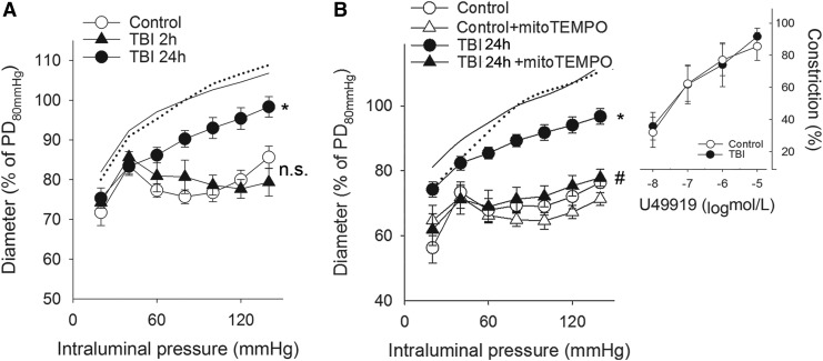
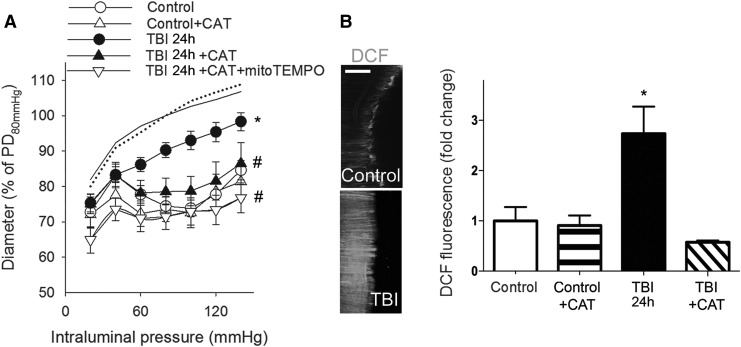
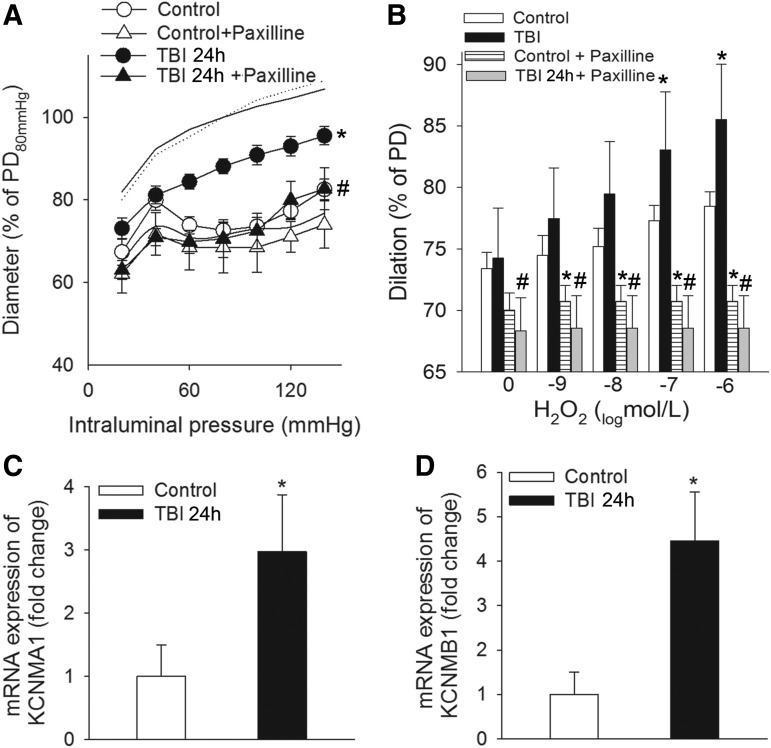
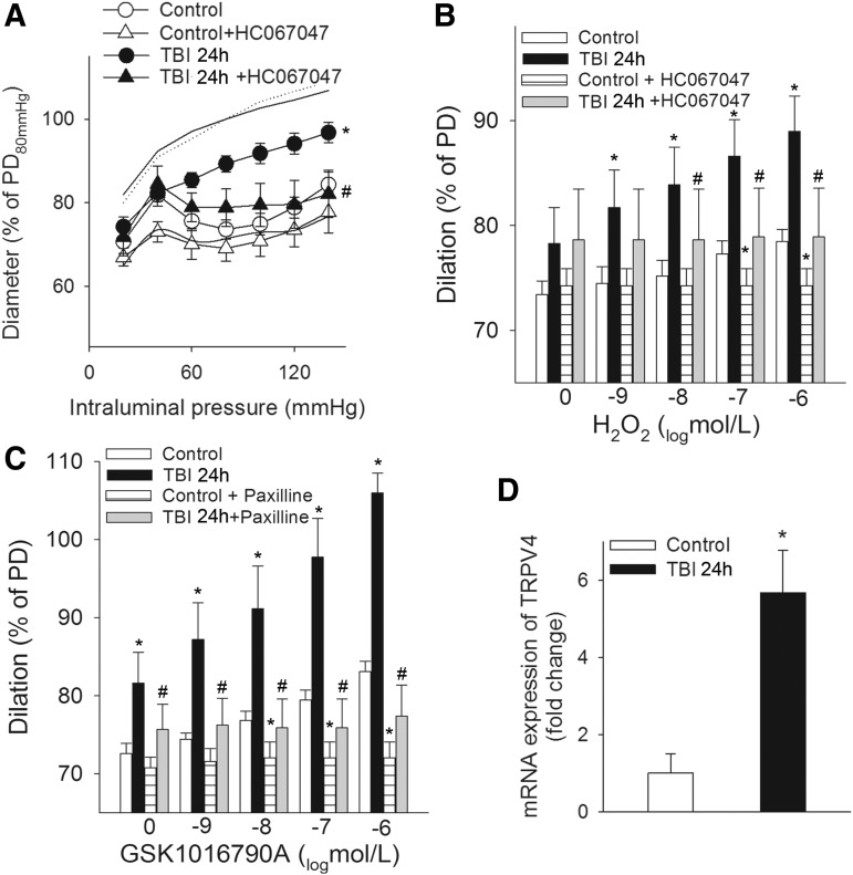
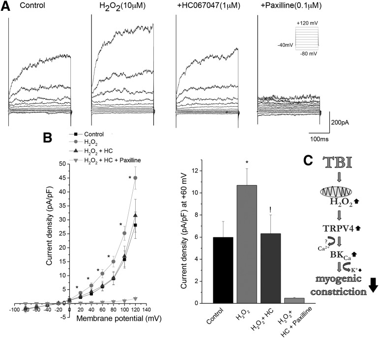
Traumatic brain injury (TBI) impairs autoregulation of cerebral blood flow, which contributes to the development of secondary brain injury, increasing mortality of patients. Impairment of pressure-induced myogenic constriction of cerebral arteries plays a critical role in autoregulatory dysfunction; however, the underlying cellular and molecular mechanisms are not well understood. To determine the role of mitochondria-derived H2O2 and large-conductance calcium-activated potassium channels (BKCa) in myogenic autoregulatory dysfunction, middle cerebral arteries (MCAs) were isolated from rats with severe weight drop-impact acceleration brain injury. We found that 24 h post-TBI MCAs exhibited impaired myogenic constriction, which was restored by treatment with a mitochondria-targeted antioxidant (mitoTEMPO), by scavenging of H2O2 (polyethylene glycol [PEG]-catalase) and by blocking both BKCa channels (paxilline) and transient receptor potential cation channel subfamily V member 4 (TRPV4) channels (HC 067047). Further, exogenous administration of H2O2 elicited significant dilation of MCAs, which was inhibited by blocking either BKCa or TRPV4 channels. Vasodilation induced by the TRPV4 agonist GSK1016790A was inhibited by paxilline. In cultured vascular smooth muscle cells H2O2 activated BKCa currents, which were inhibited by blockade of TRPV4 channels. Collectively, our results suggest that after TBI, excessive mitochondria-derived H2O2 activates BKCa channels via a TRPV4-dependent pathway in the vascular smooth muscle cells, which impairs pressure-induced constriction of cerebral arteries. Future studies should elucidate the therapeutic potential of pharmacological targeting of this pathway in TBI, to restore autoregulatory function in order to prevent secondary brain damage and decrease mortality.

Keywords: autoregulation; intracranial hypertension; oxidative stress; secondary injury.


Conflict of interest statement

No competing financial interests exist.

Figures

<b>FIG. 1.</b>
FIG. 1.
Traumatic brain injury (TBI) impairs myogenic constriction of cerebral arteries: role of mitochondrial reactive oxygen species (ROS) production. (A) Diameter responses (as percent of passive diameter [PD] at 80 mm Hg intraluminal pressure) of isolated middle cerebral arteries (MCA) are shown as a function of intraluminal pressure (myogenic response) in control rats and in rats 2 (TBI 2 h) and 24 (TBI 24 h) h after severe TBI. Note that the pressure-induced constrictor response is intact 2 h after the impact, and it is significantly attenuated 24 h post-injury. Data are mean ± S.E.M. (n = 5 for each group) *p < 0.05 versus control (lines without symbols show passive pressure-diameter curves of MCAs). (B) Myogenic responses of MCAs are depicted in control and TBI 24 h rats in the absence and presence of the mitochondrial antioxidant mitoTEMPO. Inlet depicts the constriction of basilar arteries of control and TBI 24 h rats in response to the thromboxane analogue U46619. Data are mean ± S.E.M. (n = 5 for each group) *p < 0.05 versus control; #p < 0.05 versus TBI 24 h.
<b>FIG. 2.</b>
FIG. 2.
Traumatic brain injury (TBI) impairs myogenic constriction of cerebral arteries: role of mitochondrial H2O2. (A) Diameter responses (as percent of passive diameter [PD] at 80 mm Hg intraluminal pressure) of isolated middle cerebral arteries (MCA) are shown as a function of intraluminal pressure (myogenic response) in control and TBI 24 h (24 h after the impact) rats after the administration of catalase (CAT). Note that additional administration of mitoTEMPO does not augment the effect of CAT on the diameter responses. Data are mean ± S.E.M. (n = 5 for each group) *p < 0.05 versus control; #p < 0.05 versus TBI 24 h. (B) Summary data and representative images of cerebrovascular H2O2 production in endothelium-denuded MCAs of control rats, TBI 24 h rats and control and TBI 24 h rats after incubation of the vessels in CAT shown by the fluorescence of the cell-permeant oxidative fluorescent indicator dye DCF (5 [and 6]- chloromethyl-2′,7′-dichlorodihydrofluorescein diacetate-acetyl ester). Scale bar is 50 μm. Data are mean ± S.E.M. (n = 5 for each group). *p < 0.05 versus control.
<b>FIG. 3.</b>
FIG. 3.
Traumatic brain injury (TBI) impairs myogenic constriction of cerebral arteries: role of Ca2+-activated K+ (BK) channels. (A) The effect of paxilline, a specific blocker of calcium-activated potassium (BKCa) channels on pressure-induced myogenic constriction of middle cerebral arteries (MCAs) of control rats and rats 24 h after severe TBI (TBI 24 h). Data are mean ± S.E.M. (n = 5 for each group) *p < 0.05 versus control; #p < 0.05 versus TBI 24 h. (B) Blocking BKCa channels by paxilline inhibits H2O2-induced dose-dependent dilations of MCAs of control and TBI 24 h rats. Note that H2O2-induced dilations are significantly augmented in MCAs isolated from TBI 24 h rats. Data are mean ± S.E.M. (n = 5 for each group); *p < 0.05 versus control, #p < 0.05 versus TBI 24 h. Quantitative reverse transcription polymerase chain reaction (qRT-PCR) data of mRNA expression of the α (panel C) and β (panel D) subunits of BKCa channels (KCNMA1 and KCNMB1, respectively) in MCAs of control and TBI-rats. Data are mean ± S.E.M. (n = 5 for each group) *p < 0.05 versus control.
<b>FIG. 4.</b>
FIG. 4.
Traumatic brain injury (TBI) impairs myogenic constriction of cerebral arteries: role of transient receptor potential cation channel subfamily V member 4 (TRPV4) channels. (A) Myogenic constriction of middle cerebral arteries (MCAs) of control rats and rats 24 h after TBI (TBI 24 h) in the absence and presence of the specific TRPV4 channel blocker HC 067047. Data are mean ± S.E.M. (n = 5 for each group) *p < 0.05 versus control; #p < 0.05 versus TBI 24 h. Panel B depicts the effect of the TRPV4 channel blocker HC 067047 on H2O2-induced dilations of MCAs of control and TBI rats, and C shows the effect of blocking BKCa channels on dilations of MCAs evoked by the TRPV4 agonist GSK1016790A in the same groups of animals. Note that both H2O2-induced and GSK1016790A-induced dilations of MCAs are significantly higher after TBI. Data are mean ± S.E.M. (n = 5 for each group) *p < 0.05 versus control; #p < 0.05 versus TBI 24 h. (D) Quantitative reverse transcription polymerase chain reaction (qRT-PCR) data of mRNA expression of TRPV4 channels in MCAs of control and TBI 24h rats. Data are mean ± S.E.M. (n = 5 for each group) *p < 0.05 versus control.
<b>FIG. 5.</b>
FIG. 5.
H2O2-mediated increase in calcium-activated potassium (BKCa) channel activity requires a transient receptor potential cation channel subfamily V member 4 (TRPV4) channel. Whole cell BKCa currents were recorded with 100 nM free cytosolic calcium in the presence and absence of H2O2, TRPV4 channel inhibitor (1 μM HC067047), and BKCa channel inhibitor (paxilline 100 nM). BKCa currents were elicited by 20 ms pulses from −60 to +120 mV from a Vh of −40 mV (Inset). Two to three smooth muscle cells from four Wistar–Kyoto rats were studied in each group (8–12 cells/group). Panel A represents whole cell BKCa currents before and after 10 μM H2O2, in the presence of 1μM HC 067047 and/or 100 nM paxilline. Panel B represents current voltage curves and the current density at +60 mV membrane potential. *p < 0.05 before and after application of H2O2. !p < 0.05 before and after application of 1μM HC 067047 in the presence of H2O2. Data are mean ± S.E.M. Number in the parenthesis is the number of cells studied. (C) Scheme depicting the mechanisms of impaired myogenic constriction of cerebral arteries after TBI. TBI leads to excessive cerebrovascular production of H2O2 mainly of mitochondrial origin, which activates TRPV4 on vascular smooth muscle cells. TRPV4 then activates BKCa channels leading to hyperpolarization of vascular smooth muscle cell (VSMC) membranes and subsequent dilation of cerebral vessels, which attenuates pressure-induced myogenic constriction.

Comment in

References

    1. Roozenbeek B., Maas A.I., and Menon D.K. (2013). Changing patterns in the epidemiology of traumatic brain injury. Nat. Rev. Neurol. 9, 231–236 - PubMed
    1. Langlois J.A., and Sattin R.W. (2005). Traumatic brain injury in the United States: research and programs of the Centers for Disease Control and Prevention (CDC). J. Head Trauma Rehabil. 20, 187–188 - PubMed
    1. Tagliaferri F., Compagnone C., Korsic M., Servadei F., and Kraus J. (2006). A systematic review of brain injury epidemiology in Europe. Acta Neurochir. 148, 255–268 - PubMed
    1. Sahuquillo J., Poca M.A., and Amoros S. (2001). Current aspects of pathophysiology and cell dysfunction after severe head injury. Curr. Pharm. Des. 7, 1475–1503 - PubMed
    1. Overgaard J., and Tweed W.A. (1974). Cerebral circulation after head injury. 1. Cerebral blood flow and its regulation after closed head injury with emphasis on clinical correlations. J. Neurosurg. 41, 531–541 - PubMed

LinkOut - more resources