. 2017 Nov 28;8(1):269.
doi: 10.1186/s13287-017-0717-9.

Laser-modified titanium surfaces enhance the osteogenic differentiation of human mesenchymal stem cells

Affiliations

Laser-modified titanium surfaces enhance the osteogenic differentiation of human mesenchymal stem cells

Tatiana A B Bressel et al. Stem Cell Res Ther. .

Abstract

Background: Titanium surfaces have been modified by various approaches with the aim of improving the stimulation of osseointegration. Laser beam (Yb-YAG) treatment is a controllable and flexible approach to modifying surfaces. It creates a complex surface topography with micro and nano-scaled patterns, and an oxide layer that can improve the osseointegration of implants, increasing their usefulness as bone implant materials.

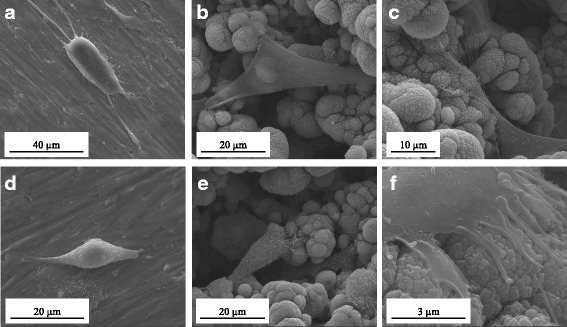
Methods: Laser beam irradiation at various fluences (132, 210, or 235 J/cm2) was used to treat commercially pure titanium discs to create complex surface topographies. The titanium discs were investigated by scanning electron microscopy, X-ray diffraction, and measurement of contact angles. The surface generated at a fluence of 235 J/cm2 was used in the biological assays. The behavior of mesenchymal stem cells from an umbilical cord vein was evaluated using a 3-(4,5-dimethylthiazol-2-yl)-2,5-diphenyltetrazolium bromide (MTT) assay, a mineralization assay, and an alkaline phosphatase activity assay and by carrying out a quantitative real-time polymerase chain reaction for osteogenic markers. CHO-k1 cells were also exposed to titanium discs in the MTT assay.

Results: The best titanium surface was that produced by laser beam irradiation at 235 J/cm2 fluence. Cell proliferation analysis revealed that the CHO-k1 and mesenchymal stem cells behaved differently. The laser-processed titanium surface increased the proliferation of CHO-k1 cells, reduced the proliferation of mesenchymal stem cells, upregulated the expression of the osteogenic markers, and enhanced alkaline phosphatase activity.

Conclusions: The laser-treated titanium surface modulated cellular behavior depending on the cell type, and stimulated osteogenic differentiation. This evidence supports the potential use of laser-processed titanium surfaces as bone implant materials, and their use in regenerative medicine could promote better outcomes.

Keywords: Biocompatibility; Human umbilical cord; Laser beam (Yb-YAG); Mesenchymal stem cells; Osteoinduction; Surface modification; Titanium.


Conflict of interest statement

Ethics approval and consent to participate

This work was submitted to and approved by the Ethics Committee of the Federal University of Rio Grande do Norte (FR132464). Umbilical cord specimens were obtained after written informed consent was signed by mothers.

Consent for publication

Not applicable.

Competing interests

The authors declare that they have no competing interests.

Publisher’s Note

Springer Nature remains neutral with regard to jurisdictional claims in published maps and institutional affiliations.

Figures

Fig. 1
Fig. 1
Scanning electron microscopy images of Ti control (a), laser-processed titanium (LPT) produced using laser radiation at 132 J/cm2 fluence (b), LPT produced using laser radiation at 210 J/cm2 fluence (c), and LPT produced using laser radiation at 235 J/cm2 fluence (d). Surfaces at × 100, ×500, ×1000, ×50,000, and × 200,000 magnification
Fig. 2
Fig. 2
X-ray diffraction spectra of Ti control (a), laser-processed titanium (LPT) produced using laser radiation at 132 J/cm2 fluence (b), LPT produced using laser radiation at 210 J/cm2 fluence (c), and LPT produced using laser radiation at 235 J/cm2 fluence (d). cps counts per second, Ti titanium
Fig. 3
Fig. 3
Scanning electron microscopy micrographs of hUC-MSCs cultured after 24 h on Ti control (a) and laser-processed titanium (LPT) produced using laser radiation at 235 J/cm2 fluence (b, c); surfaces at × 3000, ×5000, and × 7000 magnification. CHO-k1 cells after 24 h of culture on Ti control (d) and LPT produced using laser radiation at 235 J/cm2 fluence (e, f). Surfaces at × 6000, ×5000, and × 40,000 magnification
Fig. 4
Fig. 4
Scanning electron microscopy micrographs after culture for 7 days. hUC-MSCs on Ti control (a) and laser-processed titanium (LPT) (b, c) surfaces. CHO-k1 cells on Ti control (d) and LPT (e, f) surfaces. a x1000, b x500, c x800, d x600, e x400, f x800
Fig. 5
Fig. 5
MTT cell metabolic activity assay. hUC-MSC and CHO-k1 cell adhesion and proliferation results on the two different types of titanium discs (laser-processed titanium (LPT) and Ti control) at different times (3 h, and 1, 3, and 7 days). ***Statistically significant differences between cell types at p < 0.001; ### represents statistically significant differences between Ti surfaces p < 0.001. Data represent means of three independent experiments and SD. MTT, 3-(4,5-dimethylthiazol-2-yl)-2,5-diphenyltetrazolium bromide, OD optical density
Fig. 6
Fig. 6
Alkaline phosphatase (ALP) activity. ALP activity in hUC-MSCs after culture in Dulbecco's modified Eagle’s medium on Ti control and laser-processed titanium (LPT) surfaces after 3 and 7 days (a); culture of hUC-MSCs in osteogenic medium for the same times on Ti control and LPT surfaces (b). Statistically significant differences between Ti surfaces at: ***p < 0.001, *p < 0.05. Values are mean ± SD of two independent experiments. Ti titanium
Fig. 7
Fig. 7
Calcium deposition assay. Alizarin Red S staining of hUC-MSCs on Ti control and laser-processed titanium (LPT) after 7 and 14 days of culture in (a) basal medium and (b) osteogenic medium.***Statistically significant differences between Ti surfaces at p < 0.001. N = 3 ± SD. Ti titanium
Fig. 8
Fig. 8
Gene expression of osteogenic markers ALPL, RUNX2, BMP2, OCN, and OPN in hUC-MSCs following culture on laser-processed titanium (LPT) for 7 days in basal medium (BM) and osteogenic medium (OM). Gene expression evaluated by ΔΔCt method normalized to glyceraldehyde-3-phosphate dehydrogenase (GAPDH) expression and reported as fold changes in relation to the Ti control. Statistically significant differences between BM and OM at: **p < 0.01, *p < 0.05. Data presented as mean ± SD (n = 2). ALPL alkaline phosphatase, RUNX2 run-related transcription factor 2, BMP2 bone morphogenetic protein 2, OCN osteocalcin, OPN osteopontin

References

    1. Geetha M, Singh AK, Asokamani R, Gogia AK. Ti based biomaterials, the ultimate choice for orthopaedic implants—a review. Prog Mater Sci. 2009;54:397–425. doi: 10.1016/j.pmatsci.2008.06.004. - DOI
    1. Jacchetti E, Di Rienzo C, Meucci S, Nocchi F, Beltram F, Cecchini M. Wharton’s Jelly human mesenchymal stem cell contact guidance by noisy nanotopographies. Sci Rep. 2014;4:3830. doi: 10.1038/srep03830. - DOI - PMC - PubMed
    1. Schiavi J, Keller L, Morand D-N, De Isla N, Huck O, Lutz JC, et al. Active implant combining human stem cell microtissues and growth factors for bone-regenerative nanomedicine. Nanomedicine. 2015;10:753–63. doi: 10.2217/nnm.14.228. - DOI - PubMed
    1. Sisti KE, de Andrés MC, Johnston D, Almeida-Filho E, Guastaldi AC, Oreffo ROC. Skeletal stem cell and bone implant interactions are enhanced by LASER titanium modification. Biochem Biophys Res Commun. 2016;473:719–25. doi: 10.1016/j.bbrc.2015.10.013. - DOI - PubMed
    1. Novin M, Faghihi S. Mouse bone marrow-derived mesenchymal stem cell response to nanostructured titanium substrates produced by high-pressure torsion. Surf Interface Anal. 2013;45:619–27. doi: 10.1002/sia.5101. - DOI

MeSH terms