. 2018 Jan 11;131(2):236-246.
doi: 10.1182/blood-2017-07-798728. Epub 2017 Nov 27.

Inhibition of heme oxygenase ameliorates anemia and reduces iron overload in a β-thalassemia mouse model

Affiliations

Inhibition of heme oxygenase ameliorates anemia and reduces iron overload in a β-thalassemia mouse model

Daniel Garcia-Santos et al. Blood. .

Abstract

Thalassemias are a heterogeneous group of red blood cell disorders, considered a major cause of morbidity and mortality among genetic diseases. However, there is still no universally available cure for thalassemias. The underlying basis of thalassemia pathology is the premature apoptotic destruction of erythroblasts causing ineffective erythropoiesis. In β-thalassemia, β-globin synthesis is reduced causing α-globin accumulation. Unpaired globin chains, with heme attached to them, accumulate in thalassemic erythroblasts causing oxidative stress and the premature cell death. We hypothesize that in β-thalassemia heme oxygenase (HO) 1 could play a pathogenic role in the development of anemia and ineffective erythropoiesis. To test this hypothesis, we exploited a mouse model of β-thalassemia intermedia, Th3/+ We observed that HO inhibition using tin protoporphyrin IX (SnPP) decreased heme-iron recycling in the liver and ameliorated anemia in the Th3/+ mice. SnPP administration led to a decrease in erythropoietin and increase in hepcidin serum levels, changes that were accompanied by an alleviation of ineffective erythropoiesis in Th3/+ mice. Additionally, the bone marrow from Th3/+ mice treated with SnPP exhibited decreased heme catabolism and diminished iron release as well as reduced apoptosis. Our results indicate that the iron released from heme because of HO activity contributes to the pathophysiology of thalassemia. Therefore, new therapies that suppress heme catabolism may be beneficial in ameliorating the anemia and ineffective erythropoiesis in thalassemias.


Figures

Figure 1.
Figure 1.
HO-1 expression is increased in the liver and decreased in Ter119+cells of the bone marrow and the spleen of Th3/+mice. (A) Quantitative reverse transcription polymerase chain reaction (qRT-PCR) analysis of HO-1 mRNA expression in the liver of Wt and Th3/+ mice. The results are presented as fold of change relative to the Wt sample (n = 3). (B) Western blot analysis of HO-1 protein expression in the liver of Wt and Th3/+ mice (n = 3). (C) Representative pictures (20× magnification; scale bar, 200 µM) of immunohistochemical staining against HO-1 in the liver from Wt and Th3/+ mice. (D) qRT-PCR analysis of HO-1 mRNA expression in Ter119 and Ter119+ cell populations isolated from the bone marrow of Wt and Th3/+ mice. The results are presented as fold change relative to Wt Ter119 cells (n = 3). (E) Western blot analysis of HO-1 protein expression in Ter119+ bone marrow samples (n = 3). (F) Total heme levels in Ter119 and Ter119+ cells isolated from the bone marrow of Wt and Th3/+ mice (n = 6). (G) Western blot analysis of HO-1 protein expression in Ter119+ spleen samples (n = 3). (H) Total heme levels in Ter119 and Ter119+ cells isolated from the spleen of Wt and Th3/+ mice (n = 6). *P < .05, ***P < .001.
Figure 2.
Figure 2.
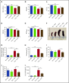
SnPP injections rescue anemia and reduce spleen size, reticulocyte levels, serum iron concentration, transferrin saturation, and EPO levels in Th3/+mice. (A) RBC, (B) Hb, (C) HCT, (D) MCV, and (E) MCH indices were determined on Wt + PBS (n = 12), Wt + SnPP (n = 12), Th3/+ + PBS (n = 11), and Th3/+ + SnPP (n = 12) mouse groups by automated analysis. (F) Representative images of spleens isolated from Wt and Th3/+ mice injected either with PBS or SnPP. (G) Spleen weight index of Wt + PBS (n = 9), Wt + SnPP (n = 9), Th3/+ + PBS (n = 8), and Th3/+ + SnPP (n = 7) mice. (H) Flow cytometry analysis of thiazole orange staining of peripheral blood obtained from Wt and Th3/+ mice injected either with PBS or SnPP (n = 6). Analysis of (I) serum iron and (J) transferrin saturation in Wt + PBS (n = 5), Wt + SnPP (n = 5), Th3/+ + PBS (n = 5), and Th3/+ + SnPP (n = 4) mice. (L) Enzyme-linked immunosorbent assay of EPO in serum from Wt and Th3/+ mice injected with PBS or SnPP (n = 11). *P < .05, ***P < .001.
Figure 3.
Figure 3.
SnPP injections decrease nonheme iron levels and heme-iron recycling in the liver of Th3/+mice. (A) Representative pictures (20× magnification; scale bar, 200 µM) of Perls’ Prussian blue staining in Wt and Th3/+ mice injected with either PBS or SnPP. (B) Ferrozine assay of nonheme iron content in the liver isolated from mice treated as follows: Wt + PBS (n = 6), Wt + SnPP (n = 6), Th3/+ + PBS (n = 5), or Th3/+ + SnPP (n = 5). (C) qRT-PCR analysis of HAMP mRNA expression in the liver of Wt and Th3/+ mice injected with either PBS or SnPP (n = 3). (D) Enzyme-linked immunosorbent assay of hepcidin in the serum of Wt and Th3/+ mice injected with either PBS or SnPP (n = 6). (E) Western blot analysis of FPN protein expression in samples isolated from the liver of Wt and Th3/+ mice injected with either PBS or SnPP (n = 3). (F) Representative images (20× magnification; scale bar, 200 µM) of immunohistochemical staining of FPN in livers from Wt or Th3/+ mice injected with PBS or SnPP. (G) Percentage of 59Fe in the liver of Wt and Th3/+ mice injected with either PBS or SnPP (n = 3). (H) Percentage of 59Fe in the heme fraction in the liver of Wt and Th3/+ mice injected with either PBS or SnPP (n = 3) (I) Percentage of 59Fe in total peripheral blood of Wt and Th3/+ mice injected with either PBS or SnPP (n = 3). Percentage was calculated based on the total 59Fe recovered for each mouse. *P < .05, ***P < .001.
Figure 4.
Figure 4.
HO-1 suppression rescues erythroid cell differentiation, suppresses erythroblast expansion, and reduces Fam132b expression in the bone marrow of Th3/+mice. (A) Representative dot plots of flow cytometry analysis of CD71 (y-axis) and Ter119 (x-axis) expression and its quantification in the bone marrow and the spleen from Wt and Th3/+ mice injected with either PBS or SnPP (n = 3). (B) Flow cytometry analysis of Ter119 and Ter119+ cells present in the bone marrow (n = 3) and the spleen (n = 5) of Wt and Th3/+ mice injected with either PBS or SnPP. (C) qRT-PCR analysis of Fam132b mRNA expression in bone marrow isolated from Wt and Th3/+ mice injected with either PBS or SnPP. The results are presented as fold change relative to Wt + PBS samples (n = 3). *P < .05, ***P < .001.
Figure 5.
Figure 5.
SnPP administration causes a decrease in α-globin precipitation and improves erythroid cell survival in Th3/+mice. (A) Representative image and densitometry quantification of a Coomassie blue-stained triton-acetic acid-urea gel loaded with membrane samples isolated from RBC from Wt and Th3/+ mice treated with either PBS or SnPP. (B) Flow cytometry analysis of CFSE-positive RBC derived from Wt + PBS, Wt + SnPP, Th3/+ + PBS, and Th3+ + SnPP groups and injected into Wt animals (n = 4). *P < .05, ***P < .001.
Figure 6.
Figure 6.
HO-1 inhibition increases the heme-regulatory pool, but does not affect in vitro erythroid colony formation in Th3/+mice bone marrow cells. (A) Western blot analysis of ferritin, phosphorylated eIF2α (eIF2α-P), and total eIF2α protein levels in Ter119+ cells isolated from the bone marrow of Th3/+ mice injected with either PBS or SnPP. Results were normalized to the total levels of eIF2α (n = 3). Bone marrow cells from Wt (n = 3) and Th3/+ mice (n = 3) were plated in methylcellulose media supplemented with EPO in the presence or absence of SnPP. (B) Number of BFU-E colonies detected in cultures of bone marrow samples derived from Wt and Th3/+ mice. (C) Number of CFU-E colonies detected in cultures of bone marrow samples derived from Wt and Th3/+ mice. *P < .05, ***P < .001.

References

    1. Musallam KM, Rivella S, Vichinsky E, Rachmilewitz EA. Non-transfusion-dependent thalassemias. Haematologica. 2013;98(6):833-844. - PMC - PubMed
    1. Yang B, Kirby S, Lewis J, Detloff PJ, Maeda N, Smithies O. A mouse model for beta 0-thalassemia. Proc Natl Acad Sci USA. 1995;92(25):11608-11612. - PMC - PubMed
    1. Drakesmith H, Nemeth E, Ganz T. Ironing out ferroportin. Cell Metab. 2015;22(5):777-787. - PMC - PubMed
    1. Maines MD. The heme oxygenase system: a regulator of second messenger gases. Annu Rev Pharmacol Toxicol. 1997;37(1):517-554. - PubMed
    1. Maines MD. The heme oxygenase system: update 2005. Antioxid Redox Signal. 2005;7(11-12):1761-1766. - PubMed

Publication types

MeSH terms