. 2017 Dec 12;114(50):13266-13271.
doi: 10.1073/pnas.1716022114. Epub 2017 Nov 27.

Synaptic homeostasis requires the membrane-proximal carboxy tail of GluA2

Affiliations

Synaptic homeostasis requires the membrane-proximal carboxy tail of GluA2

Samantha G Ancona Esselmann et al. Proc Natl Acad Sci U S A. .

Abstract

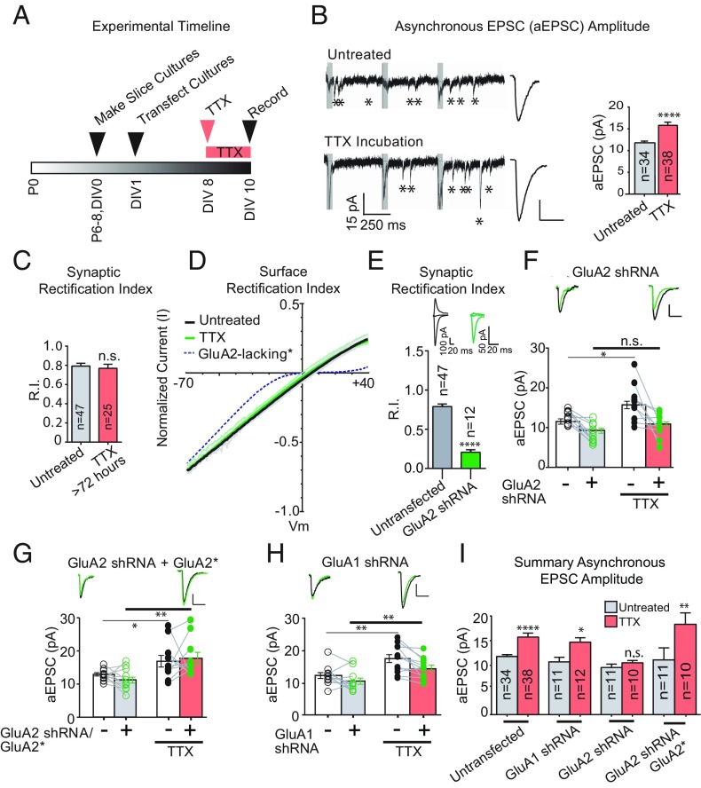
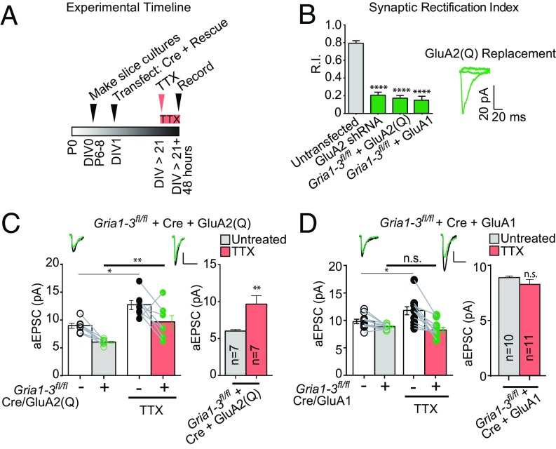
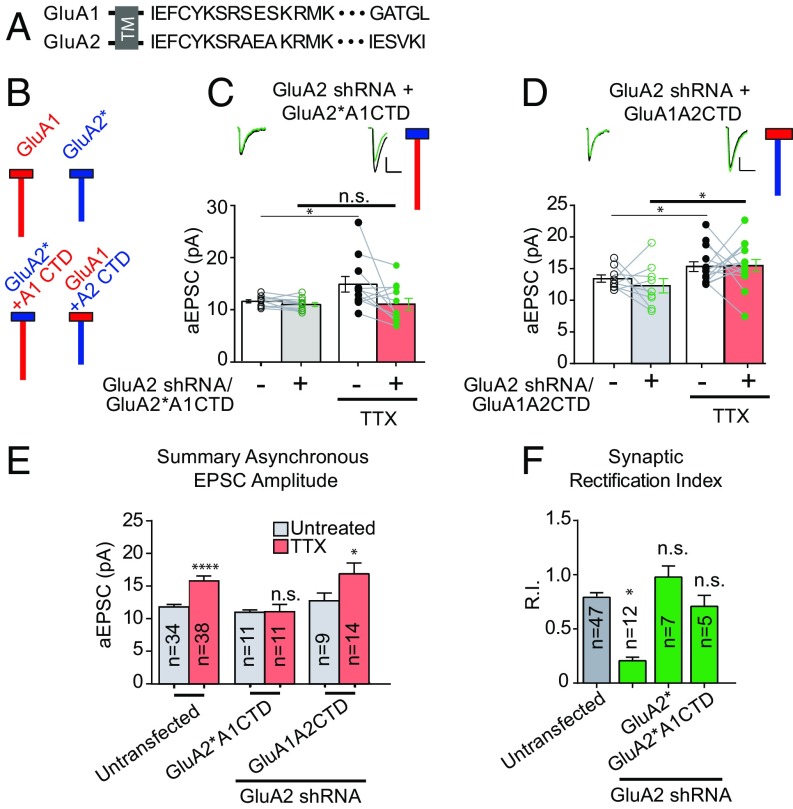
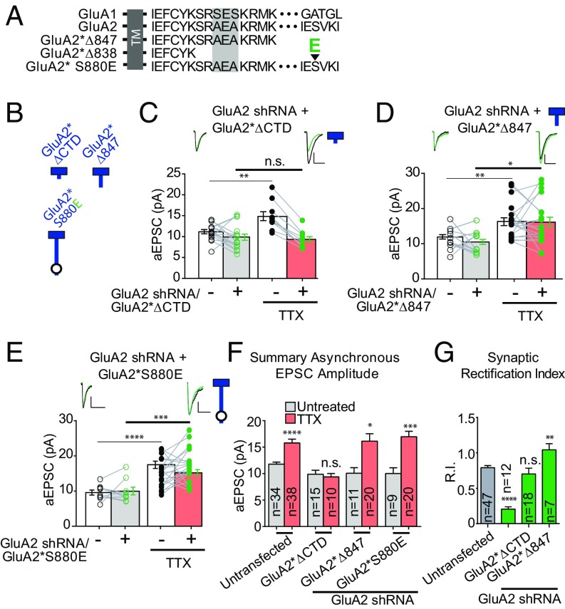
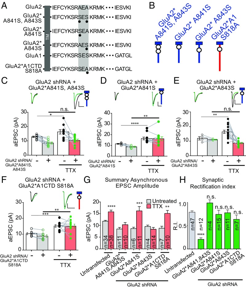
Bidirectional scaling of synaptic transmission, expressed as a compensatory change in quantal size following chronic activity perturbation, is a critical effector mechanism underlying homeostatic plasticity in the brain. An emerging model posits that the GluA2 AMPA receptor (AMPAR) subunit may be important for the bidirectional scaling of excitatory transmission; however, whether this subunit plays an obligatory role in synaptic scaling, and the identity of the precise domain(s) involved, remain controversial. We set out to determine the specific AMPAR subunit required for scaling up in CA1 hippocampal pyramidal neurons, and found that the GluA2 subunit is both necessary and sufficient. In addition, our results point to a critical role for a single amino acid within the membrane-proximal region of the GluA2 cytoplasmic tail, and suggest a distinct model for the regulation of AMPAR trafficking in synaptic homeostasis.

Keywords: AMPAR; GluA2; homeostatic plasticity; synaptic plasticity; synaptic scaling.


Conflict of interest statement

The authors declare no conflict of interest.

Figures

Fig. 1.
Fig. 1.
GluA2, not GluA1, is necessary for homeostatic synaptic scaling of quantal size. (A) Timeline of experiments. (B, Left) Sample traces of aEPSCs showing scaling of aEPSC amplitude with TTX treatment. The gray box shows the synchronous component of EPSC that is not analyzed. (B, Right) Averaged aEPSCs from TTX-treated and untreated control neurons. The bar graph shows averaged aEPSC amplitudes. (C) Synaptic rectification of AMPARs with or without TTX treatment (>72 h; Methods). (D) Surface rectification of AMPARs with and without TTX treatment. (E) Synaptic rectification of paired AMPA EPSCs in control neurons and neighboring neurons transfected with an shRNA against GluA2. (F) Paired asynchronous recordings without and with TTX treatment in control neurons and neighboring neurons transfected with GluA2 shRNA. Black, control; green, transfected. Untreated sample traces are at the top left, and TTX-treated sample traces are at the top right. (G) shRNA-insensitive GluA2 rescue under the same treatment conditions as in F. *Indicates shRNA resistance. (H) GluA1 shRNA under the same treatment conditions as in G. (I) Summary graph indicating unpaired scaling data, within the same transfection conditions. Significance was measured across treatment conditions. (Scale bars: 5 pA and 20 ms unless noted otherwise.) *P < 0.05; **P < 0.01; ****P < 0.0001; n.s., not significant.
Fig. 2.
Fig. 2.
GluA2 is sufficient to support synaptic scaling in the absence of other AMPAR subunits. (A) Timeline of organotypic slice culture preparation, TTX incubation, and recording for GluA2(Q) replacement experiments. (B) Synaptic rectification of paired AMPA EPSCs in GRIA1–3fl/fl control neurons and neighboring neurons transfected with Cre and GluA2(Q) or Cre and GluA1. GluA2 shRNA synaptic rectification is shown for comparison. (C) Paired asynchronous recordings without and with preceding chronic TTX treatment in GRIA1–3fl/fl control neurons and neighboring GRIA1–3fl/fl neurons transfected with Cre + GluA2(Q). (Right) Within-transfection condition summary bar graph. (D) Paired asynchronous recordings without and with preceding chronic TTX treatment in GRIA1–3fl/fl control neurons and neighboring GRIA1–3fl/fl neurons transfected in utero with Cre + GluA1. *P < 0.05; **P < 0.01; ****P < 0.0001; n.s., not significant.
Fig. 3.
Fig. 3.
The C-tail of the GluA2 subunit is critical for homeostatic synaptic scaling. (A) Endogenous GluA1 and GluA2 C-tail amino acid sequences. TM, transmembrane. (B) Schematic diagram of endogenous GluA1 and GluA2 AMPAR subunits next to schemata of chimeric AMPARs with swapped C-tails. Red indicates GluA1 subunit origin; blue, GluA2 subunit origin. Boxes indicate amino terminal domains and transmembrane regions of AMPARs, and vertical lines indicate intracellular C-tails. (C) Paired asynchronous recordings without and with preceding chronic TTX treatment in control neurons and neighboring neurons transfected with GluA2 shRNA and shRNA-insensitive AMPAR chimeric subunit GluA2*A1CTD. (D) GluA2 shRNA + shRNA-insensitive AMPAR chimeric subunit GluA1A2CTD. Treatment conditions are the same as in C. (E) Summary bar graph indicating unpaired scaling data under the same transfection conditions. Significance was measured across treatment conditions. (F) Comparison of synaptic rectification of chimeric AMPAR GluA2*A1CTD (with pore residue conferring calcium and intracellular polyamine block present) with cells transfected with GluA2 shRNA for comparison and cells transfected with GluA2 shRNA + full-length shRNA-insensitive GluA2. (Scale bars for aEPSC sample traces: 5 pA and 20 ms unless indicated otherwise.) *P < 0.05; ****P < 0.0001; n.s., not significant.
Fig. 4.
Fig. 4.
The membrane-proximal cytoplasmic tail of GluA2 is critical for synaptic scaling. (A) Endogenous GluA1 and GluA2 C-tail amino acid sequences, and GluA2 C-tail sequences with truncations (ΔCTD and Δ847) or point mutation (S880E; large arrowhead indicates location of phosphomimetic point mutation). The shaded-gray box in the membrane-proximal sequence illustrates the region with divergent sequences in the first 14 amino acids of AMPAR C-tails. TM, transmembrane. (B) Schematic diagram of truncated or mutated GluA2 subunits. Blue indicates the origin of the GluA2 subunit, and the circle indicates the location of the specific S880E point mutation. (C) Paired asynchronous recordings without and with preceding chronic TTX treatment in control neurons and neighboring neurons transfected with GluA2 shRNA and the shRNA-insensitive AMPAR chimeric subunit GluA2*ΔCTD. (D) GluA2 shRNA + shRNA-insensitive AMPAR chimeric subunit GluA2*Δ847. Treatment conditions are the same as in C. (E) GluA2 shRNA + shRNA-insensitive AMPAR chimeric subunit GluA2*S880E. Treatment conditions are the same as in C. (F) Summary bar graph indicating unpaired scaling data under the same transfection conditions. Significance was measured across treatment conditions (color-coded from CF). (G) Comparison of synaptic rectification of truncated or mutated GluA2 subunits with cells transfected with GluA2 shRNA. (Scale bars for aEPSC sample traces: 5 pA and 20 ms unless indicated otherwise.) *P < 0.05; **P < 0.01; ***P < 0.001; ****P < 0.0001; n.s., not significant.
Fig. 5.
Fig. 5.
A specific residue in the membrane-proximal CTD of the GluA2 AMPAR subunit is necessary for scaling. (A) Endogenous GluA1 and GluA2 C-tail amino acid sequences, and GluA2 or chimeric AMPAR C-tail sequences with point mutations. The shaded-gray box in the membrane-proximal sequence shows the region with divergent sequences in the first 14 amino acids of AMPAR C-tails, as well as the location of mutations. TM, transmembrane. (B) Schematic diagram of truncated or mutated GluA subunits. Blue and red indicate GluA2 and GluA1 subunit origin, respectively. (C) Paired asynchronous recordings without and with previous chronic TTX treatment in control neurons and neighboring neurons transfected with GluA2 shRNA and shRNA-insensitive AMPAR chimeric subunit GluA2*A841S, A843S. The schematic of the GluA2 replacement receptor repeated in the top right. (D) GluA2 shRNA + shRNA-insensitive AMPAR chimeric subunit GluA2*A841S. Treatment conditions were the same as in C. (E) GluA2 shRNA + shRNA-insensitive AMPAR chimeric subunit GluA2*A843S. Treatment conditions were the same as in C. (F) GluA2 shRNA + shRNA-insensitive AMPAR chimeric subunit GluA2*A1CTD S818A. Treatment conditions are the same as in C. (G) Summary bar graph indicating unpaired scaling data, under the same transfection conditions. Significance was measured across treatment conditions. (H) Comparison of synaptic rectification of mutated GluA2 or chimeric subunits to cells transfected with GluA2 shRNA alone. (Scale bars for aEPSC sample traces: 5 pA and 20 ms unless indicated otherwise.) *P < 0.05; **P < 0.01; ***P < 0.001; ****P < 0.0001; n.s., not significant.

Similar articles

Cited by

References

    1. Huganir RL, Nicoll RA. AMPARs and synaptic plasticity: The last 25 years. Neuron. 2013;80:704–717. - PMC - PubMed
    1. Malinow R, Malenka RC. AMPA receptor trafficking and synaptic plasticity. Annu Rev Neurosci. 2002;25:103–126. - PubMed
    1. Collingridge GL, Isaac JT, Wang YT. Receptor trafficking and synaptic plasticity. Nat Rev Neurosci. 2004;5:952–962. - PubMed
    1. Sheng M, Kim MJ. Postsynaptic signaling and plasticity mechanisms. Science. 2002;298:776–780. - PubMed
    1. Granger AJ, Shi Y, Lu W, Cerpas M, Nicoll RA. LTP requires a reserve pool of glutamate receptors independent of subunit type. Nature. 2013;493:495–500. - PMC - PubMed

Publication types

Substances