. 2017 Dec 12;114(50):13248-13253.
doi: 10.1073/pnas.1718592114. Epub 2017 Nov 27.

Protective effects of agonists of growth hormone-releasing hormone (GHRH) in early experimental diabetic retinopathy

Affiliations

Protective effects of agonists of growth hormone-releasing hormone (GHRH) in early experimental diabetic retinopathy

Menaka C Thounaojam et al. Proc Natl Acad Sci U S A. .

Abstract

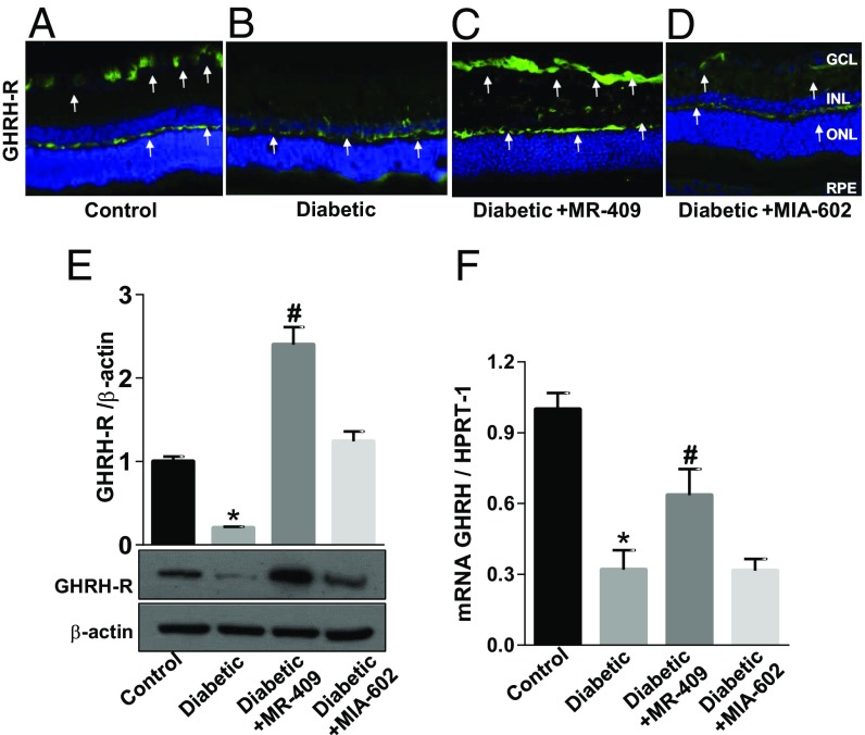
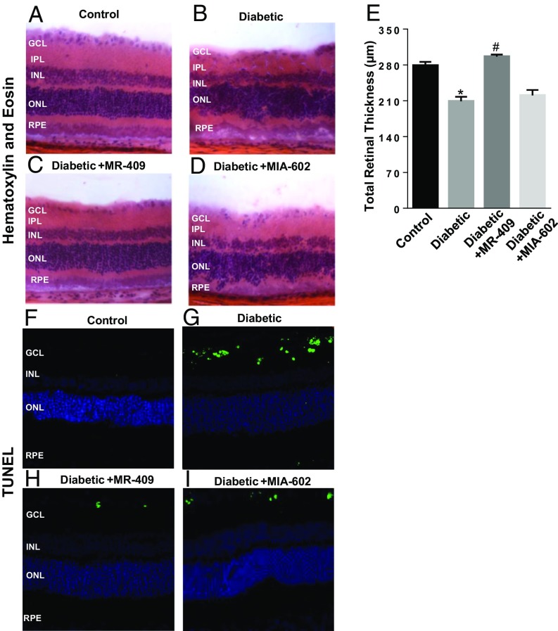
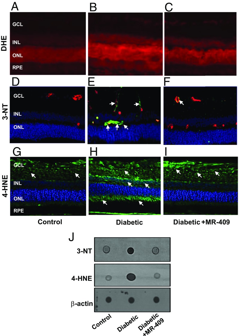
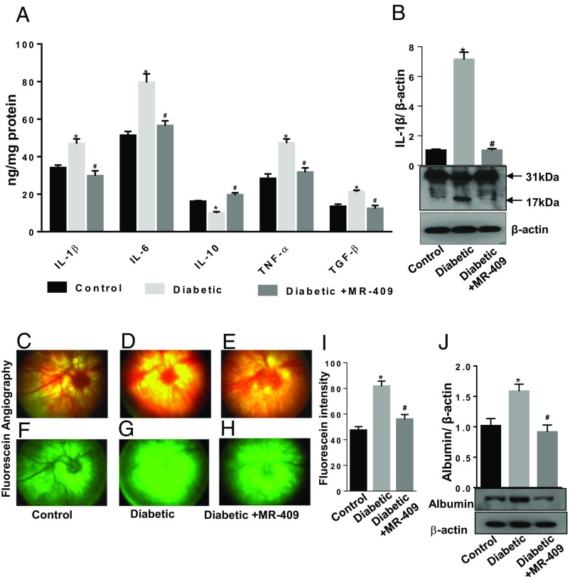
The potential therapeutic effects of agonistic analogs of growth hormone-releasing hormone (GHRH) and their mechanism of action were investigated in diabetic retinopathy (DR). Streptozotocin-induced diabetic rats (STZ-rats) were treated with 15 μg/kg GHRH agonist, MR-409, or GHRH antagonist, MIA-602. At the end of treatment, morphological and biochemical analyses assessed the effects of these compounds on retinal neurovascular injury induced by hyperglycemia. The expression levels of GHRH and its receptor (GHRH-R) measured by qPCR and Western blotting were significantly down-regulated in retinas of STZ-rats and in human diabetic retinas (postmortem) compared with their respective controls. Treatment of STZ-rats with the GHRH agonist, MR-409, prevented retinal morphological alteration induced by hyperglycemia, particularly preserving survival of retinal ganglion cells. The reverse, using the GHRH antagonist, MIA-602, resulted in worsening of retinal morphology and a significant alteration of the outer retinal layer. Explaining these results, we have found that MR-409 exerted antioxidant and anti-inflammatory effects in retinas of the treated rats, as shown by up-regulation of NRF-2-dependent gene expression and down-regulation of proinflammatory cytokines and adhesion molecules. MR-409 also significantly down-regulated the expression of vascular endothelial growth factor while increasing that of pigment epithelium-derived factor in diabetic retinas. These effects correlated with decreased vascular permeability. In summary, our findings suggest a neurovascular protective effect of GHRH analogs during the early stage of diabetic retinopathy through their antioxidant and anti-inflammatory properties.

Keywords: GH; GHRH; GHRH-R; diabetic retinopathy; type 1 diabetes.


Conflict of interest statement

Conflict of interest statement: N.L.B. owns equity in Biscayne Pharmaceuticals. A.V.S. is a coinventor on the patent for GHRH agonist, assigned to the University of Miami and the Veterans Affairs Medical Center, Miami, FL. However, the investigation of the effects of GHRH agonist MR-409 was an academic endeavor without any commercial interests. The other authors declare no conflict of interest.

Figures

Fig. 1.
Fig. 1.
Effects of GHRH agonist MR-409 or antagonist MIA-602 on GHRH-R and GHRH expression levels. (AD) Microimages illustrating GHRH-R-specific immunoreactivity (green fluorescence and white arrows) in retinal cryosections of STZ-rats (Diabetic) (BD) compared with age-matched normal rats (Control, A). In C and D, the STZ-rats received, every other day, s.c. injections of 15 μg/kg MR-409 or MIA-602 (respectively). Treatment was started 2 wk after the onset of diabetes and prolonged for 6 wk (6 wk treatment in a total of 8 wk hyperglycemia). These images show that GHRH-R is down-regulated in the diabetic retina, particularly in the GCL (Diabetic) (B), but treatment with MR-409 (Diabetic+MR-409) drastically increased GHRH-R-specific immunoreactivity in the GCL and neurofibrillary layer (C, green fluorescence, white arrows). (E) Representative immunoblotting showing GHRH-R-specific immunoreactivity measured in retinal extracts of rats subjected to different treatment conditions. Bar histograms represent the optical density values normalized to the loading control, β-actin. Diabetes suppressed GHRH-R protein levels in comparison with normal age-matched controls (*P < 0.001 vs. Control; n = 8). MR-409 stimulated a significant increase in levels of GHRH-R protein in STZ-rats at 8 wk of hyperglycemia (Diabetic+MR-409; #P < 0.001 vs. Diabetic; n = 8). (F) qPCR analysis measuring GHRH mRNA expression levels in retinas of rats from the different experimental groups. These data show that GHRH-specific mRNA levels are down-regulated in the diabetic retina (*P < 0.001 vs. Control; n = 8), and treatment with MR-409 partially rescues GHRH mRNA (#P < 0.05 vs. Diabetic), whereas the antagonist, MIA-602, has no effect.
Fig. 2.
Fig. 2.
Effects of MR-409 and MIA-602 on retinal morphological changes and apoptosis in diabetic retinas. Histopathological analysis assessing retinal morphology in response to the different treatment conditions was conducted by using H&E staining of retinal cryosections (AD). In D, a pronounced distortion of the outer nuclear layer (ONL) is evidenced. Morphometric analysis assessing total retinal thickness (E) values measured in H&E retinal cryosections obtained from the different treatment groups. The sum of the results obtained from these analyses demonstrates a significant preservation of the retinal inner nuclear layer (INL) in the STZ-rats. (FI) TUNEL staining to measure apoptosis in retinal cryosections of the different groups. Higher number of TUNEL-positive cells evident in diabetic retina (G) compared with control rats (F); whereas treatment with MR-409 (H) reduced the number of apoptotic cells, MIA-602 (I) treatment did not alter the rate of apoptosis in STZ-rats.
Fig. 3.
Fig. 3.
Oxidative/nitrative markers. (AC) Dihydroethidium (DHE) staining to evaluate superoxide production. Retinal sections from diabetic rats (B) show higher intensity of DHE staining indicating increased superoxide levels compared with control rats (A). Treatment with MR-409 (C) reduced diabetes-associated superoxide production. (DF) Immunostaining for 3-NT and double staining with isolectin B4 for double labeling with retinal vessels. Immunoreactivity for 3-NT is evidenced around blood vessels (green, 3-NT; red, isolectin B4; yellow, merging; white arrows), particularly in the diabetic retina (E, Diabetic). Treatment with GHRH agonist (F, Diabetic+MR-409) prevents this effect. (GI) 4-HNE immunostaining (green fluorescence) in retinal cryosections shows increased immunoreactivity in the diabetic retina (H, Diabetic) versus normoglycemic age-matched control (G, Control). Treatment of STZ-rats with MR-409 (I) decreased retinal immunoreactivity of 4-HNE (green fluorescence, white arrows, at 20× magnification). (J) Dot blot analysis assessing 3-NT and 4-HNE and showing increased immunoreactivity for both these markers in diabetic retina (Diabetic), which is prevented by treatment of STZ-rats with MR-409 (Diabetic+MR-409). Beta-actin was used as loading control.
Fig. 4.
Fig. 4.
Effects of MR-409 on retinal expression of cytokines and blood retinal barrier integrity. (A) Expression of IL-1β, IL-6, IL-10, TNF-α, and TGF-β were evaluated in retinal tissue using a customized ELISA kit. Bar histograms showing a significant up-regulation of retinal levels of IL-1β, IL-6, TNF-α, and TGF-β and decreased levels of IL-10 in diabetic rats (light gray bars) compared with control (black bars). Treatment with MR-409 normalized the levels of measured cytokines (dark gray bars). (B) Western blot analysis of IL-1β in the different treatment conditions showing the mature (active) 17-kDa form of IL-1β and the pro-IL-1β isoform (31 kDa). Representative images of fundus photographs (CE) and fluorescein angiography (FH) of rat eyes of control, diabetic, and diabetic treated with MR-409. Pictures were taken at constant interval for every rat studied in each experimental group. The fluorescence intensity per rat retina was calculated by Image J software and expressed as arbitrary units of fluorescence intensity (I). This analysis shows increased fluorescence intensity (extravasation) in diabetic rat retinas (D and G) compared with control normoglycemic rats (C and F) (Diabetic, *P < 0.01 vs. Control; n = 8). Treatment of STZ-rats with the GHRH agonist, MR-409, significantly decreases fluorescein extravasation (Diabetic+MR-409; #P < 0.01 vs. Diabetic; n = 8) (E and H). (J) Western blot analysis assessing albumin protein levels in retinal extracts of perfused rats from the different experimental groups. This analysis shows increased albumin immunoreactivity in the retinal lysates of STZ-rats after perfusion (Diabetic, *P < 0.05 vs. Control; n = 8). Albumin-specific immunoreactivity is significantly reduced in retinal extracts of (perfused) STZ-rats treated with MR-409 (Diabetic+MR-409, #P < 0.01 vs. Diabetic; n = 8). The data are expressed as arbitrary units of optical density and normalized for the loading control β-actin.

Similar articles

Cited by

References

    1. Sivaprasad S, Gupta B, Crosby-Nwaobi R, Evans J. Prevalence of diabetic retinopathy in various ethnic groups: A worldwide perspective. Surv Ophthalmol. 2012;57:347–370. - PubMed
    1. Tan GS, Cheung N, Simó R, Cheung GC, Wong TY. Diabetic macular oedema. Lancet Diabetes Endocrinol. 2017;5:143–155. - PubMed
    1. Tolentino MS, Tolentino AJ, Tolentino MJ. Current and investigational drugs for the treatment of diabetic retinopathy. Expert Opin Investig Drugs. 2016;25:1011–1022. - PubMed
    1. Antonetti DA, Klein R, Gardner TW. Diabetic retinopathy. N Engl J Med. 2012;366:1227–1239. - PubMed
    1. Murray PG, Higham CE, Clayton PE. 60 years of neuroendocrinology: The hypothalamo-GH axis: The past 60 years. J Endocrinol. 2015;226:T123–T140. - PubMed

Publication types

MeSH terms

Substances

LinkOut - more resources