. 2018 Jan 19;293(3):941-952.
doi: 10.1074/jbc.M117.813428. Epub 2017 Nov 27.

A neutralizing antibody that blocks delivery of the enzymatic cargo of Clostridium difficile toxin TcdB into host cells

Affiliations

A neutralizing antibody that blocks delivery of the enzymatic cargo of Clostridium difficile toxin TcdB into host cells

Heather K Kroh et al. J Biol Chem. .

Abstract

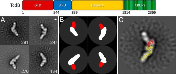
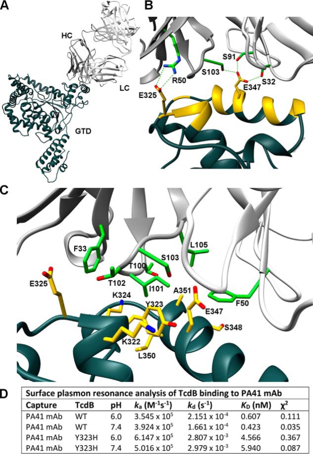
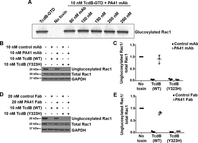
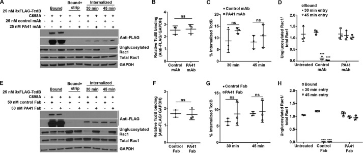
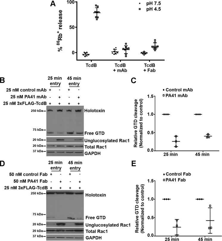
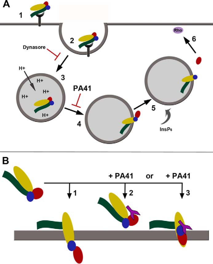
Clostridium difficile infection is the leading cause of hospital-acquired diarrhea and is mediated by the actions of two toxins, TcdA and TcdB. The toxins perturb host cell function through a multistep process of receptor binding, endocytosis, low pH-induced pore formation, and the translocation and delivery of an N-terminal glucosyltransferase domain that inactivates host GTPases. Infection studies with isogenic strains having defined toxin deletions have established TcdB as an important target for therapeutic development. Monoclonal antibodies that neutralize TcdB function have been shown to protect against C. difficile infection in animal models and reduce recurrence in humans. Here, we report the mechanism of TcdB neutralization by PA41, a humanized monoclonal antibody capable of neutralizing TcdB from a diverse array of C. difficile strains. Through a combination of structural, biochemical, and cell functional studies, involving X-ray crystallography and EM, we show that PA41 recognizes a single, highly conserved epitope on the TcdB glucosyltransferase domain and blocks productive translocation and delivery of the enzymatic cargo into the host cell. Our study reveals a unique mechanism of C. difficile toxin neutralization by a monoclonal antibody, which involves targeting a process that is conserved across the large clostridial glucosylating toxins. The PA41 antibody described here provides a valuable tool for dissecting the mechanism of toxin pore formation and translocation across the endosomal membrane.

Keywords: X-ray crystallography; antibody; bacterial pathogenesis; bacterial toxin; electron microscopy (EM); membrane transport; neutralization; toxin.


Conflict of interest statement

These studies were partially funded by MedImmune through a collaborative project awarded to D. B. L. MedImmune had the right to review the manuscript but otherwise had no role in the preparation or decision to publish. K. R., R. W., X. J., A. C. N., G. J. R., and P. W. are employees of MedImmune, a unit of AstraZeneca. K. R., R. W., X. J., A. C. N., G. J. R., and P. W. own stock in AstraZeneca. G. J. R. is now employed by MabVax Therapeutics

Figures

Figure 1.
Figure 1.
PA41 binds a single site on TcdB as evaluated by negative-stain electron microscopy. Top, domain organization of TcdB holotoxin, including residue ranges for each domain (74). A, panel of four representative two-dimensional class averages of TcdB(1–1810) bound by PA41 Fab (with number of particles in each class in white). B, schematic for TcdB (white) and PA41 Fab (red) for each of the representative classes. C, overlay of a model of TcdB(1–1810) onto one of the class averages from A (indicated with an asterisk) with the GTD in red, autoprotease domain (APD) in blue, and delivery domain in yellow.
Figure 2.
Figure 2.
X-ray crystal structure of TcdB-027-GTD + PA41 Fab and structural determinants of epitope interaction. A, wide view of complex with TcdB-027-GTD in dark gray, PA41-HC in gray, and PA41-LC in white. B, residues within the epitope on the GTD are colored yellow with critical PA41 residues in green. Side-chain carbons (red) and nitrogens (blue) are also indicated. A salt bridge is located at one end of the epitope (Glu-325–Arg-50) with GTD Glu-347 potentially mediating hydrogen bonds with both Fab chains. C, hydrophobic pocket accommodating Ile-101 of PA41-HC with residues forming all sides of site labeled (GTD, yellow; Fab, green). D, summary of kinetic rate and dissociation constants for the interactions of TcdB(WT) and TcdB(Y323H) with PA41 mAb measured by SPR.
Figure 3.
Figure 3.
PA41 inhibits TcdB-induced Rac1 glucosylation in Caco-2 cells but not in vitro. A, in vitro glucosylation of GST-Rac1 (2 μm) by TcdB-027-GTD (10 nm) in the presence of [14C]UDP-glucose (24 μm) and increasing concentrations of PA41 mAb. Controls are shown in lane 1 (GTD with Rac1) and lane 2 (Rac1 only). B, Rac1 glucosylation by TcdB(WT) or TcdB(Y323H) in the presence of isotype control or PA41 mAb was assessed in Caco-2 cells. Cells that did not receive toxin or antibody were used as controls. C, experiments in B were quantified by densitometry, and the extent of Rac1 glucosylation was determined by normalizing the unglucosylated and total Rac1 levels. Means ± S.D. (n = 3) are shown. D and E, similar experiment performed with Fab fragments. The antibody concentration was doubled to keep the available number of antigen-binding sites similar between the mAb and Fab experiments. Error bars represent S.D.
Figure 4.
Figure 4.
PA41 does not affect binding or entry of TcdB in Caco-2 cells, but it does inhibit Rac1 modification. A, TcdB binding and entry in the presence of isotype control or PA41 mAb were assessed in toxin entry assays. Cells that did not receive any toxin or antibody were used as controls. Comparison of bound toxin levels between −strip (lanes 2 and 3) and +strip (lanes 4 and 5) conditions confirms efficient removal of surface-bound toxin by this procedure. B, experiments in A were quantified by densitometry, and the relative binding of TcdB to cells was determined by normalizing bound TcdB levels to that of GAPDH (lanes 2 and 3). Means ± S.D. (n = 3) are shown and were analyzed using two-tailed t test. ns, not significant. C, the internalized TcdB signal (lanes 6–9) was normalized to the corresponding GAPDH levels to obtain the relative entry signal, which was then normalized to the relative bound toxin levels and expressed as the percentage of internalized TcdB. Means ± S.D. (n = 3) are shown and were analyzed using two-tailed t test. ns, not significant. D, the extent of Rac1 glucosylation by TcdB was determined by normalizing the unglucosylated and total Rac1 levels. Means ± S.D. (n = 3) are shown and were analyzed using one-way analysis of variance. p values were generated using Dunnett's multiple comparisons test. ***, p < 0.0005. E–H, similar experiments performed with the Fab fragments. Error bars represent S.D.
Figure 5.
Figure 5.
PA41 inhibits pore formation and the subsequent delivery of the GTD into the cytosol. A, pore formation on biological membranes. TcdB holotoxin alone (1 nm) or preincubated with PA41 mAb or Fab (10 nm) was applied to CHO-K1 cells preloaded with 86Rb+, and rubidium release was compared at pH 7.5 and 4.5. Means ± S.D. (n = 6) are shown. B, Caco-2 monolayers were intoxicated with 3xFLAG-TcdB (25 nm) preincubated with equimolar (25 nm) amounts of control or PA41 mAb. Toxin cleavage assays were performed as described under “Experimental procedures.” Blots were probed with antibodies against the toxin (anti-FLAG; detects both internalized holotoxin and free GTD), unglucosylated and total Rac1, and GAPDH. C, the fraction of the internalized toxin that is cleaved and released in each condition was determined by normalizing the free GTD signal to the corresponding internalized holotoxin signal. For each time point, the fractional GTD release measurements were normalized to that of the isotype mAb controls to obtain the relative GTD cleavage. D and E, similar experiments performed with the Fab fragments. Means ± S.D. (n = 3) are shown. Error bars represent S.D.
Figure 6.
Figure 6.
Proposed models of cellular intoxication by TcdB. A, schematic summarizing the individual steps involved in toxin uptake, including cell surface binding (1), endosome formation (2) and maturation (3), pH-dependent pore formation and translocation (4), toxin autoprocessing and GTD release (5), and targeting of Rho-family GTPases (6). In contrast to Dynasore, which directly blocks endocytosis of toxin, PA41 targets the processes involved in pore formation and/or translocation. B, a comparison of the two potential mechanisms through which PA41 could prevent cargo transport during intoxication (1). TcdB normally forms a channel through the endosomal membrane, allowing translocation of the enzymatic toxin domains (2). In the presence of PA41, formation of the pore could be directly blocked through steric hindrance of conformational changes within the toxin. Although an Rb+-conductive pore can be formed in the absence of the GTD (14), binding of PA41 may restrict individual domain movements or overall toxin conformation (3). PA41 could also prevent unfolding of the GTD for transport, which would trap the GTD at some point within the toxin pore. Either of these mechanisms could result in the decreased cargo transport observed in the presence of PA41. TcdB domains are colored as shown previously (GTD, red; autoprotease domain, blue; delivery domain, yellow; CROPs, green) with PA41 in purple.

References

    1. Smits W. K., Lyras D., Lacy D. B., Wilcox M. H., and Kuijper E. J. (2016) Clostridium difficile infection. Nat. Rev. Dis. Primers 2, 16020 10.1038/nrdp.2016.20 - DOI - PMC - PubMed
    1. Lessa F. C., Mu Y., Bamberg W. M., Beldavs Z. G., Dumyati G. K., Dunn J. R., Farley M. M., Holzbauer S. M., Meek J. I., Phipps E. C., Wilson L. E., Winston L. G., Cohen J. A., Limbago B. M., Fridkin S. K., et al. (2015) Burden of Clostridium difficile infection in the United States. N. Engl. J. Med. 372, 825–834 10.1056/NEJMoa1408913 - DOI - PMC - PubMed
    1. Carter G. P., Chakravorty A., Pham Nguyen T. A., Mileto S., Schreiber F., Li L., Howarth P., Clare S., Cunningham B., Sambol S. P., Cheknis A., Figueroa I., Johnson S., Gerding D., Rood J. I., et al. (2015) Defining the roles of TcdA and TcdB in localized gastrointestinal disease, systemic organ damage, and the host response during Clostridium difficile infections. MBio 6, e00551.10.1128/mBio.00551-15 - DOI - PMC - PubMed
    1. Steele J., Mukherjee J., Parry N., and Tzipori S. (2013) Antibody against TcdB, but not TcdA, prevents development of gastrointestinal and systemic Clostridium difficile disease. J. Infect. Dis. 207, 323–330 10.1093/infdis/jis669 - DOI - PMC - PubMed
    1. Drudy D., Fanning S., and Kyne L. (2007) Toxin A-negative, toxin B-positive Clostridium difficile. Int. J. Infect. Dis. 11, 5–10 10.1016/j.ijid.2006.04.003 - DOI - PubMed

Publication types

MeSH terms