. 2018 Jan 15;78(2):572-583.
doi: 10.1158/0008-5472.CAN-17-2040. Epub 2017 Nov 27.

Vitamin C Sensitizes Melanoma to BET Inhibitors

Affiliations

Vitamin C Sensitizes Melanoma to BET Inhibitors

Sushmita Mustafi et al. Cancer Res. .

Abstract

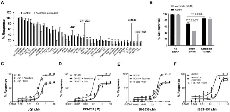
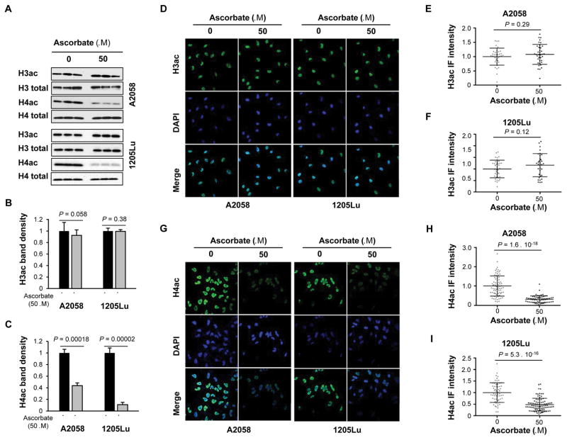
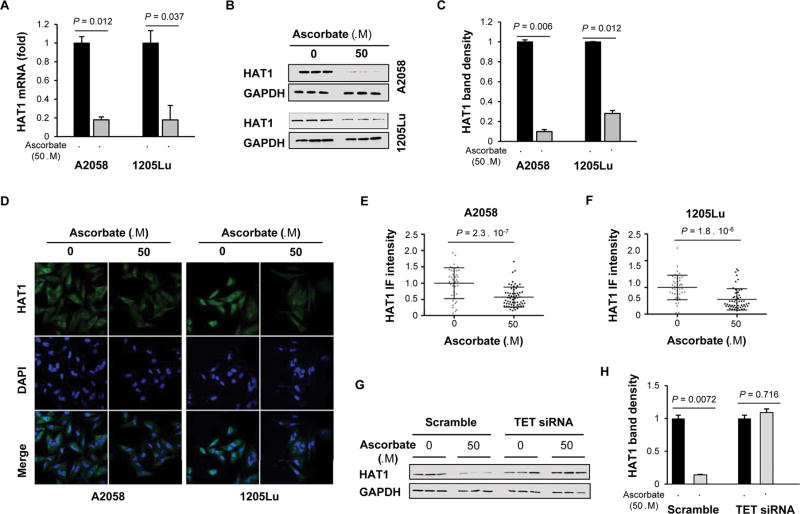
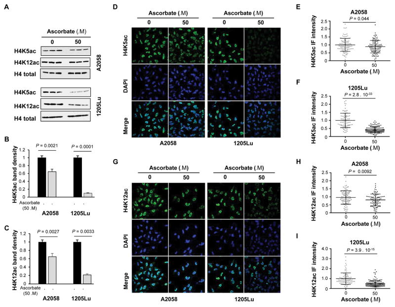
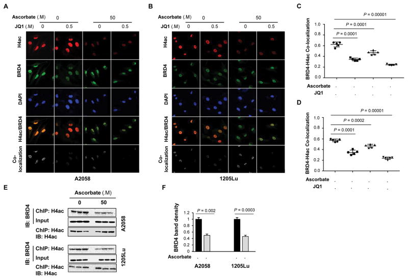
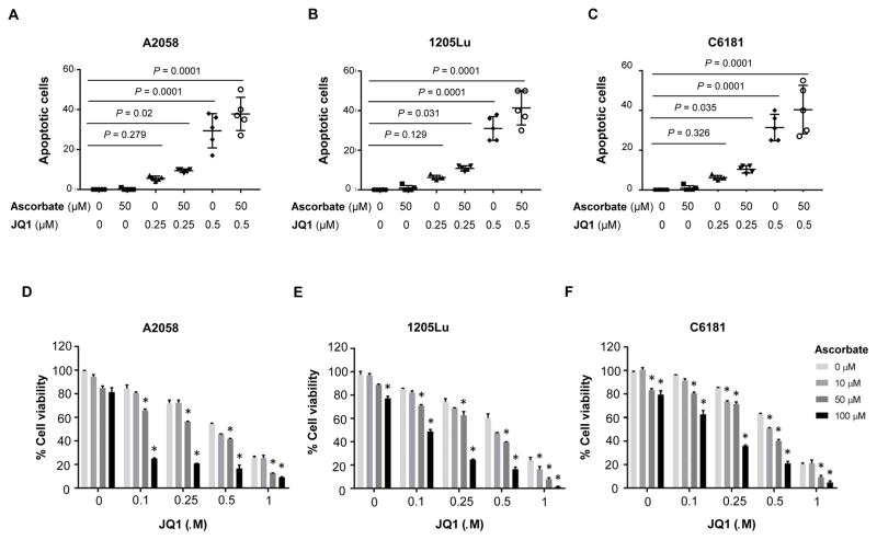
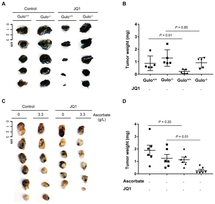
Bromodomain and extraterminal inhibitors (BETi) are promising cancer therapies, yet prominent side effects of BETi at effective doses have been reported in phase I clinical trials. Here, we screened a panel of small molecules targeting epigenetic modulators against human metastatic melanoma cells. Cells were pretreated with or without ascorbate (vitamin C), which promotes DNA demethylation and subsequently changes the sensitivity to drugs. Top hits were structurally unrelated BETi, including JQ1, I-BET151, CPI-203, and BI-2536. Ascorbate enhanced the efficacy of BETi by decreasing acetylation of histone H4, but not H3, while exerting no effect on the expression of BRD proteins. Histone acetyltransferase 1 (HAT1), which catalyzes H4K5ac and H4K12ac, was downregulated by ascorbate mainly via the TET-mediated DNA hydroxymethylation pathway. Loss of H4ac, especially H4K5ac and H4K12ac, disrupted the interaction between BRD4 and H4 by which ascorbate and BETi blocked the binding of BRD4 to acetylated histones. Cotreatment with ascorbate and JQ1 induced apoptosis and inhibited proliferation of cultured melanoma cells. Ascorbate deficiency as modeled in Gulo-/- mice diminished the treatment outcome of JQ1 for melanoma tumorgraft. In contrast, ascorbate supplementation lowered the effective dose of JQ1 needed to successfully inhibit melanoma tumors in mice. On the basis of our findings, future clinical trials with BETi should consider ascorbate levels in patients. Furthermore, ascorbate supplementation might help reduce the severe side effects that arise from BETi therapy by reducing the dosage necessary for treatment.Significance: This study shows that ascorbate can enhance the efficacy of BET inhibitors, providing a possible clinical solution to challenges arising in phase I trials from the dose-dependent side effects of this class of epigenetic therapy. Cancer Res; 78(2); 572-83. ©2017 AACR.


Conflict of interest statement

Disclosure of potential conflicts of interest: The authors declare no actual or potential conflicts of interest in relation to this article.

Figures

Figure 1
Figure 1
Ascorbate treatment enhances the efficacy of BETi. A, Representative data of the compound screen (n = 164) targeting epigenetic modulators in A2058 cells. The responses of JQ1, CPI-203, BI-2536 and I-BET151 were drastically increased by pretreatment with 50 μM ascorbate. B, The survival rate of A2058 cells was decreased after BRD4 knockdown by siRNA compared to scramble siRNA (P = 0.0008). Ascorbate (50 μM) further decreased the survival rate (P = 0.0002). C–E, Dose response curve of BETi. Ascorbate (50 μM) increased the response of A2058 cells to JQ1, CPI-203, BI-2536 and I-BET151 while GSH (50 μM) had no effect (n= 3). All data are mean ± SEM.
Figure 2
Figure 2
Ascorbate treatment reduces H4ac but not H3ac in melanoma cells. A, Immunoblot of H3ac and H4ac in A2058 and 1205Lu cells treated with or without ascorbate (50 μM) for 72 hours. Total H3 and H4 were used for loading controls. B, Densitometry analysis of H3ac normalized by total H3. No significant change in H3ac was observed after ascorbate treatment. C, Densitometry analysis of H4ac normalized by total H4. The levels of H4ac were reduced by ascorbate treatment in A2058 and1205Lu cells. D–F, Immunofluorescence and quantification of H3ac in A2058 and 1205Lu cells treated with or without ascorbate (50 μM) for 72 hours. No significant change in H3ac was observed in A2058 and 1205Lu cells. G–I, Immunofluorescence and quantification of H4ac in A2058 and 1205Lu cells treated with or without ascorbate (50 μM) for 72 hours. H4ac was decreased by ascorbate treatment in A2058 and 1205Lu cells. All data are mean ± SEM.
Figure 3
Figure 3. Downregulation of HAT1 by ascorbate treatment
A, qRT-PCR of HAT1. HAT1 mRNA were reduced by ascorbate (50 μM) treatment for 72 hours in A2058 cells (P = 0.012) and in 1205Lu cells (P = 0.037). B, Immunoblot of HAT1 in A2058 and 1205Lu cells. C, Densitometry analysis of HAT1 normalized by GAPDH shows that HAT1 was decreased by ascorbate treatment in A2058 cells and 1205Lu cells. D, Immunofluorescence of HAT1 in A2058 and 1205Lu cells treated with or without ascorbate (50 μM) for 72 hours. E–F, Quantification of HAT1 immunofluorescence. The level of HAT1 protein was decreased by ascorbate treatment in A2058 and 1205Lu cells. G–H, HAT1 protein remained unchanged by ascorbate treatment in TETs knockdown A2058 cells as shown by immunoblot and quantification.
Figure 4
Figure 4
H4K5ac and H4K12ac are decreased by ascorbate treatment. A, Immunoblot of H4K5ac and H4K12ac in A2058 and 1205Lu cells treated with or without ascorbate (50 μM) for 72 hours. B–C, Densitometry analysis of H4K5ac and H4K12ac normalized by total H4. H4K5ac and H4K12ac were decreased by ascorbate treatment in A2058 and 1205Lu cells. D–F, Immunofluorescence and quantification of H4K5ac in A2058 and 1205Lu cells treated with or without ascorbate (50 μM) for 72 hours. H4K5ac was decreased by ascorbate treatment in A2058 and 1205Lu cells. G–I, Immunofluorescence and quantification of H4K12ac in A2058 and 1205Lu cells treated with or without ascorbate (50 μM) for 72 hours. H4K12ac was decreased by ascorbate treatment in A2058 and 1205Lu cells. All data are mean ± SEM.
Figure 5
Figure 5
The binding of BRD4 to acetylated H4 is interrupted by ascorbate treatment. A–B, Confocal microscopy images of H4ac, BRD4, and their co-localization in A2058 and 1205Lu cells. C–D, Quantification of H4ac and BRD4 co-localization in A2058 and 1205Lu cells. The co-localization coefficient of H4ac and BRD4 was reduced by JQ1. Ascorbate treatment alone reduced the signal of H4ac and subsequently the H4ac-BRD4 co-localization. The co-localization coefficient of H4ac and BRD4 was further decreased by co-treatment with ascorbate and JQ1. E, ChIP assay of H4ac and BRD4 binding in A2058 cells and 1205Lu cells. F, Densitometry analysis of BRD4 in the ChIP assay. The binding between H4ac and BRD4 was reduced by ascorbate treatment in A2058 cells and 1205Lu cells. All data are mean ± SEM (n = 3).
Figure 6
Figure 6
Reduction of melanoma cell malignancy by ascorbate and JQ1. A–C, Induction of apoptosis in A2058, 1205Lu and C6181 cells by combination treatment of ascorbate and JQ1. Ascorbate alone at 50 μM did not have obvious consequence on apoptosis but enhanced the effect of JQ1 at 0.25 μM and0.5 μM to induce apoptosis. D–F, Cell viability after combination treatment of ascorbate and JQ1. Ascorbate at (50 and 100 μM) reinforced the inhibitory effect of JQ1 (0.1, 0.25, 0.5 and 1 μM) on the viability of A2058, 1205Lu and C6181 cells. * P < 0.001. All data are mean ± SEM.
Figure 7
Figure 7
Inhibition of melanoma tumorgraft by ascorbate and JQ1. A, Photograph of murine B16-F10 melanoma syngenic tumorgraft from Gulo−/− and Gulo+/+ (wild type) mice treated with or without JQ1. B, Quantification of B16-F10 melanoma tumor weights. Without JQ1 treatment, tumors in ascorbate sufficient mice are smaller (P = 0.001) compared to ascorbate deficient mice. In ascorbate sufficient mice, tumors were drastically reduced by JQ1 (50 mg/kg body weight) (P = 0.01). However, in ascorbate deficient mice, tumors were not significantly reduced by the same dose of JQ1 treatment (P = 0.85). C, Photograph of human A2058 melanoma xenografts treated with or without JQ1 from nude mice supplemented with or without ascorbate. D, Quantification of A2058 melanoma xenograft weights. Without ascorbate supplements, xenografts remained at similar sizes after JQ1 (50 mg/kg body weight) injection (P = 0.20). However, xenografts were reduced in mice treated with JQ1 when supplemented with ascorbate compared to the mice supplemented with ascorbate but no JQ1 treatment (P = 0.01), or in comparison to the mice treated only with JQ1 (P = 0.02).

References

    1. Dutta A, Abmayr SM, Workman JL. Diverse activities of histone acetylation connect metabolism to chromatin function. Mol Cell. 2016;63:547–52. - PMC - PubMed
    1. Jain AK, Barton MC. Bromodomain Histone Readers and Cancer. J Mol Biol. 2017;429:2003–10. - PubMed
    1. Patino WD, Susa J. Epigenetics of cutaneous melanoma. Adv Dermatol. 2008;24:59–70. - PubMed
    1. Filippakopoulos P, Knapp S. Targeting bromodomains: epigenetic readers of lysine acetylation. Nat Rev Drug Discov. 2014;13:337–56. - PubMed
    1. Segura MF, Fontanals-Cirera B, Gaziel-Sovran A, Guijarro MV, Hanniford D, Zhang G, et al. BRD4 sustains melanoma proliferation and represents a new target for epigenetic therapy. Cancer Res. 2013;73:6264–76. - PMC - PubMed

Publication types

MeSH terms