. 2018 Jan 29;38(4):e00364-17.
doi: 10.1128/MCB.00364-17. Print 2018 Feb 15.

mRNA Processing Factor CstF-50 and Ubiquitin Escort Factor p97 Are BRCA1/BARD1 Cofactors Involved in Chromatin Remodeling during the DNA Damage Response

Affiliations

mRNA Processing Factor CstF-50 and Ubiquitin Escort Factor p97 Are BRCA1/BARD1 Cofactors Involved in Chromatin Remodeling during the DNA Damage Response

Danae Fonseca et al. Mol Cell Biol. .

Abstract

The cellular response to DNA damage is an intricate mechanism that involves the interplay among several pathways. In this study, we provide evidence of the roles of the polyadenylation factor cleavage stimulation factor 50 (CstF-50) and the ubiquitin (Ub) escort factor p97 as cofactors of BRCA1/BARD1 E3 Ub ligase, facilitating chromatin remodeling during the DNA damage response (DDR). CstF-50 and p97 formed complexes with BRCA1/BARD1, Ub, and some BRCA1/BARD1 substrates, such as RNA polymerase (RNAP) II and histones. Furthermore, CstF-50 and p97 had an additive effect on the activation of the ubiquitination of these BRCA1/BARD1 substrates during DDR. Importantly, as a result of these functional interactions, BRCA1/BARD1/CstF-50/p97 had a specific effect on the chromatin structure of genes that were differentially expressed. This study provides new insights into the roles of RNA processing, BRCA1/BARD1, the Ub pathway, and chromatin structure during DDR.

Keywords: BRCA1/BARD1; CstF-50; DNA damage response; RNA polymerase II; chromatin remodeling; histones; p97.


Figures

FIG 1
FIG 1
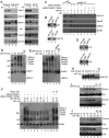
CstF-50 interacts with p97 to form a protein complex, and both can activate BRCA1/BARD1 Ub ligase activity in vitro in an additive manner. (A) p97, CstF-50, BRCA1, and BARD1 can form complexes in NEs of HeLa cells independently of UV treatment. Cells were exposed to UV irradiation (40 J m−2) and allowed to recover for 2 h before NEs were prepared. The NEs were immunoprecipitated with anti-CstF-50 (left) or anti-p97 (right). Equivalent amounts of the pellets (IP) were resolved by SDS-PAGE, and proteins were detected by immunoblotting using antibodies against the indicated proteins. Antibody against Topo II was used as a control for specificity. The positions of Topo II, RNAP II, BRCA1, BARD1, p97, and CstF-50 are indicated. Five percent of the NE used in the IP reaction is shown as input. Representative IP reactions from three independent assays are shown. (B) CstF-50 interacts directly with p97. (Top) Immobilized GST–CstF-50 or GST on glutathione beads was incubated with 1 μg of His-p97. (Bottom) Immobilized His-p97 on nickel beads was incubated with GST–CstF-50 or GST. Bound proteins were eluted, resolved by SDS-PAGE, and detected by Western blotting with anti-p97 or anti-CstF-50. Five percent His-p97 or GST–CstF-50 used in the reaction is shown as input. Recombinant proteins were treated with RNase A. Representative pulldown reactions from three independent assays are shown. (C) CstF-50 bridges the interaction between BARD1 and p97. Pulldown assays were conducted in the presence of truncated (residues 1 to 304) BRCA1 and full-length BARD1 (WT or BARD1-Q564H) heterodimer. Either ΔBRCA1/BARD1-wt or ΔBRCA1/BARD1-Q564H was incubated with GST–CstF-50 and/or His-p97. Samples were immunoprecipitated with anti-BARD1 or preimmune sera. Bound proteins were eluted, resolved by SDS-PAGE, and detected by Western blotting with CstF-50, BARD1, BRCA1, or p97 antibodies; 5% His-p97 or GST–CstF-50 used in the reaction is shown as input. (D) CstF-50 increases the autoubiquitination of the BRCA1/BARD1 heterodimer. In vitro ubiquitination reactions were conducted in the presence of limiting amounts (10 ng) of ΔBRCA1/BARD1-wt, E1, His-E2, His-Ub, and ATP and increasing amounts of recombinant GST–CstF-50. Samples were resolved by SDS-PAGE and immunoblotted with anti-BARD1. Representative autoubiquitination reactions from three independent assays are shown. (E) p97 further increases CstF-50-mediated activation of BRCA1/BARD1 autoubiquitination. In vitro ubiquitination reactions were carried out as for panel D but using limiting amounts of GST–CstF-50 (25 ng) and increasing amounts of His-p97. Samples were resolved by SDS-PAGE and immunoblotted with anti-BARD1. Representative autoubiquitination reactions from three independent assays are shown. (F) CstF-50/p97-dependent enhancement of BRCA1/BARD1 E3 Ub ligase activity depends on the ability of CstF-50 to bind the heterodimer. Autoubiquitination assays were carried out as for panel D but also including ΔBRCA1/BARD1-Q564H and increasing amounts of the p97 inhibitor DBeQ. (G) CstF-50 interacts directly with H2A (top) and H2B (bottom). Immobilized GST–CstF-50 on glutathione-agarose beads was incubated with commercially available H2A and H2B. Bound proteins were eluted, resolved by SDS-PAGE, and detected with anti-histones; 5% histones used in the reaction were loaded as input. Representative pulldown reactions from three independent assays are shown. (H) CstF-50 and p97 activate RNAP IIO ubiquitination by BRCA1/BARD1. In vitro ubiquitination reactions were performed as described for panel D in the presence of purified RNAP IIO. Proteins were detected with RNAP IIO-specific antibody (H5; Covance). Representative ubiquitination reactions from three independent assays are shown. (I) CstF-50 activates the monoubiquitination of H2A and H2B by BRCA1/BARD1. In vitro ubiquitination reactions were performed as described for panel D in the presence of commercially available H2A and H2B and increasing amounts of GST–CstF-50. Proteins were detected by Western blotting with anti-histones. (J) p97 further increases CstF-50-mediated activation of histone monoubiquitination by BRCA1/BARD1. In vitro ubiquitination reactions were performed as for panel I but using limiting amounts of GST–CstF-50 and increasing amounts of His-p97. Representative ubiquitination reactions from three independent assays are shown.
FIG 2
FIG 2
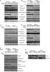
Monoubiquitination of histones H2A and H2B and polyubiquitination of RNAP IIO by BRCA1/BARD1 are affected by the functional interaction of CstF-50 and p97. CstF-50 and p97 activate UV-induced monoubiquitination of histones. (A to E) HeLa cells were transfected with siRNAs for either control (CNTL), CstF-50 (A and D), or BRCA1/BARD1 (B) or concomitantly for BRCA1/BARD1/CstF-50 (E). The cells were also transfected with HA-Ub and either FLAG-H2A or FLAG-H2B constructs. The cells were treated with UV (40 J m−2) and allowed to recover for 2 h before NEs were prepared. (C to E) Alternatively, cells were also treated with DBeQ (10 μM) during the 2-h recovery. Soluble NEs were immunoprecipitated with anti-FLAG M2 magnetic beads (Sigma), followed by Western blotting with the indicated antibodies. Antibody against Topo II was used as a control; 5% of the cell extracts used in the IP reaction is shown as input. Representative IP reactions from three independent assays are shown. (F) CstF-50 and p97 activate UV-induced polyubiquitination of RNAP IIO. HeLa cells were transfected with either control or CstF-50 siRNAs concomitantly with an HA-Ub construct. The cells were treated with UV (40 J m−2). The proteasomal inhibitor MG132 (2 μM) and DBeQ (10 μM) were added to the cells immediately after exposure to UV light, and the cells were allowed to recover for 2 h before soluble NEs were prepared. The soluble NEs were immunoprecipitated with anti-HA–agarose beads, and equivalent amounts of the pellets (IP) were analyzed by immunoblotting using antibodies against Topo II and RNAP IIO (H5). Antibody against Topo II was used as a control; 5% of the extracts used in the IP reaction are shown as input. Representative IP reactions from three independent assays are shown.
FIG 3
FIG 3
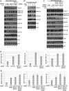
Knockdown of the BRCA1/BARD1/CstF-50/p97 complex induces changes in the localization of some of their substrates upon UV treatment in chromatin-bound fractions. (A) Chromatin-bound fractions were prepared from HeLa cells treated with control (CTRL), CstF-50, or BRCA1/BARD1 siRNA and analyzed by Western blotting with the indicated antibodies. The cells were also exposed to UV treatment (40 J m−2) and allowed to recover for 2 h. (B) Chromatin-bound fractions from HeLa cells treated with UV irradiation (40 J m−2) and the p97 inhibitor DBeQ (10 μM) during the 2-h recovery were analyzed by immunoblotting with the indicated antibodies. (C) Chromatin-bound fractions were prepared from cells treated with control and BRCA1/BARD1/CstF-50 siRNAs. The cells were also exposed to UV (40 J m−2) and DBeQ (10 μM) treatment during the 2-h recovery. Samples were analyzed by Western blotting with the indicated antibodies. (A to C) Representative Western blots from three independent assays are shown. (D and E) Quantification of the blots shown in panels A and C, respectively. Error bars represent the standard deviations derived from three independent experiments.
FIG 4
FIG 4
The content of ubiquitinated histones in the chromatin of differentially expressed genes changes in a BRCA1/BARD1- and CstF-50/p97-dependent manner during DDR. Chromatin extracts from HeLa cells treated with control, CstF-50, BRCA1/BARD1, or CstF50/BRCA1/BARD1 siRNA were prepared. The cells were also treated with UV irradiation (40 J m−2) and the p97 inhibitor DBeQ (10 μM) during the 2-h recovery. Chromatin extracts were immunoprecipitated with either Ub-H2A-specific (A) or Ub-H2B-specific (B) antibodies, followed by qPCR analysis of the immunoprecipitated DNA fragments using primers for differentially expressed genes. The error bars indicate standard deviations (n = 3).
FIG 5
FIG 5
Model for the role of the polyadenylation factor CstF-50, the Ub escort factor p97, and the BRCA1/BARD1 Ub ligase during the progression of DDR. After exposure to UV treatment, CstF-50 associated with the elongating RNAP IIO recruits p97 and a BRCA1/BARD1-containing complex, inducing RNAP II and histone H2A and H2B ubiquitination. In that scenario, p97-mediated escorting allows the displacement of Ub-H2A, Ub-H2B, and poly-Ub–RNAP II from the chromatin, leading to the proteasome degradation of poly-Ub–RNAP II. The displacement of Ub-H2A and Ub-H2B allows the opening of the chromatin at the damage site, facilitating DNA repair. After DNA damage is repaired, chromatin structure reconstitution occurs. This is in agreement with the previously described “access-repair-restore” model (49).

Similar articles

Cited by

References

    1. Warmerdam DO, Kanaar R. 2010. Dealing with DNA damage: relationships between checkpoint and repair pathways. Mutat Res 704:2–11. doi:10.1016/j.mrrev.2009.12.001. - DOI - PubMed
    1. Khobta A, Epe B. 2012. Interactions between DNA damage, repair, and transcription. Mutat Res 736:5–14. doi:10.1016/j.mrfmmm.2011.07.014. - DOI - PubMed
    1. Cevher MA, Kleiman FE. 2010. Connections between 3′-end processing and DNA damage response. Wiley Interdiscip Rev RNA 1:193–199. doi:10.1002/wrna.20. - DOI - PMC - PubMed
    1. Baer R, Ludwig T. 2002. The BRCA1/BARD1 heterodimer, a tumor suppressor complex with ubiquitin E3 ligase activity. Curr Opin Genet Dev 12:86–91. doi:10.1016/S0959-437X(01)00269-6. - DOI - PubMed
    1. Kleiman FE, Manley JL. 2001. The BARD1-CstF-50 interaction links mRNA 3′ end formation to DNA damage and tumor suppression. Cell 104:743–753. doi:10.1016/S0092-8674(01)00270-7. - DOI - PubMed

Publication types

MeSH terms

LinkOut - more resources