. 2018 Jan 8;145(1):dev155929.
doi: 10.1242/dev.155929.

Plasticity within the niche ensures the maintenance of a Sox2+ stem cell population in the mouse incisor

Affiliations

Plasticity within the niche ensures the maintenance of a Sox2+ stem cell population in the mouse incisor

Maria Sanz-Navarro et al. Development. .

Abstract

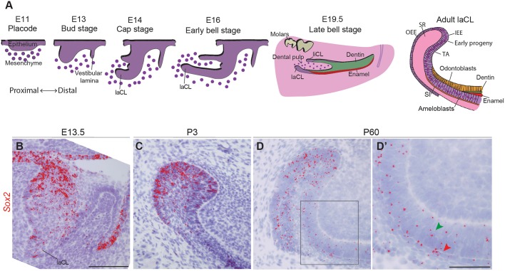
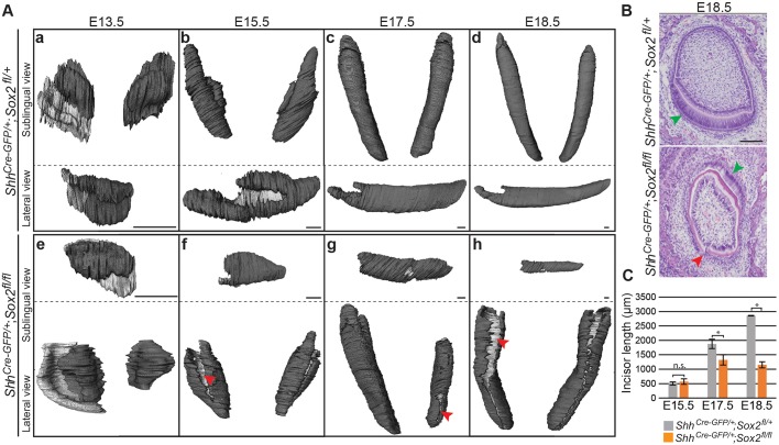
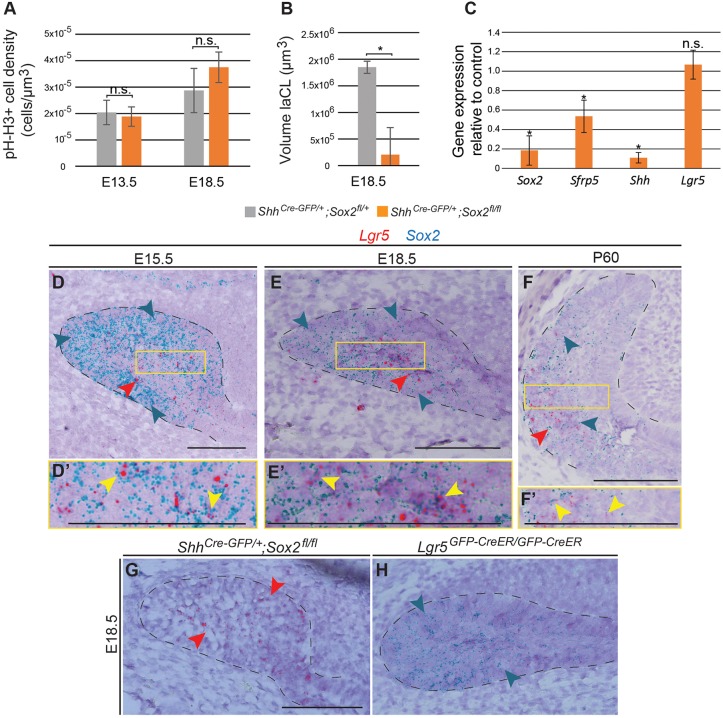
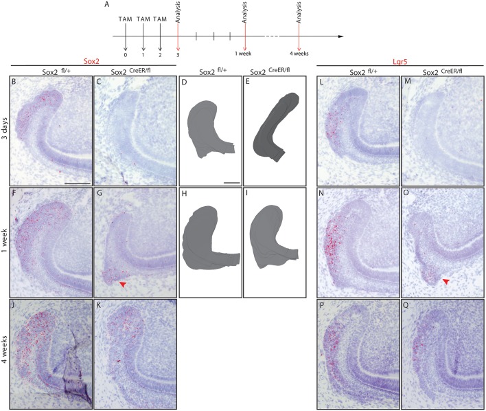
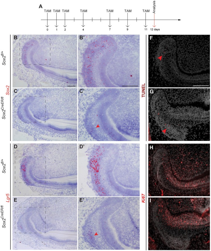
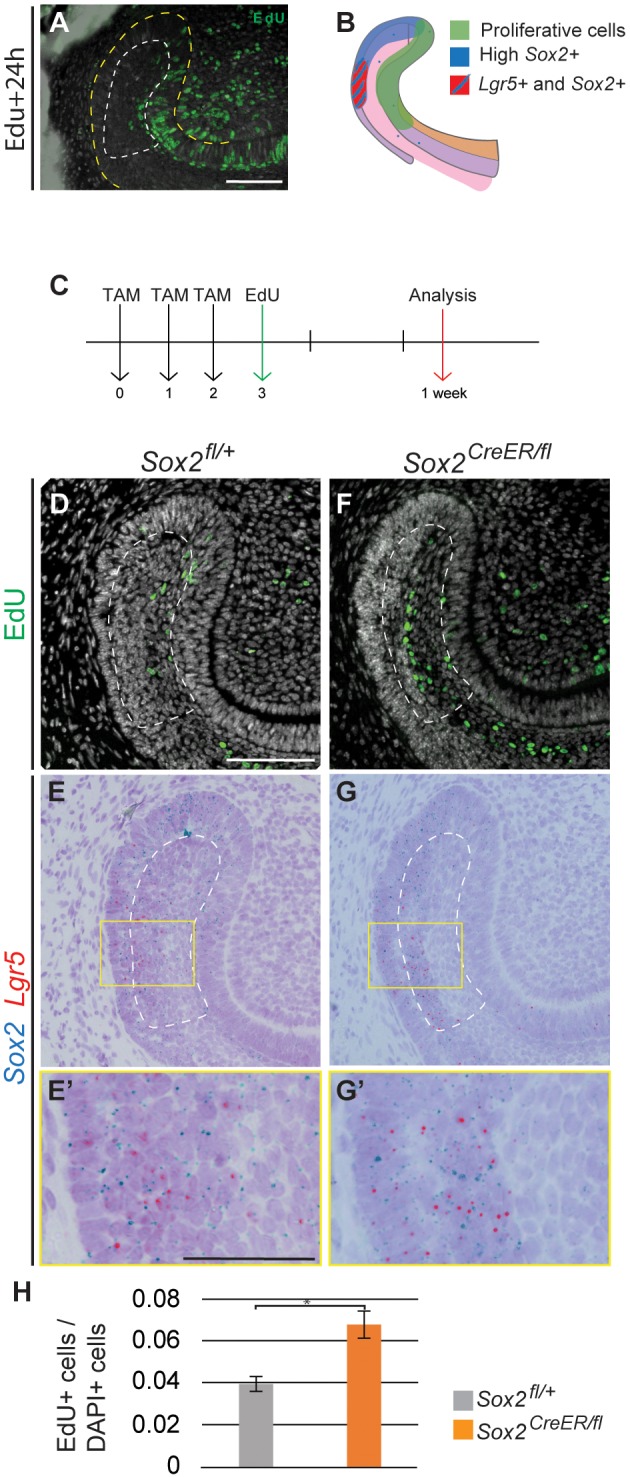
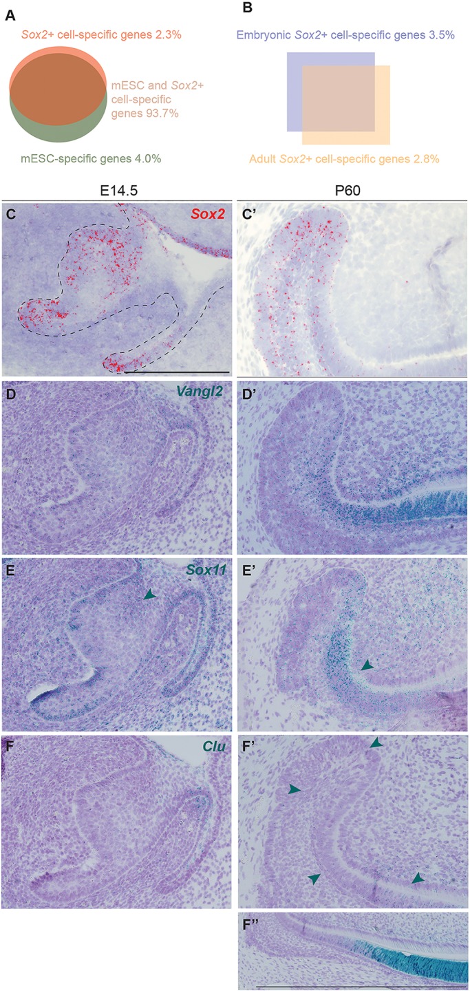
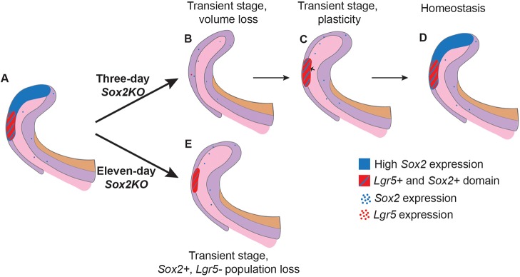
In mice, the incisors grow throughout the animal's life, and this continuous renewal is driven by dental epithelial and mesenchymal stem cells. Sox2 is a principal marker of the epithelial stem cells that reside in the mouse incisor stem cell niche, called the labial cervical loop, but relatively little is known about the role of the Sox2+ stem cell population. In this study, we show that conditional deletion of Sox2 in the embryonic incisor epithelium leads to growth defects and impairment of ameloblast lineage commitment. Deletion of Sox2 specifically in Sox2+ cells during incisor renewal revealed cellular plasticity that leads to the relatively rapid restoration of a Sox2-expressing cell population. Furthermore, we show that Lgr5-expressing cells are a subpopulation of dental Sox2+ cells that also arise from Sox2+ cells during tooth formation. Finally, we show that the embryonic and adult Sox2+ populations are regulated by distinct signalling pathways, which is reflected in their distinct transcriptomic signatures. Together, our findings demonstrate that a Sox2+ stem cell population can be regenerated from Sox2- cells, reinforcing its importance for incisor homeostasis.

Keywords: Hierarchy; Incisor; Lgr5; Morphogenesis; Renewal; Sox2; Stem cells.


Conflict of interest statement

Competing interestsThe authors declare no competing or financial interests.

Figures

Fig. 1.
Fig. 1.
Sox2 expression during incisor morphogenesis. (A) Illustration of mouse incisor development, representing the morphological steps from placode stage to adult. At E14, the dental lingual epithelium gives rise to the lingual cervical loop (liCL), while the labial side originates a larger structure: the labial cervical loop (laCL). The adult laCL is composed of the stellate reticulum (SR), the outer enamel epithelium (OEE) and inner enamel epithelium (IEE). The latter gives rise to the stem cell early progeny, namely the transient-amplifying cells (TA), the stratum intermedium (SI) and the ameloblasts. (B) Sox2 expression (red) is present throughout the entire dental epithelium at E13.5, at highest levels in the lingual side. (C) At P3, Sox2 expression is more sparse, and restricted to the laCL. (D,D′) In adult mice (P60), Sox2 is expressed in the laCL (SR, IEE, OEE and TA cells), as well as in the pre-ameloblasts, ameloblasts (green arrowhead) and SI (red arrowhead). The boxed region in D is magnified in D′. Scale bars: B-D, 100 µm; D′, 50 µm.
Fig. 2.
Fig. 2.
Mouse lower incisor shape and length are regulated by Sox2. (A) 3D reconstructions from micro-CT scans showing the dental epithelium in dark grey. The internal layer of the dental epithelium (b-d,f-h) or vestibular lamina (a,e) appears in light grey. Sox2cKO incisors exhibit an aberrant morphology at all embryonic stages. At E13.5, the tooth domain is broader, and at E15.5 clefts (red arrowhead) appear. The defects can vary within the same individual (g). (B) Histological staining of frontal sections shows that the well-organised ameloblast layer seen in control incisor (green arrowhead) is not visible in Sox2cKO individuals. A cleft is visible on the labial side of the mutant incisor (red arrowhead). (C) The length of the epithelial compartment is similar in the control and Sox2cKO at E15.5. The incisor length increases over 2 mm from E15.5 to E18.5 in controls, but only 0.6 mm in mutants. E17.5, *P=0.016; E18.5, *P=1×10⁻⁵; n.s., not significant. n=3. Scale bars: A, 50 µm; B, 100 µm.
Fig. 3.
Fig. 3.
Sox2cKO impacts laCL volume and the expression of different differentiation markers. (A) Quantification of phospho-histone H3 (pH-H3)+ cell density in the dental epithelium reveals no significant defect in cell proliferation in Sox2cKO. (B) The volume of the E18.5 laCL is drastically decreased in Sox2cKO. *P=0.026. (C) qPCR analysis demonstrates that Sox2cKO induces a decrease in Sfrp5 and Shh expression. Lgr5 expression remains unaffected. *P<0.05. (A-C) n=3. (D) Sox2 transcripts (blue arrowheads in all images) at E15.5 are detected in all cells of the laCL (outlined). Lgr5 transcripts (red arrowheads in all images) mark a subset of the Sox2+ population in the SR. (E) At E18.5 the Sox2 expression domain is smaller than at previous stages. Lgr5 expression is localised to the SR. The laCL houses the expression of both Sox2 (EE and SR) and Lgr5 (SR). (F) In the adult incisor, Sox2 transcripts are localised in different areas of the laCL, while Lgr5 expression is confined to the most proximal part. (D′,E′,F′) Magnifications of the boxed regions in D,E,F. Yellow arrowheads indicate cells expressing both Lgr5 and Sox2 transcripts. (G,H) Sox2cKO shows no changes in Lgr5 expression, as Lgr5KO displays a normal Sox2 expression pattern. Scale bars: 100 µm. qPCR.
Fig. 4.
Fig. 4.
Sox2 and Lgr5 expression are lost then restored in Sox2CreER/fl laCL. (A) The experimental setup. (B,C) Sox2 and (L,M) Lgr5 expression are almost abolished after three Tamoxifen injections in Sox2CreER/fl mice. (D,E) The Sox2CreER/fl laCL is narrower than that of controls. One week after the first Tamoxifen administration, a faint Sox2 signal is detected in the laCL (F,G, arrowhead), while the Lgr5 expression pattern appears to be normal in the mutant (N,O, arrowhead). (H,I) At this stage, the morphology of the laCL appears to be normal. (J,K,P,Q) One month after Cre recombinase activation, the mouse incisor SC niche of Sox2CreER/fl mice is indistinguishable from that of control littermates. Scale bars: 100 µm.
Fig. 5.
Fig. 5.
A day after an 11-day Sox2 ablation, the Sox2 expression pattern, proliferation and cell differentiation appear disturbed. (A) The experimental setup. (B-E′) A small amount of Sox2 transcripts is detected in the proximal area of the SR (arrowhead), where faint expression of Lgr5 is seen (arrowhead). (F,G) TUNEL assay confirms that there is no increase in apoptosis in the laCL, where only a few positive cells were found (arrowheads). (H,I) The domain of proliferating cells, visualised with Ki67 staining, appears similar to the control. Scale bars: 100 µm.
Fig. 6.
Fig. 6.
Expression of Sox2 and Lgr5 is rescued from the SR. (A) Pattern of proliferative cells (EdU+) 24 h after being administered to the mouse. (B) Schematic representation of the proliferative region and the Sox2+ and Lgr5+ domains. (C) The experimental setup. EdU+ cells (D,F) and Sox2 and Lgr5 mRNA detection (E,G) performed in identical sections. (E′,G′) Higher magnifications of the boxed regions in E,G. (H) Quantification of EdU+ cells in the SR of control and Sox2CreER/fl mice. The area quantified is delineated by the dashed line in D-G. Results are expressed as the fraction of EdU+ cells among total cells (number of nuclei, DAPI) compared with the control. *P=0.007. Scale bars: D-G, 100 µm; E′,G′, 50 µm.
Fig. 7.
Fig. 7.
Transcriptomic changes between Sox2+ embryonic progenitors and Sox2+ dental SC. (A) Signatures of Sox2+ cells and mESCs overlap by 93.7%. (B) Comparison of embryonic and adult Sox2+ cells: 3.5% of genes are specific to embryonic Sox2+ cells, and 2.8% specific to the renewing incisor Sox2+ SCs. (C,C′) Sox2 expression pattern at E14.5 and adult stage. (D,D′) Vangl2 is enriched in Sox2+ cells compared with mESCs. It is expressed in the embryonic incisor and in the adult tooth. (E,E′) Sox11 is highly expressed in the embryonic incisor (arrowhead) and in the surrounding mesenchyme. In the adult, expression is mostly localised to the TA cells (arrowhead). (F,F′) Clu expression is seen in the adult incisor (arrowheads), with the majority of transcripts found in the differentiated epithelial cells (F″). Scale bars: 100 µm.
Fig. 8.
Fig. 8.
Model for the effects of short- and long-term Sox2 ablation. (A) Summary of Sox2 and Lgr5 expression domains in normal conditions within the laCL. (B) Upon conditional deletion of Sox2 in Sox2-expressing cells for 3 days, the laCL becomes narrower, and almost all Sox2 and Lgr5 transcripts are lost. (C) Shortly after, the volume of the laCL is back to normal due to an increase in cell proliferation in the SR (small arrow). Overlapping expression of Sox2 and Lgr5 is found in the distal side of the laCL. (D) Eventually, the laCL reaches homeostasis and returns to its original state. (E) In the event of Sox2 ablation for 11 days, the laCL maintains a small Lgr5+ Sox2+ cell population.

Similar articles

Cited by

References

    1. Arnold K., Sarkar A., Yram M. A., Polo J. M., Bronson R., Sengupta S., Seandel M., Geijsen N. and Hochedlinger K. (2011). Sox2+ adult stem and progenitor cells are important for tissue regeneration and survival of mice. Cell Stem Cell 9, 317-329. 10.1016/j.stem.2011.09.001 - DOI - PMC - PubMed
    1. Barker N., van Es J. H., Kuipers J., Kujala P., van den Born M., Cozijnsen M., Haegebarth A., Korving J., Begthel H., Peters P. J. et al. (2007). Identification of stem cells in small intestine and colon by marker gene Lgr5. Nature 449, 1003-1007. 10.1038/nature06196 - DOI - PubMed
    1. Biehs B., Hu J. K.-H., Strauli N. B., Sangiorgi E., Jung H., Heber R.-P., Ho S., Goodwin A. F., Dasen J. S., Capecchi M. R. et al. (2013). BMI1 represses Ink4a/Arf and Hox genes to regulate stem cells in the rodent incisor. Nat. Cell Biol. 15, 846-852. 10.1038/ncb2766 - DOI - PMC - PubMed
    1. Blanpain C. and Fuchs E. (2014). Plasticity of epithelial stem cells in tissue regeneration. Science 344, 1242281-1242281 10.1126/science.1242281 - DOI - PMC - PubMed
    1. Cao H., Wang J., Li X., Florez S., Huang Z., Venugopalan S. R., Elangovan S., Skobe Z., Margolis H. C., Martin J. F. et al. (2010). MicroRNAs play a critical role in tooth development. J. Dent. Res. 89, 779-784. 10.1177/0022034510369304 - DOI - PMC - PubMed

Publication types

Substances