. 2017 Nov 27;7(1):16424.
doi: 10.1038/s41598-017-16144-4.

Differential prognostic value of MYC immunohistochemistry in subtypes of papillary renal cell carcinoma

Affiliations

Differential prognostic value of MYC immunohistochemistry in subtypes of papillary renal cell carcinoma

Julia Bellut et al. Sci Rep. .

Abstract

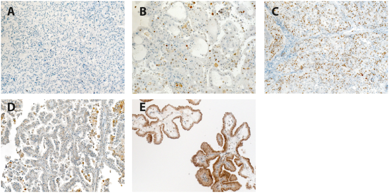
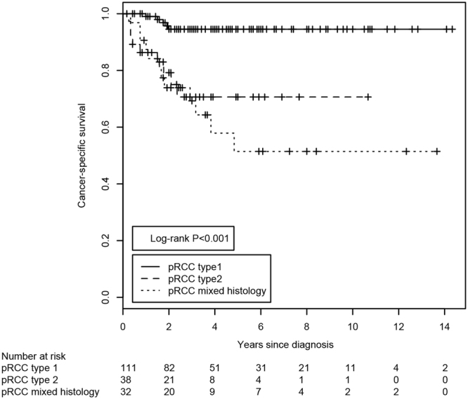
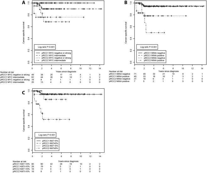
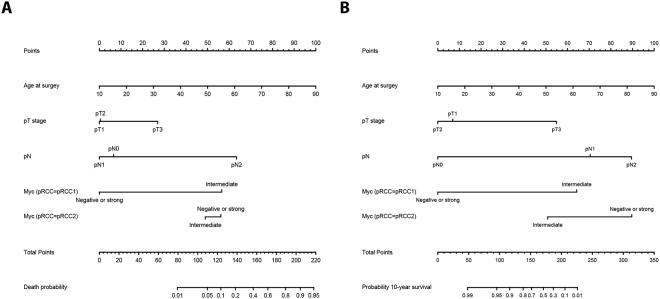
The histomorphological subtyping of papillary renal cell carcinomas (pRCCs) has improved the predictions of patients' long-term survival. Based on our previous results, we hypothesized that the MYC proto-oncogene would show differential expression in pRCC subtypes. Using a multi-institutional cohort of 204 pRCC patients we assessed the additional value of the immunohistochemical markers MYC, MINA53, and Ki67 in predicting patient's long-term survival. The clinical endpoints were overall survival (OS) and cancer-specific survival (CSS). Nomograms were constructed to predict each patient's risk of death (OS). The incorporation of the MYC staining patterns allowed the stratification of pRCC type 1 patients into better and worse prognostic groups. None of the patients with pRCC type 1 tumors and favorable MYC staining patterns died from tumor-related causes. This prognostic value was independent of the patient's age at surgery, the pathological tumor stage and presence of lymph node invasion. we could show that the immunohistochemical assessment of MYC and the histomorphological subtyping of pRCC stratifies pRCC type 1 tumors with regard to OS and CSS. The determination of the histomorphologic pRCC subtype in combination with the MYC immunohistochemical staining patterns allows a more accurate prediction of patients' individual risk of death.


Conflict of interest statement

The authors declare that they have no competing interests.

Figures

Figure 1
Figure 1
Immunohistochemical evaluation. Representative pRCC tissue sections with absent MYC staining (A), intermediate MYC staining (B) and strong MYC staining (C) are shown. Representative pRCC tissue sections with negative MINA53 staining (D) and positive MINA53 staining (E) are shown. Final magnification 200x.
Figure 2
Figure 2
Kaplan-Meier estimates of the cancer-specific survival of patients with pRCC stratified according to the histological classification of the tumor. The P-value was derived from the log-rank test. The number of subjects at risk at the displayed 2-year intervals is indicated below the Kaplan-Meier graph.
Figure 3
Figure 3
Kaplan-Meier estimates of the cancer-specific survival of patients with pRCC stratified according to the combination of histological classification and immunohistochemical staining patterns. The histomorphological subtype was combined with the (A) MYC staining patterns, (B) MINA53 staining patterns or (C) Ki67 labeling index. The P-values were derived from the log-rank test. The number of subjects at risk at the displayed 2-year intervals is indicated below the Kaplan-Meier graph.
Figure 4
Figure 4
Nomograms predicting the patients’ (A) individual probability of death (OS) and (B) individual probability of a 10-year overall survival, depending on their age at surgery, the tumor stage, lymph node invasion and the MYC staining patterns within both histological pRCC subtypes.

References

    1. Ferlay J, et al. Cancer incidence and mortality worldwide: sources, methods and major patterns in GLOBOCAN 2012. Int. J. Cancer. 2015;136:E359–386. doi: 10.1002/ijc.29210. - DOI - PubMed
    1. Moch, H., Humphrey, P. A., Ulbright, T. M. & Reuter, V. E. Pathology and genetics of tumours of the urinary system and male genital organs. 4 edn, (IARC Press, 2016). - PubMed
    1. Nelson EC, Evans CP, Lara PN., Jr. Renal cell carcinoma: current status and emerging therapies. Cancer Treat. Rev. 2007;33:299–313. doi: 10.1016/j.ctrv.2006.12.005. - DOI - PubMed
    1. Shuch B, et al. Understanding pathologic variants of renal cell carcinoma: distilling therapeutic opportunities from biologic complexity. Eur. Urol. 2015;67:85–97. doi: 10.1016/j.eururo.2014.04.029. - DOI - PubMed
    1. Delahunt B, et al. Morphologic typing of papillary renal cell carcinoma: comparison of growth kinetics and patient survival in 66 cases. Hum. Pathol. 2001;32:590–595. doi: 10.1053/hupa.2001.24984. - DOI - PubMed

Publication types