. 2017 Nov 27;7(1):16381.
doi: 10.1038/s41598-017-16777-5.

Transition metal binding selectivity in proteins and its correlation with the phylogenomic classification of the cation diffusion facilitator protein family

Affiliations

Transition metal binding selectivity in proteins and its correlation with the phylogenomic classification of the cation diffusion facilitator protein family

Shiran Barber-Zucker et al. Sci Rep. .

Abstract

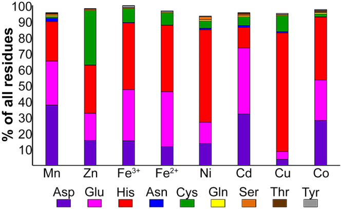
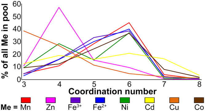
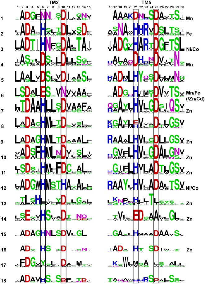
Divalent d-block metal cations (DDMCs), such as Fe, Zn and Mn, participate in many biological processes. Understanding how specific DDMCs are transported to and within the cell and what controls their binding selectivity to different proteins is crucial for defining the mechanisms of metalloproteins. To better understand such processes, we scanned the RCSB Protein Data Bank, performed a de novo structural-based comprehensive analysis of seven DDMCs and found their amino acid binding and coordination geometry propensities. We then utilized these results to characterize the correlation between metal selectivity, specific binding site composition and phylogenetic classification of the cation diffusion facilitator (CDF) protein family, a family of DDMC transporters found throughout evolution and sharing a conserved structure, yet with different members displaying distinct metal selectivity. Our analysis shows that DDMCs differ, at times significantly, in terms of their binding propensities, and that in each CDF clade, the metal selectivity-related binding site has a unique and conserved sequence signature. However, only limited correlation exists between the composition of the DDMC binding site in each clade and the metal selectivity shown by its proteins.


Conflict of interest statement

The authors declare that they have no competing interests.

Figures

Figure 1
Figure 1
Structure of YiiP A-site. (a) Full structure of YiiP (PDB code: 3H90) with A-site residues represented by purple sticks and Zn by a yellow sphere. (b) Magnification of TM2 and TM5 with A-site residues (purple) and their surrounding residues (cerulean blue) presented as sticks and numbered according to the legend of Fig. 5 (the XX-XX quartet residues are numbered as 6, 10, 21 and 25 and the five residues up- and downstream to each X of the XX-XX quartet are numbered respectively). The metal cation is presented as a yellow sphere, nitrogen atoms are in blue, oxygen atoms are in red, sulfur atoms are in yellow and carbons are in purple or cerulean blue. Images were produced using PyMol.
Figure 2
Figure 2
Number of used PDB files and metal cations in the analysis for each metal. The number of initial PDB files (light blue) refers to the number of files after primary filtration at the PDB website as described in Methods, while the numbers of used files (ocean blue) and metals (dark blue) considered in the statistical analysis refer to the final numbers after all filtration criteria were met.
Figure 3
Figure 3
Selected amino acid propensities of the different metals. For each metal, the percentage of each residue is defined as the number of times that a specific residue was bound to the metal divided by the number of all residues bound to the metal. The different residues are presented in different colors: Aspartate in purple, glutamate in pink, histidine in red, asparagine in blue, cysteine in green, glutamine in yellow, serine in orange, threonine in brown and tyrosine in gray.
Figure 4
Figure 4
Main coordination number distribution of the different metals. For each metal, the percentage of each coordination number (CN, 3 to 8) is defined as the number of times the metal was bound in a specific CN divided by the total number of this metal in the statistics (in all coordination numbers, based on the data in Supplementary Table S2). Every metal is presented in different color: Manganese in red, zinc in pink, ferric in purple, ferrous in blue, nickel in green, cadmium in yellow, copper in orange and cobalt in brown.
Figure 5
Figure 5
Most abundant quartet distribution of the different metals. For each metal, the quartet percentage is defined as the number of times the metal was bound by the specific quartet divided by the total number of quartets to which the metal was bound. The cysteine-containing quartets (first five lines) are separated from the other quartets due to the tendency of the former to participate in structural sites rather than enzyme active sites. Darker purple colors represent higher binding tendencies. As they are less relevant for enzymes active sites, the high tendencies of quartets containing more than one cysteine (first three lines) are exceptional and are presented in light purple.
Figure 6
Figure 6
LOGO presentations of all CDF clade A-sites (all at the same scale with a maximal height of 4 bits). Each 15-residue block contains the A-site residues in the TM helix in black boxes plus five residues from the N- and C-terminus. Position 6 overlaps with D45, position 10 with D49, position 21 with H153 and position 25 with D157 in YiiP. Acidic residues (E, D) are presented in red, basic residues (H, K, R) are presented in blue, small/polar (G, C, S, T, Y) are presented in green, amide residues (N, Q) are presented in pink and hydrophobic residues (A, L,V, I, M, P, F, W) are presented in black. Metals shown experimentally to be preferably transported by proteins in a clade are indicated for each clade on the right.

References

    1. Lyons, T. J., Eide, D. J. & Introduction, V. Transport and storage of metal ions in biology in Biological inorganic chemistry: structure and eeactivity (ed. Bertini, I., Gray, H., Stiefel, E., and Valentine, J.S.) 57–78 (University Science Books, 2006).
    1. Lu Y. Metalloprotein and metallo-DNA/RNAzyme design: Current approaches, success measures, and future challenges. Inorg. Chem. 2006;45:9930–9940. doi: 10.1021/ic052007t. - DOI - PMC - PubMed
    1. Dokmanić I, Sikić M, Tomić S. Metals in proteins: Correlation between the metal-ion type, coordination number and the amino-acid residues involved in the coordination. Acta Crystallogr. D. Biol. Crystallogr. 2008;64:257–263. doi: 10.1107/S090744490706595X. - DOI - PubMed
    1. Rulísek L, Vondrásek J. Coordination geometries of selected transition metal ions (Co2+, Ni2+, Cu2+, Zn2+, Cd2+, and Hg2+) in metalloproteins. J. Inorg. Biochem. 1998;71:115–127. doi: 10.1016/S0162-0134(98)10042-9. - DOI - PubMed
    1. Yamashita MM, Wesson L, Eisenmant G, Eisenberg D. Where metal ions bind in proteins (metafloprotein/protein structure/hydrophobicity contrast function) Proc. Natl. Acad. Sci. USA. 1990;87:5648–5652. doi: 10.1073/pnas.87.15.5648. - DOI - PMC - PubMed

Publication types

MeSH terms

LinkOut - more resources