. 2018 Jan;19(1):76-84.
doi: 10.1038/s41590-017-0004-z. Epub 2017 Nov 27.

Engagement of MHC class I by the inhibitory receptor LILRB1 suppresses macrophages and is a target of cancer immunotherapy

Affiliations

Engagement of MHC class I by the inhibitory receptor LILRB1 suppresses macrophages and is a target of cancer immunotherapy

Amira A Barkal et al. Nat Immunol. 2018 Jan.

Abstract

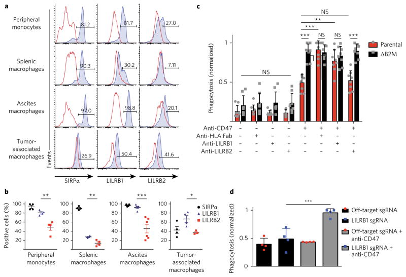
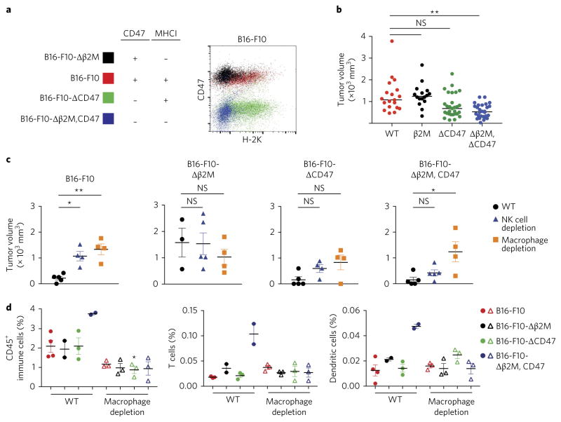
Exciting progress in the field of cancer immunotherapy has renewed the urgency of the need for basic studies of immunoregulation in both adaptive cell lineages and innate cell lineages. Here we found a central role for major histocompatibility complex (MHC) class I in controlling the phagocytic function of macrophages. Our results demonstrated that expression of the common MHC class I component β2-microglobulin (β2M) by cancer cells directly protected them from phagocytosis. We further showed that this protection was mediated by the inhibitory receptor LILRB1, whose expression was upregulated on the surface of macrophages, including tumor-associated macrophages. Disruption of either MHC class I or LILRB1 potentiated phagocytosis of tumor cells both in vitro and in vivo, which defines the MHC class I-LILRB1 signaling axis as an important regulator of the effector function of innate immune cells, a potential biomarker for therapeutic response to agents directed against the signal-regulatory protein CD47 and a potential target of anti-cancer immunotherapy.


Conflict of interest statement

Competing interests

K.W., A.M.R., I.L.W. and R.L.M. and are co-inventors on patent application PCT/US2015/057233, which is related to this work, and own stock of FortySeven, which is pursing clinical approval of antibody Hu5F9-G4, directed against human CD47.

Figures

Fig. 1
Fig. 1. Resistance to macrophage phagocytosis correlates with MHC class I expression
a, Flow cytometry–based measurement of the phagocytosis of a panel of 18 cancer cell lines (horizontal axis), including colon carcinoma (CC), small cell lung cancer (SCLC), breast carcinoma (BC), pancreatic neuroendocrine tumor (PNET), melanoma (Mel) and osteosarcoma (OS), by donor-derived human macrophages in the presence of PBS or anti-CD47 (key); results normalized to those of DLD1 cells (index control). Each symbol represents an individual donor. Error bars, s.d. of n = 3 donors (H128, H1688 and SkBr3 cells) or n = 4 donors (all other cell lines). NS, not significant (P > 0.05); *P < 0.05 (Student’s two-sided t-test without multiple-comparisons correction). b Phagocytosis induced by anti-CD47 (vertical axis; log-transformed averages of values in a plotted against mean fluorescence intensity of MHC class I (horizontal axis): each symbol represents an individual cell line (categories in key); solid diagonal line, best-fit curve; dashed lines, 95% confidence interval (R2 = 0.411; P = 0.002 (linear regression t-test)).
Fig. 2
Fig. 2. MHC class I directly protects cells from macrophage attack
a,b, Summary (left) of the presence (+ ) or absence (−) of expression of CD47 and MHC class I (above plot) by APL1 parental cells and genetically engineered sub-lines of APL1 cells lacking expression of β2M (ΔB2M) or CD47 (ΔCD47) or both (ΔB2M,CD47) (left margin) a or by DLD1 parental cells and genetically engineered sub-lines of DLD1 cells with transgenic expression of β2M (TgB2M) or lacking expression of CD47 (ΔCD47) or both (TgB2M-ΔCD47) (left margin) b, and flow cytometry (right) analyzing the expression of CD47 and MHC class I (HLA-A, HLA-B and HLA-C) by cells as at left (colors in plot match colors at left). c,d, Flow cytometry–based measurement of the phagocytosis of MHCI+ parental APL1 cells and MHCI (ΔB2M) APL1 cells (key) c or of MHCI parental DLD1 cells and MHCI+ transgenic (TgB2M) DLD1 cells (key) d by co-cultured human macrophages, in the presence (+ ) or absence (−) of anti-CD47 (horizontal axis); results normalized to the maximum phagocytosis in each independent replicate experiment. Error bars, s.d. of n = 8 donors. e,f, Flow cytometry–based measurement of the phagocytosis of genetic variants (key) of APL1 cells e or DLD1 cells f by co-cultured human macrophages, in the presence or absence of antibody to the adhesion molecule EpCam e or the epidermal growth factor receptor (EGFR) f (horizontal axis); results normalized as in c,d. Error bars, s.d. of n = 8 donors. *P < 0.05 and ***P < 0.001 (two-way analysis of variance (ANOVA) with multiple-comparisons correction). g, Flow cytometry–based measurement of phagocytosis of the MHCI DLD1 cell line (far left) and the MHCI+ cell lines U2OS, SAOS2, SKMel3, NCI-H196, HCT11 and APL1 (middle and right) by donor-derived macrophages, in the presence of various combinations (grid below plot) of antibody to HLA or CD47; results normalized as in c,d. Error bars, s.d. of n = 4 donors. *P < 0.05 (Student’s t-test without multiple-comparisons correction). Each symbol (cg) represents an individual donor.
Fig. 3
Fig. 3. LILRB1 mediates the detection of MHC class I by human macrophages
a, Flow cytometry analyzing the expression of SIRPα, LILRB1 or LILRB2 (blue shaded curves) by primary human macrophages (left margin); red lines, fluorescence-minus-one control. Numbers above bracketed lines indicate percent cells positive for expression of molecule along horizontal axis. b, Frequency of primary monocytes (n = 4 donors), primary splenic macrophages (n = 4 donors), primary macrophages in ascites fluid (n = 6 donors) and primary tumor-associated macrophages (n = 4 donors) (below plots) positive for SIRPα, LILRB1 or LILRB2 (key), among total macrophages, as determined by fluorescence-minus-one controls. *P < 0.05, **P < 0.01 and ***P < 0.001 (one-way ANOVA with multiple-comparisons correction). c, Flow cytometry–based measurement of the phagocytosis of MHCI+ parental APL1 cells and MHCI (ΔB2M) APL1 cells (key) by donor-derived macrophages, in the presence of various combinations of antibodies (grid below plot); results normalized to the maximum phagocytosis in a replicate. Error bars, s.d. of n = 8 donors. **P < 0.01 and ***P < 0.001 (two-way ANOVA with multiple-comparisons correction). d, Flow cytometry–based measurement of the phagocytosis of APL1 cells by genetically modified donor-derived macrophages electroporated with off-target sgRNA or sgRNA targeting LILRB1, in the presence or absence of anti-CD47 (key); results normalized as in c. Error bars, s.d. of n = 4 technical replicates with n = 2 donors. ***P < 0.001 and ****P < 0.0001 (one-way ANOVA with multiple-comparisons correction). Each symbol bd represents an individual donor; small horizontal lines b indicate the mean ( ± s.d.).
Fig. 4
Fig. 4. β2M confers species-specific protection against phagocytosis by macrophages
a, Crystal structure of the LILRB1–β2M–HLA-A2 complex, generated from published structure data (Protein Data Bank accession code IP7Q); inset, residues within 5 Å of LILRB1 that differ between human β2M and mouse β2M (orange), which were altered (labels in inset) to form a human–mouse chimeric β2M. b, Flow cytometry–based measurement of the surface expression of MHC class I (left plot of each pair) and binding of LILRB1-Fc (right plot of each pair) by variants (key) of DLD1 cells (left half) or APL1 cells (right half). c, Flow cytometry–based measurement of the phagocytosis of parental MHCI (β2M) cells, MHCI+ (β2M+) cells or chimeric MHCI+ cells (key; specific identification in plot) by co-cultured primary human macrophages (vertical axis) or NSG mouse macrophages (horizontal axis). Vertical error bars, s.d. of n = 8 human donors; horizontal error bars, s.d. of n = 8 replicates of macrophages pooled from n = 5 mice.
Fig. 5
Fig. 5. Expression of MHC class I protects tumor cells from macrophages in vivo
a, Tumor bioluminescence (total flux in photons per second) of MHCI and chimeric MHCI+ APL1 cells (key) at various times (horizontal axis) after subcutaneous engraftment into the flanks of NSG mice. Each symbol represents an individual host mouse; small horizontal lines indicate the mean. *P < 0.05 and **P < 0.005 (two-way ANOVA with Tukey’s multiple-comparisons correction). b, Survival of NSG mice at various times (horizontal axis) after engraftment of MHCI or chimeric MHC I+ APL1 tumors (key). P < 0.005 (Mantel-Cox test). c, Flow cytometry analyzing the expression of HLA-A, HLA-B and HLA-C in mixed-chimeric MHC I+ and MHC I DLD1 tumor cells before injection (far left) and in dissociated tumors at days 7, 14, and 28 after injection (above plots) as in a. Numbers in outlined areas indicate percent cells in each. d, Whole-mount fluorescence microscopy of mixed-chimeric MHC I+ and MHC I APL1 tumors on days 7, 14, and 28 after injection (left margin) as in a. GFP, green fluorescent protein; RFP, red fluorescent protein. Scale bars, 1 mm (top two rows) or 5 mm (bottom row). e, Flow cytometry–based quantification of the frequency of chimeric MHC I+ DLD1 cells and MHC I DLD1 cells (key) on days 7, 14, and 28 after injection (horizontal axis) as in a. Each symbol represents an individual host mouse. Error bars, s.d. of n = 10 mice, in two independent experiments. **P < 0.005 (two-way ANOVA with Sidak’s multiple-comparisons correction).
Fig. 6
Fig. 6. Expression of MHC class I protects tumor cells from macrophages in a syngeneic, immunocompetent setting
a, Summary (left) of the expression of CD47 and H-2K (MHCI) (above plot) by B16-F10 cells and four genetically engineered sub-lines of B16-F10 cells lacking expression of β2M or CD47 or both (left margin), and flow cytometry (right) analyzing the expression of CD47 and MHC class I (H-2K) by cells as at left (colors in plot match colors at left). b, Tumor volume of MHC I+CD47+ (WT), MHCICD47+ (β2M), MHCI+CD47 (ΔCD47) and MHCICD47 (Δβ2M,CD47) B16-F10 cells (horizontal axis) 20 d after engraftment into Rag−/−Il2rg−/− BALB/c mice. **P < 0.001 (one-way ANOVA with Tukey’s multiple-comparisons correction). c, Tumor volume of B16-F10 variants (above plot) 25 d after engraftment into fully immunocompetent C57BL/6 wild-type mice (WT) or C57BL/6 mice depleted of NK cells or macrophages (host, key). *P < 0.01 and **P < 0.001 (one-way ANOVA with multiple-comparisons correction). d, Frequency of all CD45+ immune cells (left), or of T cells (middle) or dendritic cells (right) among CD45+ immune cells, in B16-F10 variant tumors (key) 25 d after engraftment into fully immunocompetent C57Bl/6 wild type mice (filled circles) or C57Bl/6 mice depleted of macrophages (open triangles) (host, horizontal axis).

Comment in

  • Another way to not get eaten.
    Zhang CC, Fu YX. Zhang CC, et al. Nat Immunol. 2018 Jan;19(1):6-7. doi: 10.1038/s41590-017-0009-7. Nat Immunol. 2018. PMID: 29242546 No abstract available.

References

    1. Weiner LM, Dhodapkar MV, Ferrone S. Monoclonal antibodies for cancer immunotherapy. Lancet. 2009;373:1033–1040. - PMC - PubMed
    1. Weiskopf K, Weissman IL. Macrophages are critical effectors of antibody therapies for cancer. MAbs. 2015;7:303–310. - PMC - PubMed
    1. Jaiswal S, et al. CD47 is upregulated on circulating hematopoietic stem cells and leukemia cells to avoid phagocytosis. Cell. 2009;138:271–285. - PMC - PubMed
    1. Oldenborg PA, et al. Role of CD47 as a marker of self on red blood cells. Science. 2000;288:2051–2054. - PubMed
    1. Majeti R, et al. CD47 is an adverse prognostic factor and therapeutic antibody target on human acute myeloid leukemia stem cells. Cell. 2009;138:286–299. - PMC - PubMed

MeSH terms

Substances