. 2017 Nov 27;7(1):16332.
doi: 10.1038/s41598-017-16689-4.

Subcutaneous white adipocytes express a light sensitive signaling pathway mediated via a melanopsin/TRPC channel axis

Affiliations

Subcutaneous white adipocytes express a light sensitive signaling pathway mediated via a melanopsin/TRPC channel axis

Katarina Ondrusova et al. Sci Rep. .

Abstract

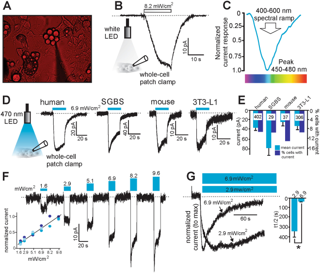
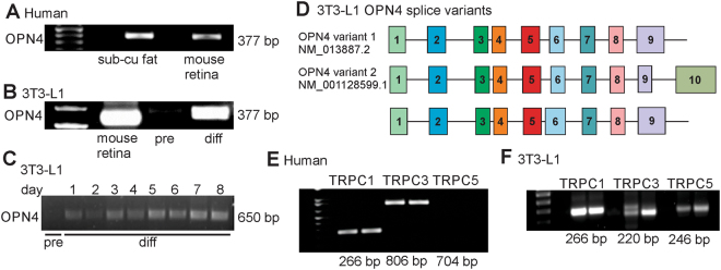
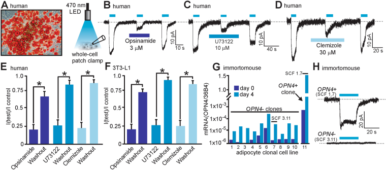
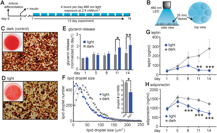
Subcutaneous white adipose tissue (scWAT) is the major fat depot in humans and is a central player in regulating whole body metabolism. Skin exposure to UV wavelengths from sunlight is required for Vitamin D synthesis and pigmentation, although it is plausible that longer visible wavelengths that penetrate the skin may regulate scWAT function. In this regard, we discovered a novel blue light-sensitive current in human scWAT that is mediated by melanopsin coupled to transient receptor potential canonical cation channels. This pathway is activated at physiological intensities of light that penetrate the skin on a sunny day. Daily exposure of differentiated adipocytes to blue light resulted in decreased lipid droplet size, increased basal lipolytic rate and alterations in adiponectin and leptin secretion. Our results suggest that scWAT function may be directly under the influence of ambient sunlight exposure and may have important implications for our current understanding of adipocyte biology. (150 words).


Conflict of interest statement

The authors declare that they have no competing interests.

Figures

Figure 1
Figure 1
Differentiated adipocytes express a light-sensitive inward current. (A) Positioning of the electrophysiological recording pipette for whole-cell patch clamp recordings from adipocytes was made under red light conditions to prevent inactivation of any light-sensitive currents. (B) Representative recording of a light-sensitive inward current from a 3T3-L1 adipocyte in response to white light stimulation at an intensity of 8.2 mW/cm2. (C) Maximal currents were obtained at between 450–480 nm wavelength in 3T3-L1 adipocytes. (D) Representative recordings of blue light-sensitive inward currents in differentiated adipocytes from (1) human primary tissue (2) human SGBS cells (3) mouse primary tissue (4) mouse 3T3-L1 cells. Cells were stimulated with 470 nm blue light at an intensity of 6.9 mW/cm2. (E) Grouped data showing mean current obtained and the % of cells tested with measurable current. Numbers inside bars are the number of cells recorded from each group. (F) Representative recordings of the light-sensitive currents to 470 nm light at different intensities in 3T3-L1 adipocytes (inset = normalized currents from 3 cells at the tested light powers). (G) Representative recordings from 3T3-L1 adipocytes of the inactivation properties of these light-sensitive currents at two different light intensities of longer duration. Inset: time to inactivate to half maximal current was 26 +/− 3 s (n = 7) and 332 +/− 69 s (n = 3) at 6.9 and 2.9 mW/cm2 respectively. **Denotes P < 0.01. Statistical significance was determined using a Student’s t test. Dashed lines denote zero current level.
Figure 2
Figure 2
PCR analysis shows the existence of OPN4 variants and TRPC channels in differentiated adipocytes. (A) Human scWAT shows message for OPN4 (mouse retinal mRNA used as positive control). (B,C) OPN4 message is present in 3T3-L1 differentiated (diff) adipocytes but not detectable in undifferentiated 3T3-L1 pre-adipocytes (pre). (D) Nested PCR analysis of exons 3 to 9, identified at least two OPN4 splice variants in 3T3-L1 differentiated adipocytes (numbered boxes = exon makeup). Variants 1 and 2 are identical to known retinal OPN4 variants (NM_013877.2 and NM_00128599.1). (E) Human scWAT shows message for TRPC1/3. (F) 3T3-L1 differentiated adipocytes shows message for TRPC1/3/5.
Figure 3
Figure 3
Pharmacological and molecular characterization of the light-sensitive current in differentiated adipocytes. (A) Human differentiated primary adipocytes stained with Oil Red-O for lipid content. (B–D) Representative whole-cell light-sensitive recordings from human primary adipocytes showing the inhibitory effects of the melanopsin inhibitor opsinamide (B), the phospholipase C inhibitor U73122 (C) and the TRPC channel inhibitor clemizole (D). (E,F) Grouped data from human primary adipocytes and 3T3-L1 adipocytes (n = 3–4 cells in human primary adipocytes and n = 4–6 cells for 3T3-L1 adipocytes). (G) Immortomouse clonal adipocyte cell line mRNA qPCR analysis reveals the presence of a single OPN4 positive clone (SCF 1.7) from 11 clonal cell lines tested. Day 0 = day of differentiation and day 4 = 4 days post differentiation. OPN4 message normalized to the 36B4 housekeeping message levels. (H) Only differentiated adipocytes from the OPN4+ clonal cell line (SCF 1.7) display light-sensitive currents. Light-sensitive currents were observed in 7 of 32 cells tested for the SCF 1.7 OPN4+ clonal line, whereas 0 of 23 cells tested displayed current in cells from the SCF 3.11 OPN4- clonal line. *Denotes P < 0.01. Statistical significance was determined using a paired one-tailed Student’s t test. (B–D & H) Blue bars above traces denote time of 470 nm light application at a power of 6.9 mW/cm2. Dashed lines denote zero current level.
Figure 4
Figure 4
Chronic blue light treatment of 3T3-L1 differentiated adipocytes alters lipid storage and adipokine release. (A) Schematic illustration of the experimental protocol used for these experiments. (B) Experimental set-up for illumination of the differentiated adipocytes in 35 mm dishes. (C) Chronic exposure to blue light (460 nm) reduces Oil Red-O lipid staining when compared to non-light treated (dark) cells (D). Inset = Oil Red-O lipid staining in 35 mm culture dishes. (E) Glycerol release is significantly increased in light treated cells at days 11 and 14. (F) Lipid droplet size and number are significantly decreased in light-treated cells. Data were plotted as a frequency distribution with a bin size of 5 μm2. Since >99% of the lipid droplet sizes were at sizes lower than or equal to 250 μm2, data was plotted with a maximum size of 250 μm2 and fitted with a Lorentizan (least squares) nonlinear regression (n = 3 experiments per group). (G,H) Leptin and adiponectin secretion is significantly reduced in light-treated cells (n = 3 experiments per group). #, *,** and *** denote significant differences of P < 0.05, <0.01, 0.001, and 0.0001 respectively. Statistical significance was determined using either a paired one-tailed Student’s t-test (F) or a two-way repeated measures ANOVA followed by a Sidak multiple comparisons test (G,H).

References

    1. Hevener AL, Febbraio MA. Stock Conference Working, G. The 2009 stock conference report: inflammation, obesity and metabolic disease. Obesity reviews: an official journal of the International Association for the Study of Obesity. 2010;11:635–644. doi: 10.1111/j.1467-789X.2009.00691.x. - DOI - PubMed
    1. Lee MJ, Wu Y, Fried SK. Adipose tissue heterogeneity: implication of depot differences in adipose tissue for obesity complications. Molecular aspects of medicine. 2013;34:1–11. doi: 10.1016/j.mam.2012.10.001. - DOI - PMC - PubMed
    1. Misra A, Vikram NK. Clinical and pathophysiological consequences of abdominal adiposity and abdominal adipose tissue depots. Nutrition. 2003;19:457–466. doi: 10.1016/S0899-9007(02)01003-1. - DOI - PubMed
    1. Wajchenberg BL. Subcutaneous and visceral adipose tissue: their relation to the metabolic syndrome. Endocrine reviews. 2000;21:697–738. doi: 10.1210/edrv.21.6.0415. - DOI - PubMed
    1. Fleury, N., Geldenhuys, S. & Gorman, S. Sun Exposure and Its Effects on Human Health: Mechanisms through Which Sun Exposure Could Reduce the Risk of Developing Obesity and Cardiometabolic Dysfunction. International journal of environmental research and public health13 (2016). - PMC - PubMed

Publication types

Grants and funding