. 2017 Jun:87:257-421.
doi: 10.1016/j.simyco.2017.09.003. Epub 2017 Sep 28.

Mycosphaerellaceae - Chaos or clarity?

Affiliations

Mycosphaerellaceae - Chaos or clarity?

S I R Videira et al. Stud Mycol. 2017 Jun.

Abstract

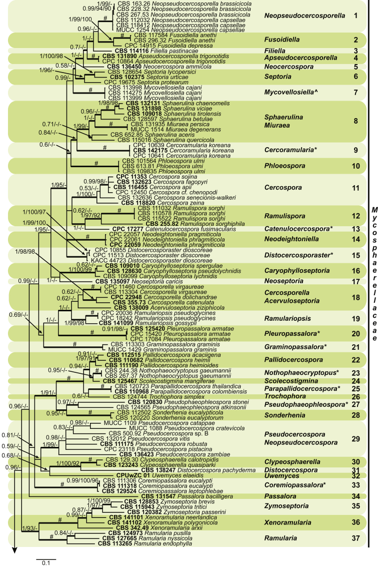
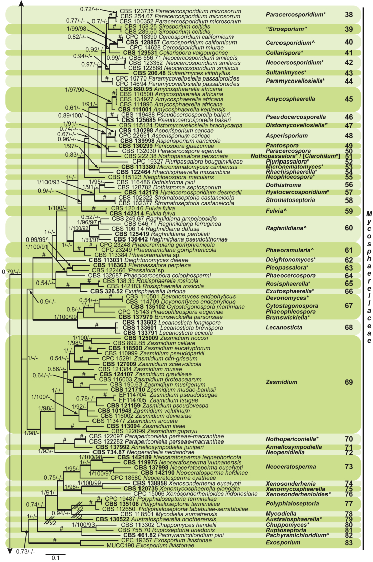
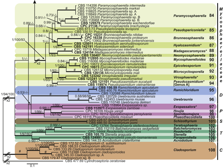
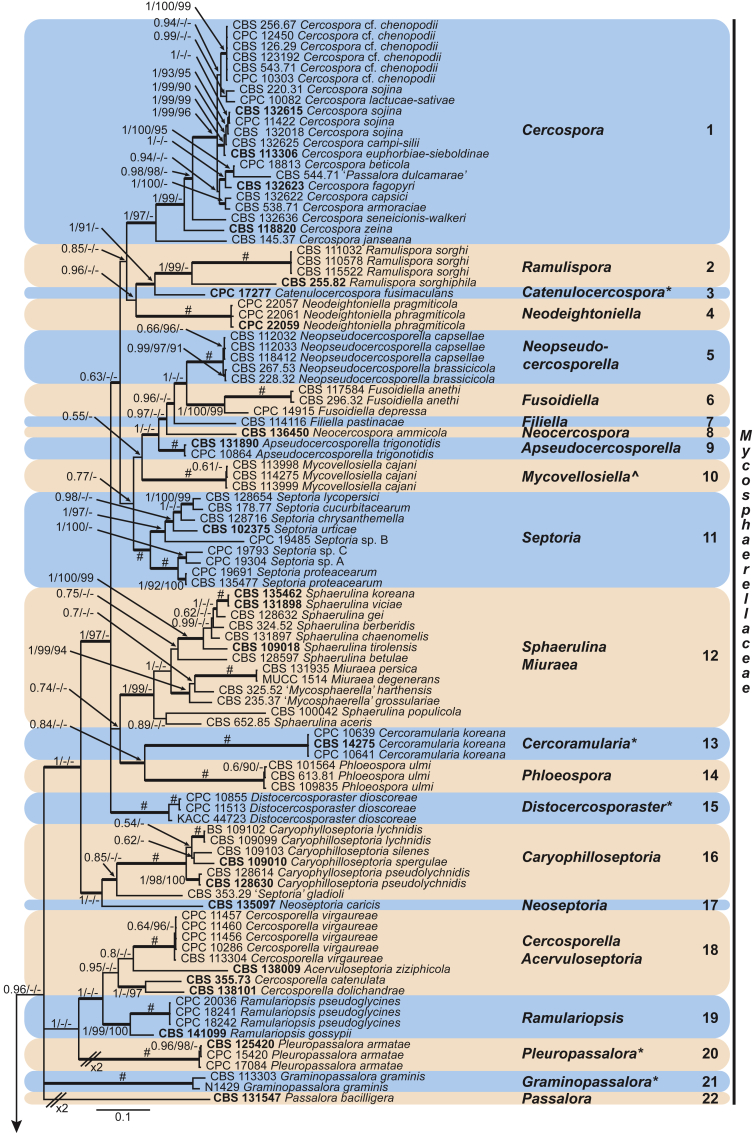
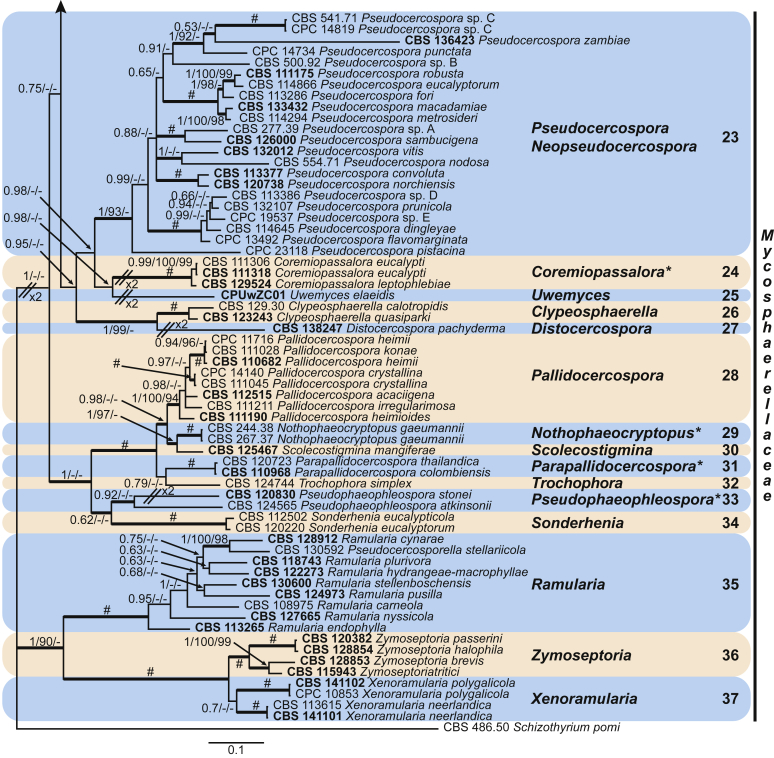
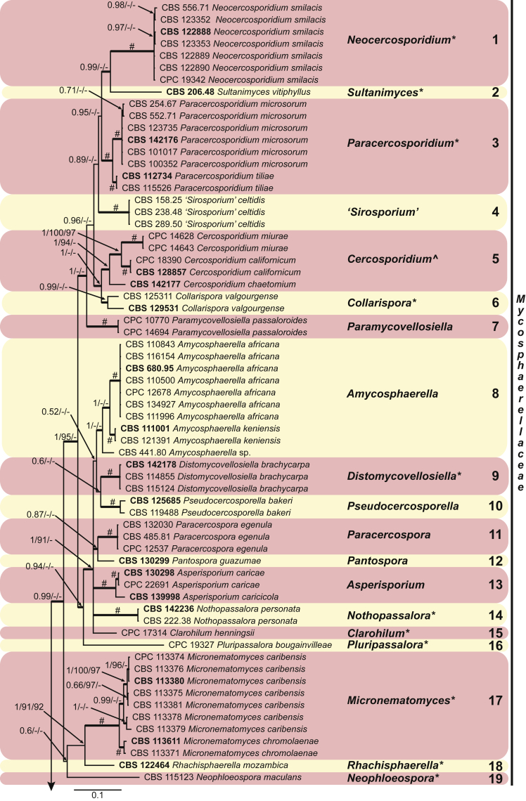
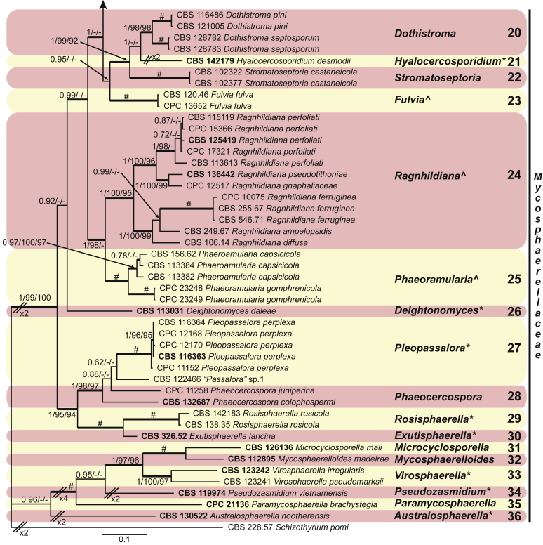
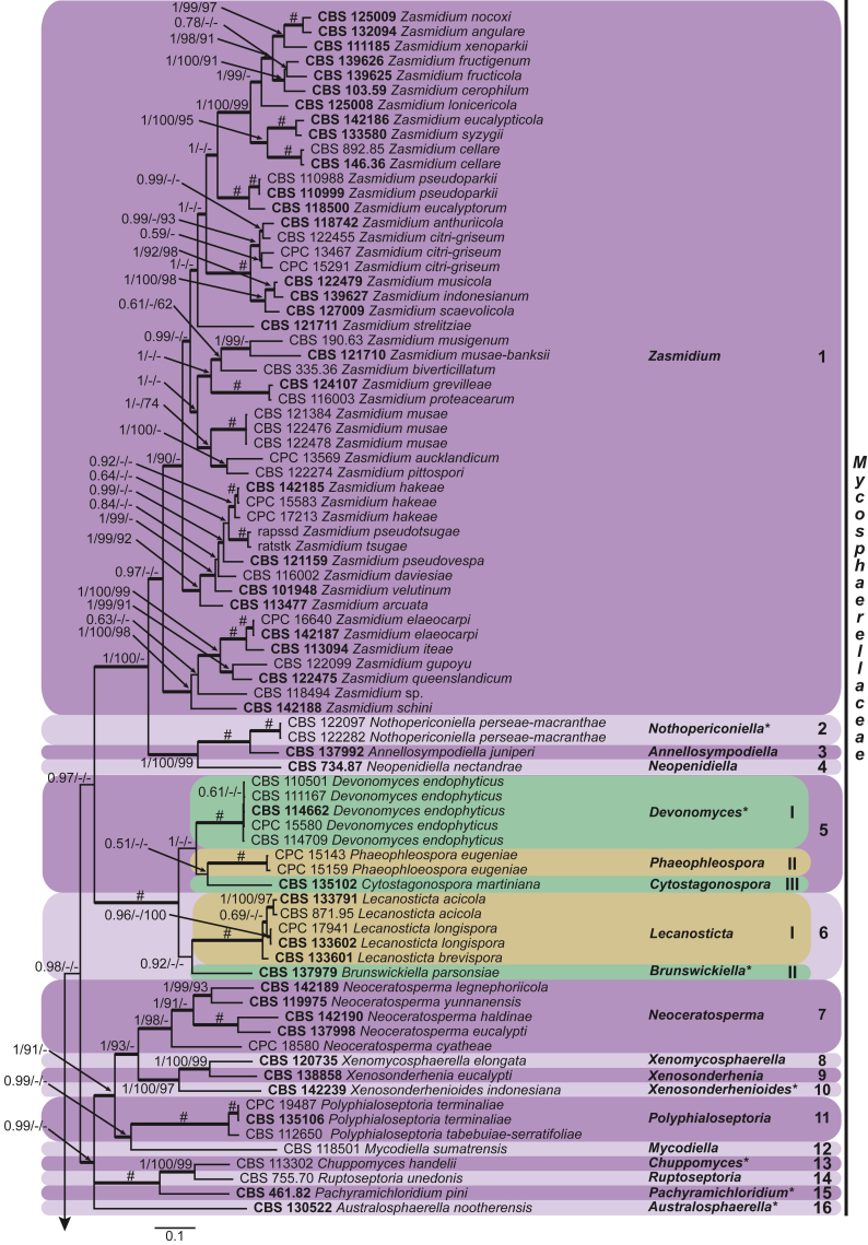
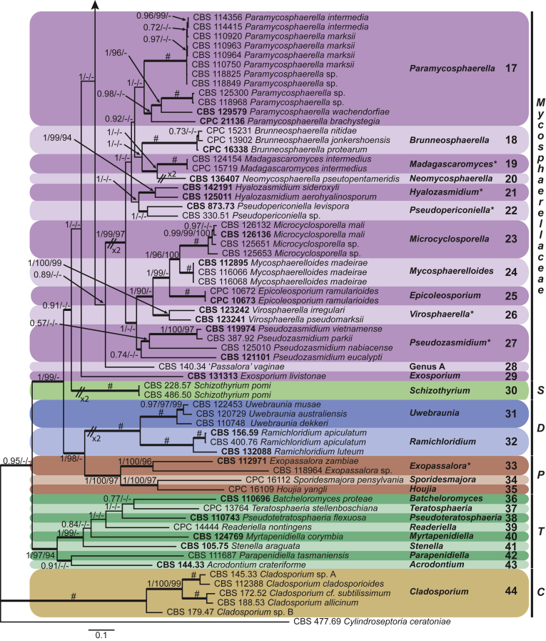
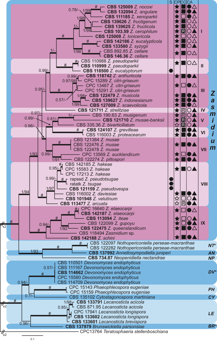
The Mycosphaerellaceae represent thousands of fungal species that are associated with diseases on a wide range of plant hosts. Understanding and stabilising the taxonomy of genera and species of Mycosphaerellaceae is therefore of the utmost importance given their impact on agriculture, horticulture and forestry. Based on previous molecular studies, several phylogenetic and morphologically distinct genera within the Mycosphaerellaceae have been delimited. In this study a multigene phylogenetic analysis (LSU, ITS and rpb2) was performed based on 415 isolates representing 297 taxa and incorporating ex-type strains where available. The main aim of this study was to resolve the phylogenetic relationships among the genera currently recognised within the family, and to clarify the position of the cercosporoid fungi among them. Based on these results many well-known genera are shown to be paraphyletic, with several synapomorphic characters that have evolved more than once within the family. As a consequence, several old generic names including Cercosporidium, Fulvia, Mycovellosiella, Phaeoramularia and Raghnildiana are resurrected, and 32 additional genera are described as new. Based on phylogenetic data 120 genera are now accepted within the family, but many currently accepted cercosporoid genera still remain unresolved pending fresh collections and DNA data. The present study provides a phylogenetic framework for future taxonomic work within the Mycosphaerellaceae.

Keywords: Adelopus gaeumannii T. Rohde; Amycosphaerella keniensis (Crous & T.A. Cout.) Videira & Crous; Australosphaerella Videira & Crous; Australosphaerella nootherensis (Carnegie) Videira & Crous; Biharia vangueriae Thirum. & Mishra; Brunswickiella Videira & Crous; Brunswickiella parsonsiae (Crous & Summerell) Videira & Crous; Catenulocercospora C. Nakash., Videira & Crous; Catenulocercospora fusimaculans (G.F. Atk.) C. Nakash., Videira & Crous; Cercoramularia Videira, H.D. Shin, C. Nakash. & Crous; Cercoramularia koreana Videira, H.D. Shin, C. Nakash. & Crous; Cercospora brachycarpa Syd.; Cercospora cajani Henn.; Cercospora desmodii Ellis & Kellerm.; Cercospora ferruginea Fuckel; Cercospora gnaphaliacea Cooke; Cercospora gomphrenicola Speg.; Cercospora henningsii Allesch.; Cercospora mangiferae Koord.; Cercospora microsora Sacc.; Cercospora rosicola Pass.; Cercospora smilacis Thüm.; Cercospora tiliae Peck; Cercosporidium californicum (S.T. Koike & Crous) Videira & Crous; Cercosporidium helleri Earle; Chuppomyces Videira & Crous; Chuppomyces handelii (Bubák) U. Braun, C. Nakash., Videira & Crous; Cladosporium bacilligerum Mont. & Fr.; Cladosporium chaetomium Cooke; Cladosporium fulvum Cooke; Cladosporium lonicericola Yong H. He & Z.Y. Zhang; Cladosporium personatum Berk. & M.A. Curtis; Clarohilum Videira & Crous; Clarohilum henningsii (Allesch.) Videira & Crous; Clasterosporium degenerans Syd. & P. Syd.; Clypeosphaerella calotropidis (Ellis & Everh.) Videira & Crous; Collarispora Videira & Crous; Collarispora valgourgensis (Crous) Videira & Crous; Coremiopassalora U. Braun, C. Nakash., Videira & Crous; Coremiopassalora eucalypti (Crous & Alfenas) U. Braun, C. Nakash., Videira & Crous; Coremiopassalora leptophlebae (Crous et al.) U. Braun, C. Nakash., Videira & Crous; Coryneum vitiphyllum Speschnew; Cryptosporium acicola Thüm.; Deightonomyces Videira & Crous; Deightonomyces daleae (Ellis & Kellerm.) Videira & Crous; Devonomyces Videira & Crous; Devonomyces endophyticus (Crous & H. Sm. Ter) Videira & Crous; Distocercosporaster Videira, H.D. Shin, C. Nakash. & Crous; Distocercosporaster dioscoreae (Ellis & G. Martin) Videira, H.D. Shin, C. Nakash. & Crous; Distomycovellosiella U. Braun, C. Nakash., Videira & Crous; Distomycovellosiella brachycarpa (Syd.) U. Braun, C. Nakash., Videira & Crous; Exopassalora Videira & Crous; Exopassalora zambiae (Crous & T.A. Cout.) Videira & Crous; Exosporium livistonicola U. Braun, Videira & Crous for Distocercospora livistonae U. Braun & C.F. Hill; Exutisphaerella Videira & Crous; Exutisphaerella laricina (R. Hartig) Videira & Crous; Fusoidiella anethi (Pers.) Videira & Crous; Graminopassalora U. Braun, C. Nakash., Videira & Crous; Graminopassalora graminis (Fuckel) U. Braun, C. Nakash., Videira & Crous; Helicoma fasciculatum Berk. & M.A. Curtis.; Hyalocercosporidium Videira & Crous; Hyalocercosporidium desmodii Videira & Crous; Hyalozasmidium U. Braun, C. Nakash., Videira & Crous; Hyalozasmidium aerohyalinosporum (Crous & Summerell) Videira & Crous; Hyalozasmidium sideroxyli U. Braun, C. Nakash., Videira & Crous; Isariopsis griseola Sacc.; Madagascaromyces U. Braun, C. Nakash., Videira & Crous; Madagascaromyces intermedius (Crous & M.J. Wingf.) Videira & Crous; Micronematomyces U. Braun, C. Nakash., Videira & Crous; Micronematomyces caribensis (Crous & Den Breeÿen) U. Braun, C. Nakash., Videira & Crous; Micronematomyces chromolaenae (Crous & Den Breeÿen) U. Braun, C. Nakash., Videira & Crous; Multi-gene phylogeny; Mycosphaerella; Neoceratosperma haldinae U. Braun, C. Nakash., Videira & Crous; Neoceratosperma legnephoricola U. Braun, C. Nakash., Videira & Crous; Neocercosporidium Videira & Crous; Neocercosporidium smilacis (Thüm.) U. Braun, C. Nakash., Videira & Crous; Neophloeospora Videira & Crous; Neophloeospora maculans (Bérenger) Videira & Crous; Nothopassalora U. Braun, C. Nakash., Videira & Crous; Nothopassalora personata (Berk. & M.A. Curtis) U. Braun, C. Nakash., Videira & Crous; Nothopericoniella Videira & Crous; Nothopericoniella perseae-macranthae (Hosag. & U. Braun) Videira & Crous; Nothophaeocryptopus Videira, C. Nakash., U. Braun, Crous; Nothophaeocryptopus gaeumannii (T. Rohde) Videira, C. Nakash., U. Braun, Crous; Pachyramichloridium Videira & Crous; Pachyramichloridium pini (de Hoog & Rahman) U. Braun, C. Nakash., Videira & Crous; Paracercosporidium Videira & Crous; Paracercosporidium microsorum (Sacc.) U. Braun, C. Nakash., Videira & Crous; Paracercosporidium tiliae (Peck) U. Braun, C. Nakash., Videira & Crous; Paramycosphaerella wachendorfiae (Crous) Videira & Crous; Paramycovellosiella Videira, H.D. Shin & Crous; Paramycovellosiella passaloroides (G. Winter) Videira, H.D. Shin & Crous; Parapallidocercospora Videira, Crous, U. Braun, C. Nakash.; Parapallidocercospora colombiensis (Crous et al.) Videira & Crous; Parapallidocercospora thailandica (Crous et al.) Videira & Crous; Phaeocercospora juniperina (Georgescu & Badea) U. Braun, C. Nakash., Videira & Crous; Plant pathogen; Pleopassalora Videira & Crous; Pleopassalora perplexa (Beilharz et al.) Videira & Crous; Pleuropassalora U. Braun, C. Nakash., Videira & Crous; Pleuropassalora armatae (Crous & A.R. Wood) U. Braun, C. Nakash., Videira & Crous; Pluripassalora Videira & Crous; Pluripassalora bougainvilleae (Munt.-Cvetk.) U. Braun, C. Nakash., Videira & Crous; Pseudocercospora convoluta (Crous & Den Breeÿen) U. Braun, C. Nakash., Videira & Crous; Pseudocercospora nodosa (Constant.) U. Braun, C. Nakash., Videira & Crous; Pseudocercospora platanigena Videira & Crous for Stigmella platani Fuckel, non Pseudocercospora platani (J.M. Yen) J.M. Yen 1979; Pseudocercospora zambiensis (Deighton) Crous & U. Braun; Pseudopericoniella Videira & Crous; Pseudopericoniella levispora (Arzanlou, W. Gams & Crous) Videira & Crous; Pseudophaeophleospora U. Braun, C. Nakash., Videira & Crous; Pseudophaeophleospora atkinsonii (Syd.) U. Braun, C. Nakash., Videira & Crous; Pseudophaeophleospora stonei (Crous) U. Braun, C. Nakash., Videira & Crous; Pseudozasmidium Videira & Crous; Pseudozasmidium eucalypti (Crous & Summerell) Videira & Crous; Pseudozasmidium nabiacense (Crous & Carnegie) Videira & Crous; Pseudozasmidium parkii (Crous & Alfenas) Videira & Crous; Pseudozasmidium vietnamense (Barber & T.I. Burgess) Videira & Crous; Ragnhildiana ampelopsidis (Peck) U. Braun, C. Nakash., Videira & Crous; Ragnhildiana diffusa (Heald & F.A. Wolf) Videira & Crous; Ragnhildiana ferruginea (Fuckel) U. Braun, C. Nakash., Videira & Crous; Ragnhildiana gnaphaliaceae (Cooke) Videira, H.D. Shin, C. Nakash. & Crous; Ragnhildiana perfoliati (Ellis & Everh.) U. Braun, C. Nakash., Videira & Crous; Ragnhildiana pseudotithoniae (Crous & Cheew.) U. Braun, C. Nakash., Videira & Crous; Ramulispora sorghiphila U. Braun, C. Nakash., Videira & Crous; Rhachisphaerella Videira & Crous; Rhachisphaerella mozambica (Arzanlou & Crous) Videira & Crous; Rosisphaerella Videira & Crous; Rosisphaerella rosicola (Pass.) U. Braun, C. Nakash., Videira & Crous; Scolicotrichum roumeguerei Briosi & Cavara; Septoria martiniana Sacc; Sphaerella araneosa Rehm; Sphaerella laricina R. Hartig; Stictosepta cupularis Petr.; Stigmella platani Fuckel; Sultanimyces Videira & Crous; Sultanimyces vitiphyllus (Speschnew) Videira & Crous; Tapeinosporium viride Bonord; Taxonomy; Utrechtiana roumeguerei (Cavara) Videira & Crous; Virosphaerella Videira & Crous; Virosphaerella irregularis (Cheew. et al.) Videira & Crous; Virosphaerella pseudomarksii (Cheew. et al.) Videira & Crous; Xenosonderhenioides Videira & Crous; Xenosonderhenioides indonesiana C. Nakash., Videira & Crous; Zasmidium arcuatum (Arzanlou et al.) Videira & Crous; Zasmidium biverticillatum (Arzanlou & Crous) Videira & Crous; Zasmidium cerophilum (Tubaki) U. Braun, C. Nakash., Videira & Crous; Zasmidium daviesiae (Cooke & Massee) U. Braun, C. Nakash., Videira & Crous; Zasmidium elaeocarpi U. Braun, C. Nakash., Videira & Crous; Zasmidium eucalypticola U. Braun, C. Nakash., Videira & Crous; Zasmidium grevilleae U. Braun, C. Nakash., Videira & Crous; Zasmidium gupoyu (R. Kirschner) U. Braun, C. Nakash., Videira & Crous; Zasmidium hakeae U. Braun, C. Nakash., Videira & Crous; Zasmidium iteae (R. Kirschner) U. Braun, C. Nakash., Videira & Crous; Zasmidium musae-banksii Videira & Crous for Ramichloridium australiense Arzanlou & Crous, non Zasmidium australiense (J.L. Mulder) U. Braun & Crous 2013; Zasmidium musigenum Videira & Crous for Veronaea musae Stahel ex M.B. Ellis, non Zasmidium musae (Arzanlou & Crous) Crous & U. Braun 2010; Zasmidium proteacearum (D.E. Shaw & Alcorn) U. Braun, C. Nakash. & Crous; Zasmidium pseudotsugae (V.A.M. Mill. & Bonar) Videira & Crous; Zasmidium pseudovespa (Carnegie) U. Braun, C. Nakash., Videira & Crous; Zasmidium schini U. Braun, C. Nakash., Videira & Crous; Zasmidium strelitziae (Arzanlou et al.) Videira & Crous; Zasmidium tsugae (Dearn.) Videira & Crous; Zasmidium velutinum (G. Winter) Videira & Crous.


Figures

Fig. 1
Fig. 1
Phylogenetic tree (50 % majority rule consensus) resulting from a Bayesian analysis of the combined LSU and rpb2 sequence alignment (dataset 1). Bayesian posterior probabilities (PP), maximum likelihood bootstrap support values (≥ 90 %; ML-BS) and maximum parsimony bootstrap support values (≥ 90 %; MP-BS) are indicated at the nodes (PP/ML-BS/MP-BS; a hash (#) symbol denotes fully-supported branches) and the scale bar represents the expected number of changes per site. Branches in a thicker stroke represent the branches present in the strict consensus parsimony tree. Genera clades are delimited in coloured boxes, with the genus name and clade number indicated to the right. All taxa names are written in black, ex-type strains are represented in bold, novel genera denoted with an asterisk (*) and resurrected genera with a circumflex (ˆ). A vertical bar is used to the right of the coloured boxes and encompasses all genera within their respective families. The family name Mycosphaerellaceae is unabbreviated while the rest are abbreviated as follows: D = Dissoconiaceae, P = Phaeothecoidiellaceae, S = Schizothyriaceae, T = Teratosphaeriaceae, C = Cladosporiaceae. The tree was rooted to Cylindroseptoria ceratoniae (CBS 477.69).
Fig. 1
Fig. 1
Phylogenetic tree (50 % majority rule consensus) resulting from a Bayesian analysis of the combined LSU and rpb2 sequence alignment (dataset 1). Bayesian posterior probabilities (PP), maximum likelihood bootstrap support values (≥ 90 %; ML-BS) and maximum parsimony bootstrap support values (≥ 90 %; MP-BS) are indicated at the nodes (PP/ML-BS/MP-BS; a hash (#) symbol denotes fully-supported branches) and the scale bar represents the expected number of changes per site. Branches in a thicker stroke represent the branches present in the strict consensus parsimony tree. Genera clades are delimited in coloured boxes, with the genus name and clade number indicated to the right. All taxa names are written in black, ex-type strains are represented in bold, novel genera denoted with an asterisk (*) and resurrected genera with a circumflex (ˆ). A vertical bar is used to the right of the coloured boxes and encompasses all genera within their respective families. The family name Mycosphaerellaceae is unabbreviated while the rest are abbreviated as follows: D = Dissoconiaceae, P = Phaeothecoidiellaceae, S = Schizothyriaceae, T = Teratosphaeriaceae, C = Cladosporiaceae. The tree was rooted to Cylindroseptoria ceratoniae (CBS 477.69).
Fig. 2
Fig. 2
Phylogenetic tree (50 % majority rule consensus) resulting from a Bayesian analysis of the combined LSU, rpb2 and ITS sequence alignment (dataset 2; representing clades 1–37 of Fig. 1). Bayesian posterior probabilities (PP), maximum likelihood bootstrap support values (≥ 90 %; ML-BS) and maximum parsimony bootstrap support values (≥ 90 %; MP-BS) are indicated at the nodes (PP/ML-BS/MP-BS) and the scale bar represents the expected number of changes per site. Branches in a thicker stroke represent the branches present in the strict consensus parsimony tree. Genera clades are delimited in coloured boxes, with the genus name and clade number are indicated to the right. All taxa names are written in black, ex-type species strains are represented in bold, novel genera with an asterisk (*) and resurrected genera a circumflex (ˆ). A vertical bar is used to the right of the coloured boxes and encompass all genera within their respective family, the Mycosphaerellaceae. The tree was rooted to Schizothyrium pomi (CBS 486.50).
Fig. 2
Fig. 2
Phylogenetic tree (50 % majority rule consensus) resulting from a Bayesian analysis of the combined LSU, rpb2 and ITS sequence alignment (dataset 2; representing clades 1–37 of Fig. 1). Bayesian posterior probabilities (PP), maximum likelihood bootstrap support values (≥ 90 %; ML-BS) and maximum parsimony bootstrap support values (≥ 90 %; MP-BS) are indicated at the nodes (PP/ML-BS/MP-BS) and the scale bar represents the expected number of changes per site. Branches in a thicker stroke represent the branches present in the strict consensus parsimony tree. Genera clades are delimited in coloured boxes, with the genus name and clade number are indicated to the right. All taxa names are written in black, ex-type species strains are represented in bold, novel genera with an asterisk (*) and resurrected genera a circumflex (ˆ). A vertical bar is used to the right of the coloured boxes and encompass all genera within their respective family, the Mycosphaerellaceae. The tree was rooted to Schizothyrium pomi (CBS 486.50).
Fig. 3
Fig. 3
Phylogenetic tree (50 % majority rule consensus) resulting from a Bayesian analysis of the combined LSU, rpb2 and ITS sequence alignment (dataset 3; representing clades 38–66, 79, 84 and 92–94 of Fig. 1). Bayesian posterior probabilities (PP), maximum likelihood bootstrap support values (≥ 90 %; ML-BS) and maximum parsimony bootstrap support values (≥ 90 %; MP-BS) are indicated at the nodes (PP/ML-BS/MP-BS) and the scale bar represents the expected number of changes per site. Branches in a thicker stroke represent the branches present in the strict consensus parsimony tree. Genera clades are delimited in coloured boxes, with the genus name and clade number indicated to the right. All taxa names are written in black, ex-type strains are represented in bold, novel genera with an asterisk (*) and resurrected genera with a circumflex (ˆ). A vertical bar is used to the right of the coloured boxes and encompass all genera within their respective family, the Mycosphaerellaceae. The tree was rooted to Schizothyrium pomi (CBS 228.57).
Fig. 3
Fig. 3
Phylogenetic tree (50 % majority rule consensus) resulting from a Bayesian analysis of the combined LSU, rpb2 and ITS sequence alignment (dataset 3; representing clades 38–66, 79, 84 and 92–94 of Fig. 1). Bayesian posterior probabilities (PP), maximum likelihood bootstrap support values (≥ 90 %; ML-BS) and maximum parsimony bootstrap support values (≥ 90 %; MP-BS) are indicated at the nodes (PP/ML-BS/MP-BS) and the scale bar represents the expected number of changes per site. Branches in a thicker stroke represent the branches present in the strict consensus parsimony tree. Genera clades are delimited in coloured boxes, with the genus name and clade number indicated to the right. All taxa names are written in black, ex-type strains are represented in bold, novel genera with an asterisk (*) and resurrected genera with a circumflex (ˆ). A vertical bar is used to the right of the coloured boxes and encompass all genera within their respective family, the Mycosphaerellaceae. The tree was rooted to Schizothyrium pomi (CBS 228.57).
Fig. 4
Fig. 4
Phylogenetic tree (50 % majority rule consensus) resulting from a Bayesian analysis of the combined LSU, rpb2 and ITS sequence alignment (dataset 4; representing clades 67–99 of Fig. 1). Bayesian posterior probabilities (PP), maximum likelihood bootstrap support values (≥ 90 %; ML-BS) and maximum parsimony bootstrap support values (≥ 90 %; MP-BS) are indicated at the nodes (PP/ML-BS/MP-BS) and the scale bar represents the expected number of changes per site. Branches in a thicker stroke represent the branches present in the strict consensus parsimony tree. Genera clades are delimited in coloured boxes, with the genus name and clade number indicated to the right. All taxa names are written in black, ex-type strains are represented in bold, novel genera with an asterisk (*) and resurrected genera with a circumflex (ˆ). A vertical bar is used to the right of the coloured boxes and encompass all genera within their respective families. The family name Mycosphaerellaceae is unnabreviated while the rest are abbreviated as follows: D = Dissoconiaceae, P = Phaeothecoidiellaceae, S = Schizothyriaceae, T = Teratosphaeriaceae, C = Cladosporiaceae. The tree was rooted to Cylindroseptoria ceratoniae (CBS 477.69).
Fig. 4
Fig. 4
Phylogenetic tree (50 % majority rule consensus) resulting from a Bayesian analysis of the combined LSU, rpb2 and ITS sequence alignment (dataset 4; representing clades 67–99 of Fig. 1). Bayesian posterior probabilities (PP), maximum likelihood bootstrap support values (≥ 90 %; ML-BS) and maximum parsimony bootstrap support values (≥ 90 %; MP-BS) are indicated at the nodes (PP/ML-BS/MP-BS) and the scale bar represents the expected number of changes per site. Branches in a thicker stroke represent the branches present in the strict consensus parsimony tree. Genera clades are delimited in coloured boxes, with the genus name and clade number indicated to the right. All taxa names are written in black, ex-type strains are represented in bold, novel genera with an asterisk (*) and resurrected genera with a circumflex (ˆ). A vertical bar is used to the right of the coloured boxes and encompass all genera within their respective families. The family name Mycosphaerellaceae is unnabreviated while the rest are abbreviated as follows: D = Dissoconiaceae, P = Phaeothecoidiellaceae, S = Schizothyriaceae, T = Teratosphaeriaceae, C = Cladosporiaceae. The tree was rooted to Cylindroseptoria ceratoniae (CBS 477.69).
Fig. 5
Fig. 5
Phylogenetic tree (50 % majority rule consensus) resulting from a Bayesian analysis of the combined LSU, rpb2 and ITS sequence alignment of the strains in the clades 1–6 from Fig. 4 (clades 67–72 of Fig. 1). Bayesian posterior probabilities (PP) and maximum parsimony bootstrap support values (≥ 90 %; MP-BS) are indicated at the nodes (PP/MP-BS) and the scale bar represents the expected number of changes per site. Branches in a thicker stroke represent the branches present in the strict consensus parsimony tree. Genera clades are delimited in dark and light blue boxes, with the genus name indicated to the right. The genus name Zasmidium is unnabreviated while the rest are abbreviated as follows: NT = Nothopericoniella, AN = Annellosimpodiella, NP = Neopenidiella, DV = Devonomyces, PH = Phaeophleospora, CY = Cytostagonospora, LE = Lecanosticta, BR = Brunswickiella. All taxa names are written in black, ex-type strains are represented in bold and novel genera with an asterisk (*). The dark and light pink coloured boxes, numbered with roman numerals to the right, represent a possible phylogenetic division of the genus Zasmidium based on branch support and/or taxonomic history. Within the pink boxes, the generic name of Zasmidium was abbreviated (Z. = Zasmidium) and a grid representing morphological charaters respective of each taxon is displayed to the right of the taxa and should be interpreted as: S – only sexual morph described (filled hexagon); CP – conidiophores unbranched (filled star), conidiophores branched (empty star); CC – conidiogenous cell terminal (filled square), conidiogenous cell terminal forming rachis (filled square with letter R in white), conidiogenous cell terminal and intercalary (empty square), conidiogenous cell terminal and intercalary forming rachis (empty square with letter R in black); CA – conidia long (>30 μm average; full circle), conidia short (<30 μm average; empty circle), single (full triangle), catenate (empty triangle). The tree was rooted to Teratosphaeria stellenboschiana (CPC 13764).
Fig. 6
Fig. 6
Mycovellosiella cajani (CBS 114275). A–E. Observations in vivo. F–K. Observations in vitro. A, B. Leaf spot symptoms on the host. C, D, G–I. Conidiophores, conidiogenous cells and conidia. E, J–K. Catenate conidia. F. Culture on V8. Scale bars = 10 μm.
Fig. 7
Fig. 7
Miuraea degenerans (MUCC 1514). A–D. Observations in vitro. A. Culture on MEA. B. Olivaceous conidia and short conidiophore. C. Hyaline (left) and pigmented (right) conidia. D. Microcyclic conidia. E–G. Conidiophores and conidia observed using SEM. Scale bars = 10 μm.
Fig. 8
Fig. 8
Cercoramularia koreana (CPC 10709). A–E. Observations in vivo. A, B. Leaf spot symptoms on the host. C, D. Conidiophores, conidiogenous cells and conidia. E. Catenate conidia. F–L. Observations in vitro. F. Culture on OA. G. Conidiophore and conidiogenous cell. H. Conidiophore, conidiogenous cell and conidium. I, J. Conidiogenous cell and catenate conidia. K, L. Catenate conidia. Scale bars = 10 μm.
Fig. 9
Fig. 9
Ramulispora sorghi (CBS 110578). A–F. Observations in vitro. A. Culture on OA. B–F. Conidiophore, conidiogenous cell and conidia. Scale bars = 10 μm.
Fig. 10
Fig. 10
Ramulispora sorghiphila (CBS 255.82). A–F. Observations in vitro. A. Culture on V8. B. Stromata. C. Conidiophore and conidia. D–F. Conidia. Scale bars = 10 μm.
Fig. 11
Fig. 11
Catenulocercospora fusimaculans (CPC 17277). A–E. Observations in vivo. A. Leaf spot symptoms on the host. B. Conidia sporulating on the lesion. C, D. Conidiophores, conidiogenous cells and conidia. E. Single and catenate conidia. F–J. Observations in vitro. F. Culture on OA. G–I. Conidiophores, conidiogenous cells and conidia. J. Catenate conidia. Scale bars = 10 μm.
Fig. 12
Fig. 12
Distocercosporaster dioscoreae (CPC 11513). A–E. Observations in vivo. A, B. Leaf spot symptoms on the host. C, D. Conidiophores, conidiogenous cells and conidia. E. Catenate conidia. F–L. Observations in vitro. F. Culture on V8. G. Conidiophore and conidiogenous cell. H. Conidiophore, conidiogenous cell, single and catenate conidia. I, J. Catenate conidia. Scale bars = 10 μm.
Fig. 13
Fig. 13
Graminopassalora graminis (CBS 113303). A–D. Observations in vivo. A, B. Leaf spot symptoms on the host. C. Conidiophores. D. Conidia. Scale bars = 10 μm.
Fig. 14
Fig. 14
Distocercospora pachyderma (CBS 138247). A–E. Observations in vivo. A, B. Leaf spot symptoms on the host. C. Conidiophores and conidiogenous cells. D. Conidiophores, conidiogenous cells and conidia. E. Conidia. F–J. Observations in vitro. F. Culture on OA. G. Conidiophores erect and emerging from hyphae. H–I. Conidiophore, conidiogenous cell and conidia. J. Conidia. Scale bars = 10 μm.
Fig. 15
Fig. 15
Passalora bacilligera (CBS 131547). A–G. Observations in vivo. A, B. Leaf spot symptoms on the host. C–E. Conidiophores and conidiogenous cells. F. Conidiogenous cell and conidium. G. Single conidia. Scale bars = 10 μm.
Fig. 16
Fig. 16
Paracercosporidium microsorum (CPC 15550). A–E. Observations in vivo. A, B. Leaf spot symptoms on the host. C, D. Conidiophores, conidiogenous cells and conidia. E. Conidia. F–I. Observations in vitro. F. Culture on OA. G, H. Conidiophore, conidiogenous cells and conidia. I. Conidiogenous cells and conidia. Scale bars = 10 μm.
Fig. 17
Fig. 17
Paracercosporidium tiliae (CBS 112734). A–E. Observations in vivo. A, B. Leaf spot symptoms on the host. C. Conidiophores. D. Conidiophores, conidiogenous cells and conidia. E. Conidia. F–J. Observations in vitro. F. Culture on V8. G, H. Conidiophore, conidiogenous cell and conidia. I. Conidiophore. J. Conidia. Scale bars = 10 μm.
Fig. 18
Fig. 18
Cercosporidium californicum (CBS 128857). A–D. Observations in vivo. A. Leaf spot symptoms on the host. B. Conidiophores, conidiogenous cells and conidia. C. Conidiophores and conidiogenous cells. D. Conidia. E–J. Observations in vitro. E. Culture on V8. F. Mycelium producing red pigment inside the cells and outside. G, I. Conidiophore, conidiogenous cell and conidia. H, J. Conidia. Scale bars = 10 μm.
Fig. 19
Fig. 19
Cercosporidium chaetomium (CBS 142177). A–D. Observations in vivo. A. Leaf spot symptoms on the host. B. Conidiophores, conidiogenous cells and conidia. C. Conidiophores and conidiogenous cells. D. Single conidia. E–J. Observations in vitro. E. Culture on OA. F. Conidiophore and conidiogenous cell. G–J. Conidiophore, conidiogenous cell and conidia. Scale bars = 10 μm.
Fig. 20
Fig. 20
Cercosporidium helleri (NY00945740). A–E. Observations in vivo. A. Leaf spot symptoms on the host. B, D. Conidiophores, conidiogenous cells and conidia. C. Partial conidiophore, conidiogenous cell and conidia. E. Conidia. Scale bars = 10 μm.
Fig. 21
Fig. 21
Cercosporidium miurae (CPC 14628). A–E. Observations in vitro. A. Culture on V8. B–D. Conidiophore, conidiogenous cell and conidia. E. Single conidia. Scale bars = 10 μm.
Fig. 22
Fig. 22
Neocercosporidium smilacis (CPC 19342). A–F. Observations in vitro. A. Culture on V8. B–D. Conidiophore, conidiogenous cell and conidia. E. Conidiogenous cell and conidia. F. Single conidia. Scale bars = 10 μm.
Fig. 23
Fig. 23
Paramycovellosiella passaloroides (CPC 14694). A–J. Observations in vitro. A. Culture on SNA. B, G. Conidiophore, conidiogenous cell and conidia. C, D, E. Conidiogenous cell and conidia. F, H. Single and catenate conidia. I. Culture on V8 supplemented with banana leaf promoted the development of black spermogonia and spermatia whitish in mass. J. Hyaline spermatia. Scale bars = 10 μm.
Fig. 24
Fig. 24
Distomycovellosiella brachycarpa (CPC 18381). A–D. Observations in vivo. A, B. Leaf spot symptoms on the host. C. Catenate and single conidia. D. Conidiophores synnematous-like, conidiogenous cells and conidia. E–G. Observations in vitro. E–G. Conidiophores, conidiogenous cells and conidia. Scale bars = 10 μm.
Fig. 25
Fig. 25
Pantospora guazumae (IMI 59269). A–H. Observations in vivo. A. Leaf spot symptoms on the host. B. Conidiophores and conidia. C–F. Partial conidiophore, conidiogenous cells and conidia. G–H. Conidia. Scale bars = 10 μm.
Fig. 26
Fig. 26
Nothopassalora personata (CPC 19466). A–E. Observations in vivo. A, B. Leaf spot symptoms on the host. C, D. Conidiophores and conidiogenous cells. E. Single conidia. F–K. Observations in vitro. F. Culture on SNA. G. Conidiophore and conidiogenous cell. H. Partial conidiophore and conidiogenous cell. I–K. Single conidia. Scale bars = 10 μm.
Fig. 27
Fig. 27
Clarohilum henningsii (CPC 17314). A–E. Observations in vivo. A, B. Leaf spot symptoms on the host. C, D. Conidiophores and conidiogenous cells. E. Single conidia. F–K. Observations in vitro. F. Culture on OA. G–J. Partial conidiophore, conidiogenous cell and conidia. K. Single conidia. Scale bars = 10 μm.
Fig. 28
Fig. 28
Pluripassalora bougainvilleae (CPC 19327). A–E. Observations in vivo. A, B. Leaf spot symptoms on the host. C, D. Conidiophores, conidiogenous cells and conidia. E. Single conidia. F–K. Observations in vitro. F. Culture on OA. G–K. Partial conidiophore, conidiogenous cell and conidia. Scale bars = 10 μm.
Fig. 29
Fig. 29
Neophloeospora maculans (CBS 115123). A–E. Observations in vitro. F. Culture on SNA. B, C. Conidiophore and conidia. D, E. Conidia. Scale bars = 10 μm.
Fig. 30
Fig. 30
Hyalocercosporidium desmodii (CPC 19483). A–F. Observations in vitro. A. Culture on OA. B. Mycelium. C–E. Partial conidiophore, conidiogenous cell and conidia. F. Conidia. Scale bars = 10 μm.
Fig. 31
Fig. 31
Fulvia fulva (CPC 13652). A–H. Observations in vitro. A. Culture on V8. B, D. Conidiophore reduced to conidiogenous cell and catenate conidia. C. Conidiogenous cell and catenate conidia. E, F. Conidiophore and catenate conidia. G, H. Catenate conidia. Scale bars = 10 μm.
Fig. 32
Fig. 32
Ragnhildiana ampelopsidis (CBS 249.67). A–E. Observations in vitro. A. Culture on V8. B. Partial conidiophore, conidiogenous cell and catenate conidia. C. Conidiogenous cell and conidia. D, E. Catenate conidia. Scale bars = 10 μm.
Fig. 33
Fig. 33
Ragnhildiana ferruginea (CPC 10075). A–F. Observations in vivo. A, B. Leaf spot symptoms on the host. C. Conidiophores. D, E. Partial conidiophore, conidiogenous cell and conidium. F. Conidia. G–K. Observations in vitro. G. Culture on V8. H, I. Conidiophore, conidiogenous cell and conidia. J. Partial conidiogenous cell with single and catenate conidia. K. Single conidia. Scale bars = 10 μm.
Fig. 34
Fig. 34
Ragnhildiana gnaphaliacea (CPC 12517). A–E. Observations in vivo. A, B. Leaf spot symptoms on the host. C. Conidiophores. D. Conidiophores, conidiogenous cells and conidia. E. Catenate conidia and single conidia. F–J. Observations in vitro. F. Culture on OA. G–I. Conidiophore, conidiogenous cell and conidia. J. Partial conidiophore, conidiogenous cell and conidia. Scale bars = 10 μm.
Fig. 35
Fig. 35
Phaeoramularia gomphrenicola (CPC 23248). A–E. Observations in vivo. A, B. Leaf spot symptoms on the host. C. Conidiophores. D. Partial conidiophore, conidiogenous cells and conidia. E. Catenate conidia. F–J. Observations in vitro. F. Culture on OA. G–I. Partial conidiophore, conidiogenous cell and conidia. J. Single and catenate conidia. Scale bars = 10 μm.
Fig. 36
Fig. 36
Pleopassalora perplexa (CPC 12168). A–H. Observations in vitro. A. Culture on OA. B, C. Conidiophore and conidia type II. D, E. Conidiophore and conidia type I. F. Conidiophore type I. G. Conidia type I, slightly constricted at the septa and swollen cells at the base, and type II, smaller and narrower. H. Conidia type I and type II. Scale bars = 10 μm.
Fig. 37
Fig. 37
Phaeocercospora juniperina (CPC 11258). A–F. Observations in vivo. A. Leaf spot symptoms on the host. B. Conidiophores and conidia on the lesions. C, E. Conidiogenous cells and conidia. D. Conidiophores and conidia. F. Conidia. G–K. Observations in vitro. G. Culture on SNA. H, I. Conidiophore and conidia. J, K. Conidia. Scale bars = 10 μm.
Fig. 38
Fig. 38
Rosisphaerella rosicola (CPC 12548). A–E. Observations in vivo. A, B. Leaf spot symptoms on the host. C, D. Conidiophores and conidiogenous cells. E. Single conidia. F–K. Observations in vitro. F. Culture on OA. G–I. Conidiophore, conidiogenous cells and conidia. J. Partial conidiophore, conidiogenous cells and conidia. K. Conidia. Scale bars = 10 μm.
Fig. 39
Fig. 39
Zasmidium elaeocarpi (CPC 16640). A–F. Observations in vitro. A. Culture on OA. B, C. Conidiophore, conidiogenous cells and conidia. D, E. Partial conidiophore, conidiogenous cells and conidia. F. Conidia. Scale bars = 10 μm.
Fig. 40
Fig. 40
Zasmidium eucalypticola (CPC 15149). A–D. Conidiophores and conidia observed in vivo. Scale bars = 10 μm.
Fig. 41
Fig. 41
Zasmidium hakeae (CPC 15577). A–F. Observations in vivo. A, B. Leaf spot symptoms on the host. C. Partial conidiophore and conidiogenous cells. D–F. Conidia. G–L. Observations in vitro. G. Culture on V8. H–J. Partial conidiophore, conidiogenous cells and conidia. K, L. Conidia. Scale bars = 10 μm.
Fig. 42
Fig. 42
Zasmidium schini (CPC 19516). A–F. Observations in vitro. A. Culture on V8. B–D. Conidiophore. E, F. Conidia. Scale bars = 10 μm.
Fig. 43
Fig. 43
Neoceratosperma legnephoricola (CPC 16411). A–E. Observations in vivo. A. Leaf spot symptoms on the host. B. Partial conidiophore, conidiogenous cells and conidia. C. Conidiophore and conidiogenous cells. D, E. Conidia. F–K. Observations in vitro. F. Culture on V8. G. Conidiophore and conidiogenous cell. H, I. Conidiogenous cell and conidia. J, K. Conidia. Scale bars = 10 μm.
Fig. 44
Fig. 44
Neoceratosperma haldinae (CPC 19202). A–F. Observations in vitro. A. Culture on OA. B, C. Conidiophore and conidiogenous cell. D–F. Conidia. Scale bars = 10 μm.
Fig. 45
Fig. 45
Xenosonderhenioides indonesiana (CPC 15066). A–E. Observations in vitro. A. Culture on OA. B, C. Conidiophore and conidiogenous cell. D. Conidiogenous cell and conidia. E. Conidia. Scale bars = 10 μm.
Fig. 46
Fig. 46
Chuppomyces handelii (CBS 113302). A. Disease symptoms on the host leaves. B. Drawings of the asexual morph (from Crous & Braun 2003). C. Drawings of the sexual morph (from Crous & Braun 2003).
Fig. 47
Fig. 47
Hyalozasmidium sideroxyli (CPC 23462). A– E. Observations in vitro. A. Culture on V8. B, C. Conidiophores and conidiogenous cells. D. Conidia. E. Irregular swollen conidia synanamorph. Scale bars = 10 μm.
Fig. 48
Fig. 48
Annelophora solani (E00417817). A–F. Observations in vivo. A. Symptoms on host. B, C. Conidiophores erect and developing on the host surface. D. Attachment of the conidiophore to a leaf trichome. E. Conidiogenous cell and conidium. F. Conidia. Scale bars = 10 μm.
Fig. 49
Fig. 49
Annelophragmia coonoorensis (IMI 245197). A–F. Observations in vivo. A. Conidiophores in compact fascicles, erect and emerging from the host. B. Apical area of the conidiophores, with conidiogenous cells and conidia. C, D. Conidiogenous cells and conidia. E, F. Conidiogenous cell and conidium. Scale bars = 10 μm.
Fig. 50
Fig. 50
Camptomeris leucaenae (CBS H-22884). A–F. Observations in vivo. A, B. Leaf spot symptoms on the host. C. Conidiophores emerging from the leaf host with conidiogenous cells. D. Apex of the conidiogenous cells with the conidiogenous scar. E. Conidiogenous cells and conidia. F. Conidia. Scale bars = 10 μm.
Fig. 51
Fig. 51
Cladosporiella cercosporicola (IMI 107538). A–E. Observations in vivo. A. Leaf spot symptoms on the host. B. Conidiophores and conidiogenous cells. C. Conidiophores, conidiogenous cells and conidium. D, E. Catenate and single conidia. Scale bars = 10 μm.
Fig. 52
Fig. 52
Deightoniella africana (IMI 39675a). A–E. Observations in vivo. A. Conidiophores emerging on the leaf surface. B, C. Conidiophores. D, E. Single conidia. Scale bars = 10 μm.
Fig. 53
Fig. 53
Denticularia modesta (IMI 62524). A–E. Observations in vivo. A. Leaf spot symptoms on the host. B, C. Conidiophores and conidiogenous cells. D. Conidiophores, conidiogenous cells and conidia. E. Catenate and single conidia. Scale bars = 10 μm.
Fig. 54
Fig. 54
Dictyodesmium ulmicola (NY 00838655). A, C–E. Observations in vivo. A. Leaf spot symptoms on the host. B. Conidiophores and conidia drawing on the specimen envelope. C–E. Conidia. Scale bars = 10 μm.
Fig. 55
Fig. 55
Elletevera parasitica (IMI 127995). A. Drawing in the specimen envelope. B–E. Observations in vivo. B–D. Conidiophores and conidiogenous cells. E. Conidiogenous cells and conidium. Scale bars = 10 μm.
Fig. 56
Fig. 56
Eriocercospora balladynae (IMI 5293c). A–E. Observations in vivo. A. Leaf spot symptoms on the host. B. Conidiophores, conidiogenous cells and conidia. C. Partial conidiophore, conidiogenous cells and conidium. D, E. Single conidia. Scale bars = 10 μm.
Fig. 57
Fig. 57
Eriocercosporella indica (IMI 302747). A–I. Observations in vivo. A. Leaf spot symptoms on the host. B, C. Conidiophores. D. Conidiogenous cells proliferating percurrently and conidia. E–H. Conidiophores and conidia. I. Conidia. Scale bars = 10 μm.
Fig. 58
Fig. 58
Fusicladiella aronici (IMI 371583). A–H. Observations in vivo. A, B. Leaf spot symptoms on the host. C. Conidiophores. D. Partial conidiophore and conidiogenous cell. E, F. Conidiogenous cells and conidia. G, H. Conidia. Scale bars = 10 μm.
Fig. 59
Fig. 59
Laocoön paradoxus (IMI 375866). A–D. Observations in vivo. A. Leaf spot symptoms on the host. B–D. Conidiophores, conidiogenous cells and conidia. Scale bars = 10 μm.
Fig. 60
Fig. 60
Oedothea vismiae (NY 00945740). A–E. Observations in vivo. A. Leaf spot symptoms on the host. B–D. Conidiophores and conidia. E. Conidia. Scale bars = 10 μm.
Fig. 61
Fig. 61
Pleurovularia polliniae (S F43065). A–E. Observations in vivo. A. Leaf spot symptoms on the host. B. Conidiophores, conidiogenous cells and conidium. C, D. Conidiophores and conidiogenous cells. E. Conidia. Scale bars = 10 μm.
Fig. 62
Fig. 62
Prathigada crataevae (IMI 234117 and IMI 182578). A–G. Observations in vivo. A, B. Leaf spot symptoms on the host. C, D, F. Conidiophores, conidiogenous cells and conidia. E. Conidiogenous cell and conidium. G. Conidia. Scale bars = 10 μm.
Fig. 63
Fig. 63
Pseudocercosporidium venezuelanum (S F38692). A–H. Observations in vivo. A. Leaf spot symptoms on the host. B–D. Conidiophores, conidiogenous cells and conidia. E–G. Conidiogenous cells and conidia. H. Conidia. Scale bars = 10 μm.
Fig. 64
Fig. 64
Semipseudocercospora peristrophes-acuminatae (IMI 122324). A–E. Observations in vivo. A. Leaf spot symptoms on the host. B–D. Partial conidiophore, conidiogenous cells and conidia. E. Single conidia. Scale bars = 10 μm.
Fig. 65
Fig. 65
Sirosporium antenniforme (IMI 1253). A–F. Observations in vivo. A, B. Conidiophores emerging from the host leaf. C–F. Conidia. Scale bars = 10 μm.
Fig. 66
Fig. 66
Stenellopsis fragariae (PDD 75945). A–G. Observations in vivo. A. Leaf spot symptoms on the host. B. Conidiophores. C–G. Conidiophores, conidiogenous cells and conidia. Scale bars = 10 μm.

Similar articles

  • Phylogenetic lineages in Pseudocercospora.
    Crous PW, Braun U, Hunter GC, Wingfield MJ, Verkley GJ, Shin HD, Nakashima C, Groenewald JZ. Crous PW, et al. Stud Mycol. 2013 Jun 30;75(1):37-114. doi: 10.3114/sim0005. Stud Mycol. 2013. PMID: 24014898 Free PMC article.
  • Genera of phytopathogenic fungi: GOPHY 4.
    Chen Q, Bakhshi M, Balci Y, Broders KD, Cheewangkoon R, Chen SF, Fan XL, Gramaje D, Halleen F, Jung MH, Jiang N, Jung T, Májek T, Marincowitz S, Milenković I, Mostert L, Nakashima C, Nurul Faziha I, Pan M, Raza M, Scanu B, Spies CFJ, Suhaizan L, Suzuki H, Tian CM, Tomšovský M, Úrbez-Torres JR, Wang W, Wingfield BD, Wingfield MJ, Yang Q, Yang X, Zare R, Zhao P, Groenewald JZ, Cai L, Crous PW. Chen Q, et al. Stud Mycol. 2022 Jul;101:417-564. doi: 10.3114/sim.2022.101.06. Epub 2022 Jun 2. Stud Mycol. 2022. PMID: 36059898 Free PMC article.
  • Fusarium: more than a node or a foot-shaped basal cell.
    Crous PW, Lombard L, Sandoval-Denis M, Seifert KA, Schroers HJ, Chaverri P, Gené J, Guarro J, Hirooka Y, Bensch K, Kema GHJ, Lamprecht SC, Cai L, Rossman AY, Stadler M, Summerbell RC, Taylor JW, Ploch S, Visagie CM, Yilmaz N, Frisvad JC, Abdel-Azeem AM, Abdollahzadeh J, Abdolrasouli A, Akulov A, Alberts JF, Araújo JPM, Ariyawansa HA, Bakhshi M, Bendiksby M, Ben Hadj Amor A, Bezerra JDP, Boekhout T, Câmara MPS, Carbia M, Cardinali G, Castañeda-Ruiz RF, Celis A, Chaturvedi V, Collemare J, Croll D, Damm U, Decock CA, de Vries RP, Ezekiel CN, Fan XL, Fernández NB, Gaya E, González CD, Gramaje D, Groenewald JZ, Grube M, Guevara-Suarez M, Gupta VK, Guarnaccia V, Haddaji A, Hagen F, Haelewaters D, Hansen K, Hashimoto A, Hernández-Restrepo M, Houbraken J, Hubka V, Hyde KD, Iturriaga T, Jeewon R, Johnston PR, Jurjević Ž, Karalti I, Korsten L, Kuramae EE, Kušan I, Labuda R, Lawrence DP, Lee HB, Lechat C, Li HY, Litovka YA, Maharachchikumbura SSN, Marin-Felix Y, Matio Kemkuignou B, Matočec N, McTaggart AR, Mlčoch P, Mugnai L, Nakashima C, Nilsson RH, Noumeur SR, Pavlov IN, Peralta MP, Phillips AJL, Pitt JI, Polizzi G, Quaedvlieg W, Rajeshkumar KC, Restrepo S, Rhaiem A, Robert J, Robert V, Ro… See abstract for full author list ➔ Crous PW, et al. Stud Mycol. 2021 Aug 17;98:100116. doi: 10.1016/j.simyco.2021.100116. eCollection 2021 Apr. Stud Mycol. 2021. PMID: 34466168 Free PMC article.
  • Redefining the megagenus Erica L. (Ericaceae): the contributions of E. G. H. Oliver and I. M. Oliver (née Nitzsche) to taxonomy and nomenclature.
    Nelson EC, Pirie MD, Bellstedt DU. Nelson EC, et al. PhytoKeys. 2024 Jul 5;244:39-55. doi: 10.3897/phytokeys.244.121705. eCollection 2024. PhytoKeys. 2024. PMID: 39006939 Free PMC article. Review.
  • Dr. John Cooke on the Various Species of Palsy.
    [No authors listed] [No authors listed] Med Chir Rev J Med Sci Anal Ser. 1821 Mar 1;1(4):725-743. Med Chir Rev J Med Sci Anal Ser. 1821. PMID: 29917411 Free PMC article. Review. No abstract available.

Cited by

  • Taxonomy and phylogeny of cercosporoid ascomycetes on Diospyros spp. with special emphasis on Pseudocercospora spp.
    Braun U, Nakashima C, Bakhshi M, Zare R, Shin HD, Alves RF, Sposito MB. Braun U, et al. Fungal Syst Evol. 2020 Dec;6:95-127. doi: 10.3114/fuse.2020.06.06. Epub 2020 Apr 3. Fungal Syst Evol. 2020. PMID: 32904397 Free PMC article.
  • Fungal Planet description sheets: 868-950.
    Crous PW, Carnegie AJ, Wingfield MJ, Sharma R, Mughini G, Noordeloos ME, Santini A, Shouche YS, Bezerra JDP, Dima B, Guarnaccia V, Imrefi I, Jurjević Ž, Knapp DG, Kovács GM, Magistà D, Perrone G, Rämä T, Rebriev YA, Shivas RG, Singh SM, Souza-Motta CM, Thangavel R, Adhapure NN, Alexandrova AV, Alfenas AC, Alfenas RF, Alvarado P, Alves AL, Andrade DA, Andrade JP, Barbosa RN, Barili A, Barnes CW, Baseia IG, Bellanger JM, Berlanas C, Bessette AE, Bessette AR, Biketova AY, Bomfim FS, Brandrud TE, Bransgrove K, Brito ACQ, Cano-Lira JF, Cantillo T, Cavalcanti AD, Cheewangkoon R, Chikowski RS, Conforto C, Cordeiro TRL, Craine JD, Cruz R, Damm U, de Oliveira RJV, de Souza JT, de Souza HG, Dearnaley JDW, Dimitrov RA, Dovana F, Erhard A, Esteve-Raventós F, Félix CR, Ferisin G, Fernandes RA, Ferreira RJ, Ferro LO, Figueiredo CN, Frank JL, Freire KTLS, García D, Gené J, Gêsiorska A, Gibertoni TB, Gondra RAG, Gouliamova DE, Gramaje D, Guard F, Gusmão LFP, Haitook S, Hirooka Y, Houbraken J, Hubka V, Inamdar A, Iturriaga T, Iturrieta-González I, Jadan M, Jiang N, Justo A, Kachalkin AV, Kapitonov VI, Karadelev M, Karakehian J, Kasuya T, Kautmanová I, Kruse J, Kušan I, Kuznetsova TA, Landell MF, L… See abstract for full author list ➔ Crous PW, et al. Persoonia. 2019 Jun;42:291-473. doi: 10.3767/persoonia.2019.42.11. Epub 2019 Jul 19. Persoonia. 2019. PMID: 31551622 Free PMC article.
  • 101 Dothideomycetes genomes: A test case for predicting lifestyles and emergence of pathogens.
    Haridas S, Albert R, Binder M, Bloem J, LaButti K, Salamov A, Andreopoulos B, Baker SE, Barry K, Bills G, Bluhm BH, Cannon C, Castanera R, Culley DE, Daum C, Ezra D, González JB, Henrissat B, Kuo A, Liang C, Lipzen A, Lutzoni F, Magnuson J, Mondo SJ, Nolan M, Ohm RA, Pangilinan J, Park HJ, Ramírez L, Alfaro M, Sun H, Tritt A, Yoshinaga Y, Zwiers LH, Turgeon BG, Goodwin SB, Spatafora JW, Crous PW, Grigoriev IV. Haridas S, et al. Stud Mycol. 2020 Feb 1;96:141-153. doi: 10.1016/j.simyco.2020.01.003. eCollection 2020 Jun. Stud Mycol. 2020. PMID: 32206138 Free PMC article.
  • Fungal Planet description sheets: 951-1041.
    Crous PW, Wingfield MJ, Lombard L, Roets F, Swart WJ, Alvarado P, Carnegie AJ, Moreno G, Luangsaard J, Thangavel R, Alexandrova AV, Baseia IG, Bellanger JM, Bessette AE, Bessette AR, De la Peña-Lastra S, García D, Gené J, Pham THG, Heykoop M, Malysheva E, Malysheva V, Martín MP, Morozova OV, Noisripoom W, Overton BE, Rea AE, Sewall BJ, Smith ME, Smyth CW, Tasanathai K, Visagie CM, Adamčík S, Alves A, Andrade JP, Aninat MJ, Araújo RVB, Bordallo JJ, Boufleur T, Baroncelli R, Barreto RW, Bolin J, Cabero J, Caboň M, Cafà G, Caffot MLH, Cai L, Carlavilla JR, Chávez R, de Castro RRL, Delgat L, Deschuyteneer D, Dios MM, Domínguez LS, Evans HC, Eyssartier G, Ferreira BW, Figueiredo CN, Liu F, Fournier J, Galli-Terasawa LV, Gil-Durán C, Glienke C, Gonçalves MFM, Gryta H, Guarro J, Himaman W, Hywel-Jones N, Iturrieta-González I, Ivanushkina NE, Jargeat P, Khalid AN, Khan J, Kiran M, Kiss L, Kochkina GA, Kolařík M, Kubátová A, Lodge DJ, Loizides M, Luque D, Manjón JL, Marbach PAS, Massola NS Jr, Mata M, Miller AN, Mongkolsamrit S, Moreau PA, Morte A, Mujic A, Navarro-Ródenas A, Németh MZ, Nóbrega TF, Nováková A, Olariaga I, Ozerskaya SM, Palma MA, Petters-Vandresen DAL, Piontelli E, Popov ES… See abstract for full author list ➔ Crous PW, et al. Persoonia. 2019;43:223-425. doi: 10.3767/persoonia.2019.43.06. Epub 2019 Dec 18. Persoonia. 2019. PMID: 32214501 Free PMC article.
  • Foliar pathogens of eucalypts.
    Crous PW, Wingfield MJ, Cheewangkoon R, Carnegie AJ, Burgess TI, Summerell BA, Edwards J, Taylor PWJ, Groenewald JZ. Crous PW, et al. Stud Mycol. 2019 Aug 8;94:125-298. doi: 10.1016/j.simyco.2019.08.001. eCollection 2019 Sep. Stud Mycol. 2019. PMID: 31636729 Free PMC article.

References

    1. Aly A.H., Debbab A., Proksch P. Fungal endophytes: unique plant inhabitants with great promises. Applied Microbiology Biotechnology. 2012;90:1829–1845. - PubMed
    1. Aptroot A. Mycosphaerella and its anamorphs 2. Conspectus of Mycosphaerella. CBS Biodiversity series. 2006;5:1–231.
    1. Arnold R.H. The Nectriella element of “Hyalodothis”. Mycologia. 1967;59:246–254.
    1. Arnold R.H. Melanodothis caricis, n. gen., n. sp. and 'Hyalodothis? caricis'. Canadian Journal of Botany. 1971;49(12):2187–2196.
    1. Arzanlou M., Groenewald J.Z., Fullerton R.A. Multiple gene genealogies and phenotypic characters differentiate several novel species of Mycosphaerella and related anamorphs on banana. Persoonia. 2008;20:19–37. - PMC - PubMed

LinkOut - more resources