. 2017 Nov 13:8:1539.
doi: 10.3389/fimmu.2017.01539. eCollection 2017.

Induction of Robust B Cell Responses after Influenza mRNA Vaccination Is Accompanied by Circulating Hemagglutinin-Specific ICOS+ PD-1+ CXCR3+ T Follicular Helper Cells

Affiliations

Induction of Robust B Cell Responses after Influenza mRNA Vaccination Is Accompanied by Circulating Hemagglutinin-Specific ICOS+ PD-1+ CXCR3+ T Follicular Helper Cells

Gustaf Lindgren et al. Front Immunol. .

Erratum in

Abstract

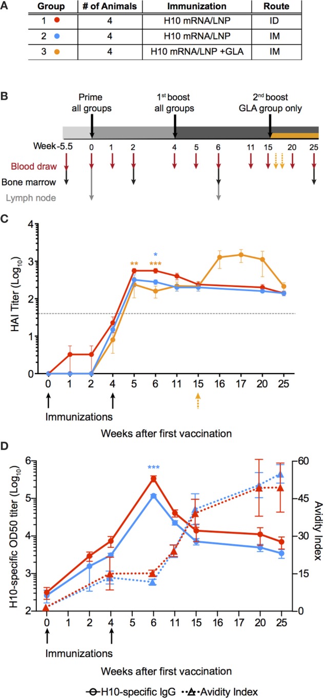
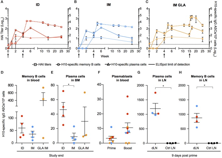
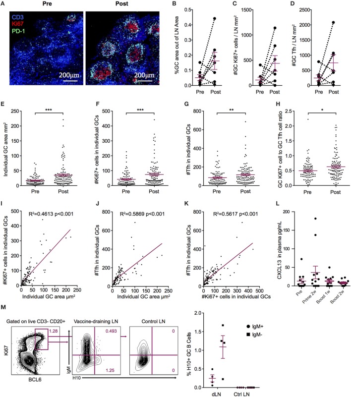
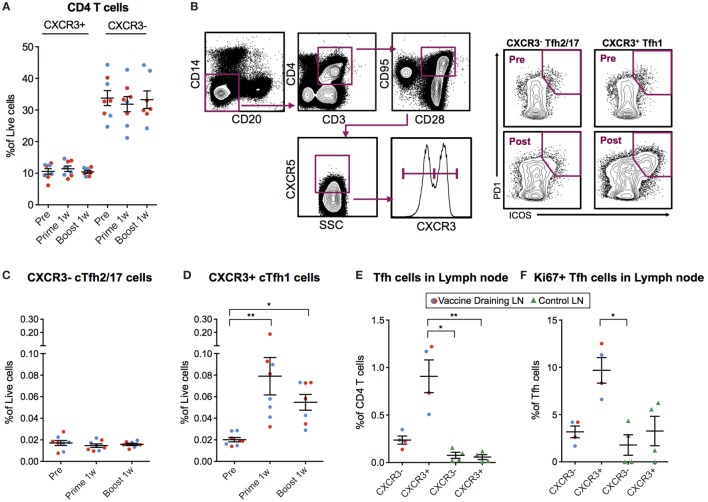
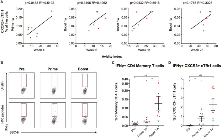
Modified mRNA vaccines have developed into an effective and well-tolerated vaccine platform that offers scalable and precise antigen production. Nevertheless, the immunological events leading to strong antibody responses elicited by mRNA vaccines are largely unknown. In this study, we demonstrate that protective levels of antibodies to hemagglutinin were induced after two immunizations of modified non-replicating mRNA encoding influenza H10 encapsulated in lipid nanoparticles (LNP) in non-human primates. While both intradermal (ID) and intramuscular (IM) administration induced protective titers, ID delivery generated this response more rapidly. Circulating H10-specific memory B cells expanded after each immunization, along with a transient appearance of plasmablasts. The memory B cell pool waned over time but remained detectable throughout the 25-week study. Following prime immunization, H10-specific plasma cells were found in the bone marrow and persisted over time. Germinal centers were formed in vaccine-draining lymph nodes along with an increase in circulating H10-specific ICOS+ PD-1+ CXCR3+ T follicular helper cells, a population shown to correlate with high avidity antibody responses after seasonal influenza vaccination in humans. Collectively, this study demonstrates that mRNA/LNP vaccines potently induce an immunological repertoire associated with the generation of high magnitude and quality antibodies.

Keywords: T follicular helper cells; adaptive immune responses; germinal centers; influenza; mRNA vaccine; non-human primates.


Figures

Figure 1
Figure 1
mRNA vaccine encoding influenza H10 elicits high antibody titers by intradermal and intramuscular immunization. Rhesus macaques were vaccinated with mRNA encoding for hemagglutinin of H10N8 influenza encapsulated in LNPs. (A) Shows the study groups and (B) shows the immunization and sampling schedule. (C) Antibody levels over time induced in the different groups as assayed by hemagglutination inhibition assay. Arrows indicate immunizations at week 0 and 4, as well as 15 for the glucopyranosyl lipid adjuvant group only. (D) The left y-axis shows antigen-specific plasma IgG titers (circles) and the right y-axis shows avidity index (triangles). Arrows indicate immunizations at week 0, 4. Titers are displayed as mean ± SEM. Dotted line depicts the accepted level of protection for seasonal influenza. Statistics was calculated by two-way ANOVA with Tukey’s multiple comparison test: *p < 0.05, **p < 0.01, ***p < 0.001.
Figure 2
Figure 2
Kinetics of vaccine-induced antibody-secreting cells (ASCs) in bone marrow and blood. (A–C) The left y-axis shows HAI titers (squares). The right y-axis shows H10-specific cultured memory B cells in blood (circles) and H10-specific plasma cells (PCs) in bone marrow (triangles) determined as ASCs per 1 × 106 cells. ASCs were plotted with HAI (squares) for comparison. Dotted line depicts the limit of detection for the ELISpot assay. (D–E) Study end values of antigen-specific memory B cells (D) and bone marrow PCs (E) between the groups. (F) Antigen-specific plasmablasts per 1 × 106 cells 7 days after prime and boost. (G,H) Naïve animals were immunized and LNs were collected after 9 days. Antigen-specific PCs (G) and memory B cells (H) in vaccine-draining LNs vs non-draining control LNs. Red colored dots corresponding to intradermal immunization, blue to IM immunization, and orange to IM immunization with glucopyranosyl lipid adjuvant adjuvant. Data show mean ± SEM. Statistics was based on Mann–Whitney U-test *p < 0.05.
Figure 3
Figure 3
Increased germinal center (GC) activation post influenza mRNA vaccine immunization. Axillary lymph nodes (LNs) were obtained before prime and 2 weeks after boost. A variety of readouts for GC activity were evaluated following the boost immunization. (A) LN sections stained for GCs before and after vaccination. Sections were stained with anti-CD3 (blue), PD-1 (green), and Ki67 (red) and follicular structures with CD3+PD-1+ T follicular helper (Tfh) and Ki67+ cells were considered GCs. Images were analyzed with CellProfiler software. (B–D) Each LN was analyzed for average GC area and total number of Ki67+ cells and Tfh cells, divided by the LN area for normalization. (E–H) Individual GCs represented by dots. (E) GC area. (F) Number of Tfh cells. (G) Number of Ki67+ cells. (H) Ki67+ to Tfh cell ratio. (I–K) GC architecture homogeneity was investigated by plotting GC area, Tfh, and Ki67+ cell numbers against each other. (L) CXCL13 in plasma at indicated time points. (M) Antigen-specific GC (CD3− CD20+ BCL6+ Ki67+) B cells in vaccine-draining LNs 9 days after prime vs non-draining control LNs by flow cytometry using an H10 tetramer probe. Data show mean ± SEM. Nonparametric Wilcoxon matched-pairs signed rank test was used for (B–D) and Mann–Whitney U-test for (E–H). *p < 0.05, **p < 0.01, ***p < 0.001.
Figure 4
Figure 4
Circulating CXCR3+ T follicular helper (cTfh) cells are detected 7 days postvaccination. (A) Peripheral blood mononuclear cells (PBMCs) from before vaccination and 1 week after prime and boost immunization were phenotyped for the frequency of CXCR3 +/− CD4 T cells. cTfh cells from the same time points were enumerated using the gating strategy as indicated in (B). Lymph node cell suspensions after prime immunizations and non-draining LNs were phenotyped for the frequency of Tfh cells and Ki67+ cells using the gating strategy as indicated in (B). (C,D) Show the percentage of CXCR3− vs CXCR3+ cTfh cells out of live cells, respectively. (E) CXCR3+/− Tfh cells out of CD4+ T Cells in vaccine- and non-draining lymph nodes (LNs). (F) Ki67+ cells out of CXCR3+/− Tfh cells in vaccine- and non-draining LNs. Data from the ID and IM group are shown as red and blue, respectively. Data show mean ± SEM. Statistics was based on Wilcoxon matched-pairs signed rank test for (C,D) and Kruskal–Wallis test with Dunn’s multiple comparison for (E,F), *p < 0.05, **p < 0.01.
Figure 5
Figure 5
Circulating CXCR3+ T follicular helper (cTfh) cells correlate with antibody avidity and are antigen-specific. (A) Correlations between circulating CXCR3+ cTfh cells after prime and boost and avidity index at indicated time points. (B) Peripheral blood mononuclear cells were stimulated overnight with an H10 overlapping peptide pool (15mers overlapping by 11 amino acids) to recall H10 specific T follicular helper (Tfh) cells within the memory T cell pool. Representative gates showing IFNγ+ cells within CXCR3+ Tfh cells as gated in Figure 4B. (C) Percentage of IFNγ+ cells within the overall CD4 T cell memory pool after background subtraction. (D) Percentage of IFNγ+ cells within the CXCR3+ cTfh cells after background subtraction. Data from the ID and IM group are shown as red and blue, respectively. Data show mean ± SEM. Statistics were based on Friedman’s test with Dunn’s multiple comparison *p < 0.05, **p < 0.01, ***p < 0.001.

Similar articles

Cited by

References

    1. Pardi N, Hogan MJ, Pelc RS, Muramatsu H, Andersen H, DeMaso CR, et al. Zika virus protection by a single low-dose nucleoside-modified mRNA vaccination. Nature (2017) 543:248–51.10.1038/nature21428 - DOI - PMC - PubMed
    1. Richner JM, Himansu S, Dowd KA, Butler SL, Salazar V, Fox JM, et al. Modified mRNA vaccines protect against Zika virus infection. Cell (2017) 168:1114–25.e10.10.1016/j.cell.2017.02.017 - DOI - PMC - PubMed
    1. Ulmer JB, Geall AJ. Recent innovations in mRNA vaccines. Curr Opin Immunol (2016) 41:18–22.10.1016/j.coi.2016.05.008 - DOI - PubMed
    1. Petsch B, Schnee M, Vogel AB, Lange E, Hoffmann B, Voss D, et al. Protective efficacy of in vitro synthesized, specific mRNA vaccines against influenza A virus infection. Nat Biotechnol (2012) 30:1210–6.10.1038/nbt.2436 - DOI - PubMed
    1. Bahl K, Senn JJ, Yuzhakov O, Bulychev A, Brito LA, Hassett KJ, et al. Preclinical and clinical demonstration of immunogenicity by mRNA vaccines against H10N8 and H7N9 influenza viruses. Mol Ther (2017) 25:1316–27.10.1016/j.ymthe.2017.03.035 - DOI - PMC - PubMed