

Effect of itraconazole on the cornea in a murine suture model and penetrating keratoplasty model
- PMID: 29181306
- PMCID: PMC5686361
- DOI: 10.18240/ijo.2017.11.03
Effect of itraconazole on the cornea in a murine suture model and penetrating keratoplasty model
Abstract
Aim: To investigate the anti-(lymph)angiogenic and/or anti-inflammatory effect of itraconazole in a corneal suture model and penetrating keratoplasty (PK) model.
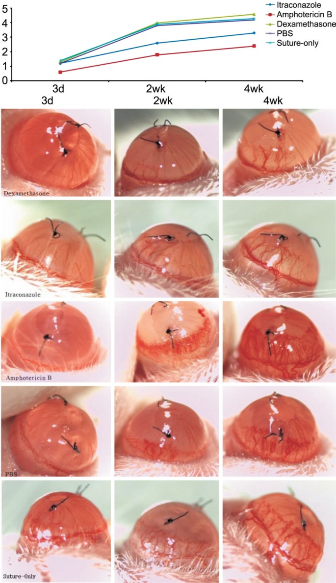
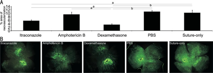
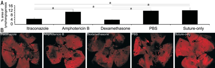
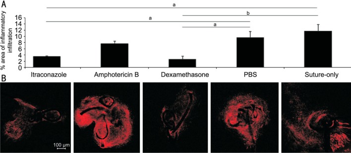
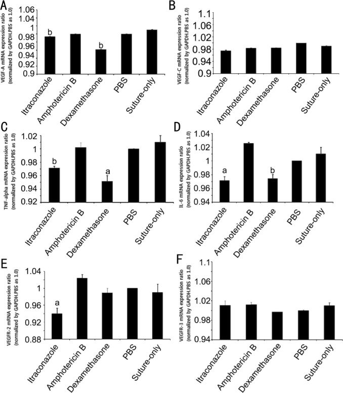
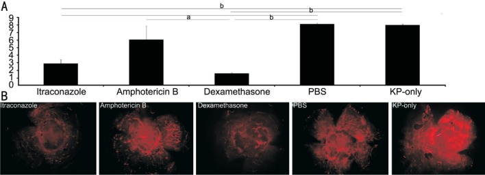
Methods: Graft survival, corneal neovascularization, and corneal lymphangiogenesis were compared among itraconazole, amphotericin B, dexamethasone, phosphate buffered saline (PBS) and surgery-only groups following subconjunctival injection in mice that underwent PK and corneal suture. Immunohistochemical staining and analysis were performed in each group. Real-time polymerase chain reaction (RT-PCR) was performed to quantify the expression of inflammatory cytokines (TNF-alpha, IL-6) and vascular endothelial growth factor (VEGF)-A, VEGF-C, VEGFR-2, and VEGFR-3.
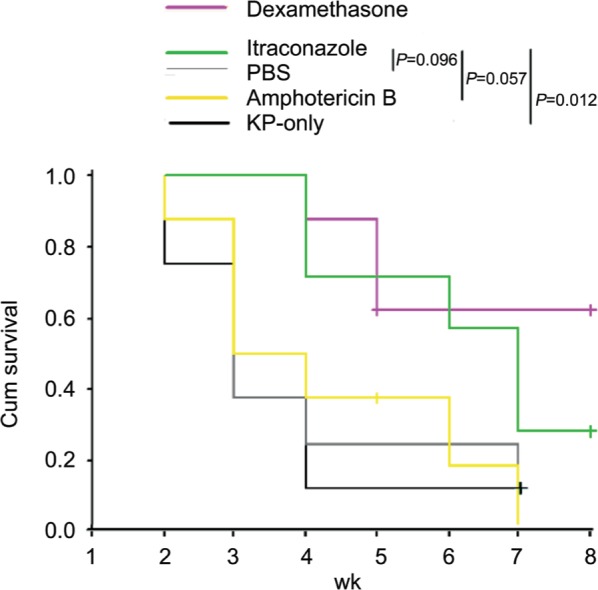
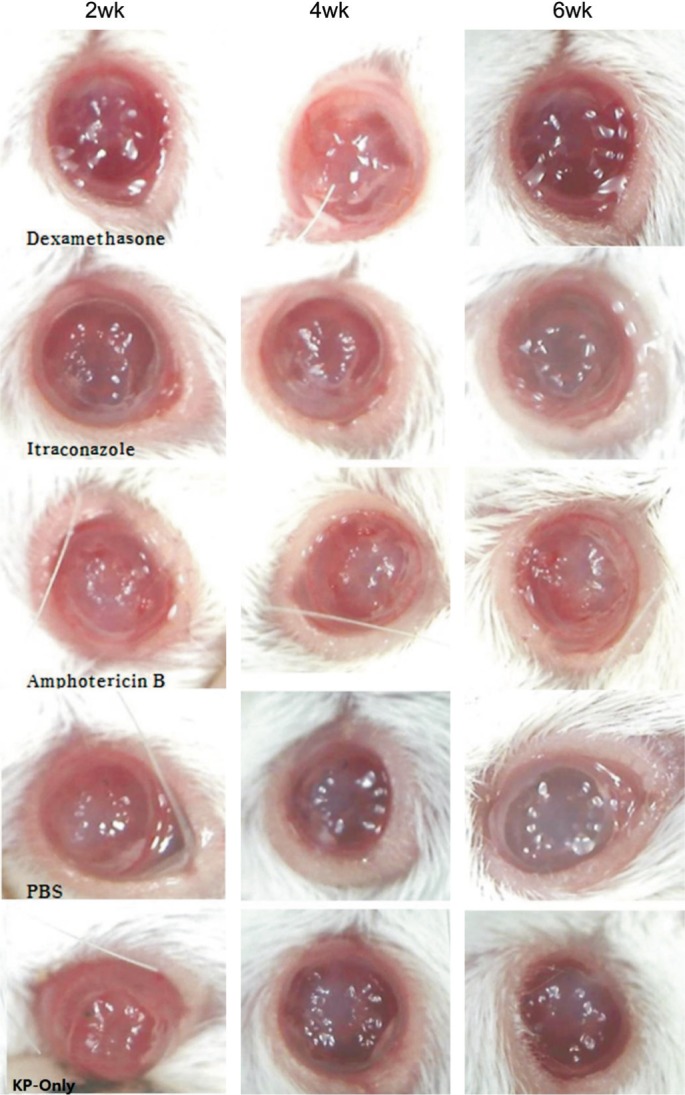
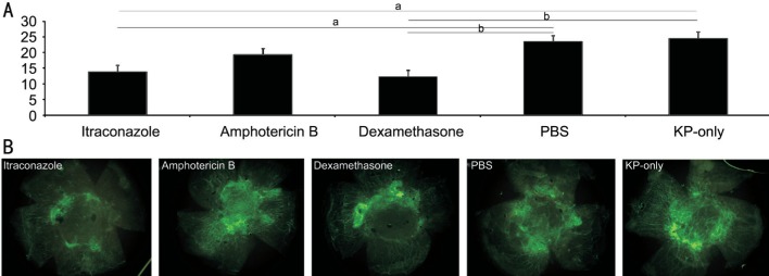
Results: In the suture model, the itraconazole group showed less angiogenesis, less lymphangiogenesis, and less inflammatory infiltration than the PBS group (all P<0.05). The itraconazole group showed reduced expression of VEGF-A, VEGFR-2, TNF-alpha, IL-6 than the PBS group (all P<0.05). In PK model, the two-month graft survival rate was 28.57% in itraconazole group, 62.50% in dexamethasone group, 12.50% in PBS group, 0 in amphotericin B group and 0 in surgery-only group. Graft survival in the itraconazole group was higher than that in the amphotericin, PBS and surgery-only group (P=0.057, 0.096, 0.012, respectively). The itraconazole group showed less total angiogenesis and lymphangiogenesis than PBS group (all P<0.05).
Conclusion: Itraconazole decrease neovascularization, lymphangiogenesis, and inflammation in both a corneal suture model and PK model. Itraconazole has anti-(lymph)-angiogenic and anti-inflammatory effects in addition to its intrinsic antifungal effect and is therefore an alternative treatment option in cases where steroids cannot be used.
Keywords: amphotericin B; dexamethasone; graft survival; itraconazole; lymphangiogenesis; neovascularization.
Figures
References
-
- Thomas PA. Fungal infections of the cornea. Eye (Lond) 2003;17(8):852–862. - PubMed
-
- Yavas GF, Ozturk F, Cusbeci T, Cetinkaya Z, Ermis SS, Kiraz N, Inan UU. Antifungal efficacy of voriconazole, itraconazole and amphotericin B in experimental fusarium solani keratitis. Graefes Arch Clin Exp Ophthalmol. 2008;246(2):275–279. - PubMed
-
- Lalitha P, Shapiro BL, Srinivasan M, Prajna NV, Acharya NR, Fothergill AW, Ruiz J, Chidambaram JD, Maxey KJ, Hong KC, McLeod SD, Lietman TM. Antimicrobial susceptibility of Fusarium, Aspergillus, and other filamentous fungi isolated from keratitis. Arch Ophthalmol. 2007;125(6):789–793. - PubMed
-
- Muller GG, Kara-Jose N, Silvestre de Castro R. Antifungals in eye infections: drugs and routes of administration. Rev Bras Oftalmol. 2013;72(2):132–141.
-
- Thomas PA, Kaliamurthy J. Mycotic keratitis: epidemiology, diagnosis and management. Clin Microbiol Infect. 2013;19(3):210–220. - PubMed
LinkOut - more resources
Full Text Sources
Other Literature Sources
Miscellaneous