. 2018 Jan 1;75(1):105-113.
doi: 10.1001/jamaneurol.2017.2065.

Neurologic Phenotypes Associated With Mutations in RTN4IP1 (OPA10) in Children and Young Adults

Affiliations

Neurologic Phenotypes Associated With Mutations in RTN4IP1 (OPA10) in Children and Young Adults

Majida Charif et al. JAMA Neurol. .

Abstract

Importance: Neurologic disorders with isolated symptoms or complex syndromes are relatively frequent among mitochondrial inherited diseases. Recessive RTN4IP1 gene mutations have been shown to cause isolated and syndromic optic neuropathies.

Objective: To define the spectrum of clinical phenotypes associated with mutations in RTN4IP1 encoding a mitochondrial quinone oxidoreductase.

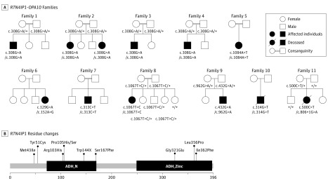
Design, setting, and participants: This study involved 12 individuals from 11 families with severe central nervous system diseases and optic atrophy. Targeted and whole-exome sequencing were performed-at Hospital Angers (France), Institute of Neurology Milan (Italy), Imagine Institute Paris (France), Helmoltz Zentrum of Munich (Germany), and Beijing Genomics Institute (China)-to clarify the molecular diagnosis of patients. Each patient's neurologic, ophthalmologic, magnetic resonance imaging, and biochemical features were investigated. This study was conducted from May 1, 2014, to June 30, 2016.

Main outcomes and measures: Recessive mutations in RTN4IP1 were identified. Clinical presentations ranged from isolated optic atrophy to severe encephalopathies.

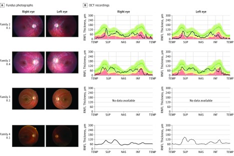
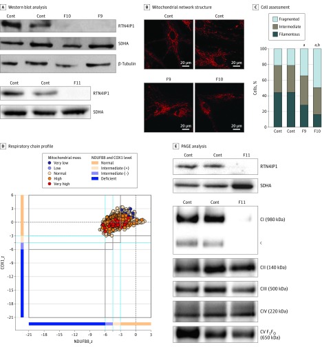
Results: Of the 12 individuals in the study, 6 (50%) were male and 6 (50%) were female. They ranged in age from 5 months to 32 years. Of the 11 families, 6 (5 of whom were consanguineous) had a member or members who presented isolated optic atrophy with the already reported p.Arg103His or the novel p.Ile362Phe, p.Met43Ile, and p.Tyr51Cys amino acid changes. The 5 other families had a member or members who presented severe neurologic syndromes with a common core of symptoms, including optic atrophy, seizure, intellectual disability, growth retardation, and elevated lactate levels. Additional clinical features of those affected were deafness, abnormalities on magnetic resonance images of the brain, stridor, and abnormal electroencephalographic patterns, all of which eventually led to death before age 3 years. In these patients, novel and very rare homozygous and compound heterozygous mutations were identified that led to the absence of the protein and complex I disassembly as well as mild mitochondrial network fragmentation.

Conclusions and relevance: A broad clinical spectrum of neurologic features, ranging from isolated optic atrophy to severe early-onset encephalopathies, is associated with RTN4IP1 biallelic mutations and should prompt RTN4IP1 screening in both syndromic neurologic presentations and nonsyndromic recessive optic neuropathies.


Conflict of interest statement

Conflict of Interest Disclosures: None reported.

Figures

Figure 1.
Figure 1.. Pedigree of the RTN4IP1 (OPA10) Families, Segregation of the Mutations, and Alterations of the Protein Primary Structure
A, Pedigrees show the affected cases in the 11 families and the segregation of the mutations. Black symbols indicate affected patients. B, Localization of RTN4IP1 residue changes in the protein: the primary structure of RTN4IP1 protein (domains and amino acid positions) is described in the figure with all of the pathogenic mutations reported in this study. ADH_N indicates alcohol dehydrogenase GroES-like domain; ADH_Zinc, zinc-binding dehydrogenase domain.
Figure 2.
Figure 2.. Ophthalmological Examination of RTN4IP1 (OPA10) Patients
Eye fundus of the right and left eyes from indicated patients (A) and the corresponding optical coherence tomographic (OCT) recordings (B) are shown, when available. INF indicates inferior; NAS, nasal; RNFL retinal nerve fiber layer; SUP, superior; and TEMP, temporal.
Figure 3.
Figure 3.. Association of Decreased RTN4IP1 Levels With Mitochondrial Network Structure Muscle of Severely Affected Patients
A, Western blot analysis of RTN4IP1 protein levels in fibroblasts from controls (Cont) and RTN4IP1 index cases of families 9 (F9), 10 (F10), and 11 (F11). Succinate dehydrogenase and β-tubulin antibodies were used as loading Cont. B, Assessment of mitochondrial network structure from 2 Cont fibroblast cell lines and from fibroblasts of the index cases of F9 and F10 (MitoTracker stain; Thermo Fisher Scientific). C, Assessment of the percentage of cells presenting a fragmented, intermediate, or filamentous network. D, Respiratory chain profile following quadruple oxidative phosphorylation immunofluorescence analysis of cryosectioned muscle from the index case of F11, confirming the presence of fibers lacking complex I (NDUFB8) protein but with normal complex IV (COX1) expression. (Experimental procedures per the protocols of Rocha et al.) Each dot represents the measurement from an individual muscle fiber, color coded according to its mitochondrial mass. Gray dashed lines indicate SD limits for the classification of fibers; lines next to x- and y-axes, the levels (SDs from the average of Cont fibers after normalization to porin/VDAC1 levels) of NDUFB8 and COX1, respectively; and _z, the z score for COX1 or NDUFB8. (For formulas and full description of statistics involved, see the Methods section of Rocha et al.) Blue dotted lines indicate the mean expression level observed in respiratory normal fibers. E, One-dimensional blue native polyacrylamide gel electrophoresis (PAGE) (4%-16% gradient) analysis showing a specific complex I assembly defect in skeletal muscle from the index case of F11 and age-matched Cont. Individual oxidative phosphorylation complexes were detected by immunoblotting using subunit-specific antibodies (complex I [C1] [NDUFB8], CII [SDHA], CIII [UQCRC2], CIV [COX1], and CV [ATP5A]). Complex V comprises the F1 and FO subcomplexes. F1FO denotes that the band is the complete assembled form of adenosine triphosphate synthase (CV). aP < .001 compared with Cont. bP < .05 compared with F9. cIndicates the presence of an additional, partially assembled CI intermediate also detected with the NDUFB8 antibody, absent in the patient sample, and likely corresponding to the approximately 650-kDa Iβ subcomplex of the hydrophobic membrane arm.

References

    1. DiMauro S. Mitochondrial diseases. Biochim Biophys Acta. 2004;1658(1-2):80-88. - PubMed
    1. Munnich A, Rustin P. Clinical spectrum and diagnosis of mitochondrial disorders. Am J Med Genet. 2001;106(1):4-17. - PubMed
    1. Angebault C, Guichet PO, Talmat-Amar Y, et al. Recessive mutations in RTN4IP1 cause isolated and syndromic optic neuropathies. Am J Hum Genet. 2015;97(5):754-760. - PMC - PubMed
    1. Hu WH, Hausmann ON, Yan MS, Walters WM, Wong PK, Bethea JR. Identification and characterization of a novel Nogo-interacting mitochondrial protein (NIMP). J Neurochem. 2002;81(1):36-45. - PubMed
    1. Lenaers G, Hamel C, Delettre C, et al. Dominant optic atrophy. Orphanet J Rare Dis. 2012;7:46. - PMC - PubMed

Publication types