Comparative Study
. 2018 Mar;39(3):538-547.
doi: 10.1007/s00246-017-1785-4. Epub 2017 Nov 27.

Comparison of 3D Echocardiogram-Derived 3D Printed Valve Models to Molded Models for Simulated Repair of Pediatric Atrioventricular Valves

Affiliations
Comparative Study

Comparison of 3D Echocardiogram-Derived 3D Printed Valve Models to Molded Models for Simulated Repair of Pediatric Atrioventricular Valves

Adam B Scanlan et al. Pediatr Cardiol. 2018 Mar.

Abstract

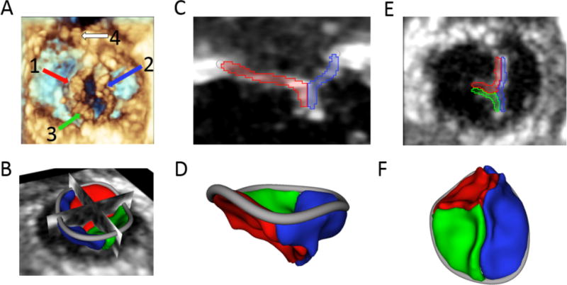
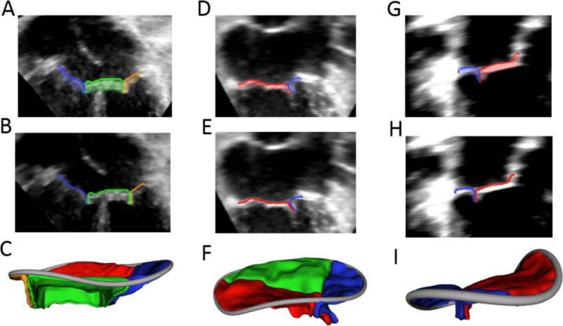
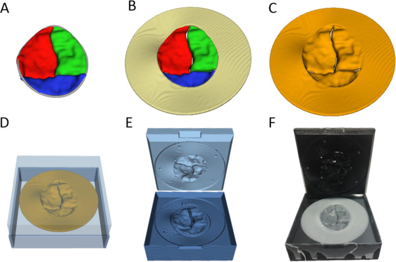
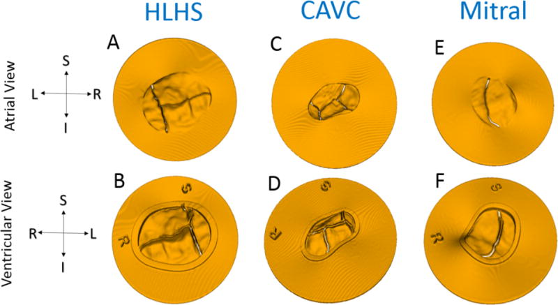
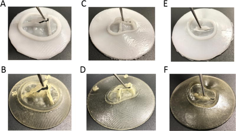
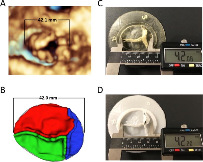
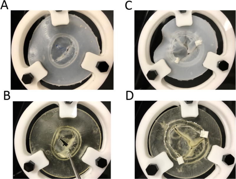
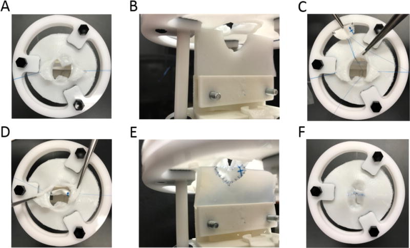
Mastering the technical skills required to perform pediatric cardiac valve surgery is challenging in part due to limited opportunity for practice. Transformation of 3D echocardiographic (echo) images of congenitally abnormal heart valves to realistic physical models could allow patient-specific simulation of surgical valve repair. We compared materials, processes, and costs for 3D printing and molding of patient-specific models for visualization and surgical simulation of congenitally abnormal heart valves. Pediatric atrioventricular valves (mitral, tricuspid, and common atrioventricular valve) were modeled from transthoracic 3D echo images using semi-automated methods implemented as custom modules in 3D Slicer. Valve models were then both 3D printed in soft materials and molded in silicone using 3D printed "negative" molds. Using pre-defined assessment criteria, valve models were evaluated by congenital cardiac surgeons to determine suitability for simulation. Surgeon assessment indicated that the molded valves had superior material properties for the purposes of simulation compared to directly printed valves (p < 0.01). Patient-specific, 3D echo-derived molded valves are a step toward realistic simulation of complex valve repairs but require more time and labor to create than directly printed models. Patient-specific simulation of valve repair in children using such models may be useful for surgical training and simulation of complex congenital cases.

Keywords: 3D echocardiography; 3D printing; Surgical simulation; Valve repair.


Conflict of interest statement

Conflict of Interest: The authors declare they have no conflict of interest.

Figures

Figure 1
Figure 1
Tricuspid valve in 4-day-old infant with HLHS: (A) 3D echo view of the ventricular face of the tricuspid valve with the (1) anterior leaflet, (2) septal leaflet, (3) posterior leaflet, and (4) native pulmonary valve visible; (B) 3D segmentation of the tricuspid valve displayed with 2D planes; (C) 2D plane showing anterior and septal leaflets; (D) 3D segmentation of the valve from anterior view; (E) 2D plane of coaptation as seen from the ventricle; (F) 3D segmentation as seen from the ventricle.
Figure 2
Figure 2
Examples of Valve Segmentations and atrial surface extraction: (A) 2D view of segmented CAVC; (B) atrial surface extraction of CAVC; (C) 3D rendering of thickened atrial surface of CAVC for modeling; (D) 2D view of segmented tricuspid; (E) atrial surface extraction of tricuspid; (F) 3D rendering of thickened atrial surface of tricuspid for modeling; (G) 2D view of segmented mitral; (H) atrial surface extraction of mitral; (I) 3D rendering of thickened atrial surface of mitral for modeling.
Figure 3
Figure 3
Creation of 3D Printed Valve Molds: (A) Valve segmentation; (B) Valve segmentation with addition of valve skirt; (C) Model Ready for direct printing; (D) Automatically generated mold around direct printing template; (E) Rendering of upper and lower mold pieces; (F) Molded valve in mold.
Figure 4
Figure 4
Models Ready for Direct 3D Printing from atrial and ventricular viewpoints: (A-B) Tricuspid in HLHS; (C-D) CAVC; (E-F) Mitral. Note preservation of coaptation with a small gap artificially created to ensure the ability to separate leaflets after printing. The coaptation gap for the tricuspid and mitral was 0.3mm and 0.2mm for the mitral.
Figure 5
Figure 5
Molded and Printed Valve Models: (A) Molded tricuspid valve; (B) Printed tricuspid valve; (C) Molded CAVC valve; (D) Printed CAVC valve; (E) Molded mitral valve; (F) Printed mitral valve. Note preservation of coaptation surfaces.
Figure 6
Figure 6
Comparison of Measurements: (A) Tricuspid valve rendered in QLab; (B) Valve segmentation in 3D Slicer; (C) Directly printed valve measured with calipers; (D) Molded valve measured with calipers.
Figure 7
Figure 7
Simulated Valve Surgery in Molded (top) and directly printed valves(bottom) (A) Molded silicone valve cuts and realistically holds suture; (B) Directly printed valve is relatively brittle when cutting and tears easily at suture sites; (C) Simulated annuloplasty on tricuspid molded valve demonstrating realistic “cinching” of the suture to reduce the annular circumference; (D) Simulated annuloplasty on directly printed valve does not cinch and excessive suture tension results in tearing through relatively brittle material.
Figure 8
Figure 8
Simulated Repair of CAVC with ventricular 3D printed ventricular septal holder (B and E); (A) Bridging leaflets are pulled back; (B) View of ventricular septal defect prior to insertion of patch; (C and E) Suturing of patch to VSD; (E and F) Final view of completed CAVC simulation from the atrial perspective (E) and ventricular perspective (F).

References

    1. Carpentier A, Chauvaud S, Mace L, Relland J, Mihaileanu S, Marino JP, Abry B, Guibourt P. A new reconstructive operation for Ebstein’s anomaly of the tricuspid valve. J Thorac Cardiovasc Surg. 1988;96(1):92–101. - PubMed
    1. Chauvaud S, Perier P, Touati G, Relland J, Kara SM, Benomar M, Carpentier A. Long-term results of valve repair in children with acquired mitral valve incompetence. Circulation. 1986;74(3 Pt 2):I104–109. - PubMed
    1. Galloway AC, Colvin SB, Baumann FG, Esposito R, Vohra R, Harty S, Freeberg R, Kronzon I, Spencer FC. Long-term results of mitral valve reconstruction with Carpentier techniques in 148 patients with mitral insufficiency. Circulation. 1988;78(3 Pt 2):I97–105. - PubMed
    1. Mahmood F, Matyal R. A quantitative approach to the intraoperative echocardiographic assessment of the mitral valve for repair. Anesth Analg. 2015;121(1):34–58. doi: 10.1213/ANE.0000000000000726. - DOI - PubMed
    1. Poelaert JI, Bouchez S. Perioperative echocardiographic assessment of mitral valve regurgitation: a comprehensive review. Eur J Cardiothorac Surg. 2016;50(5):801–812. doi: 10.1093/ejcts/ezw196. - DOI - PubMed

Publication types

MeSH terms

LinkOut - more resources