. 2018 Feb/Mar;10(2):315-324.
doi: 10.1080/19420862.2017.1409319. Epub 2017 Dec 20.

Elimination of tumor by CD47/PD-L1 dual-targeting fusion protein that engages innate and adaptive immune responses

Affiliations

Elimination of tumor by CD47/PD-L1 dual-targeting fusion protein that engages innate and adaptive immune responses

Boning Liu et al. MAbs. 2018 Feb/Mar.

Abstract

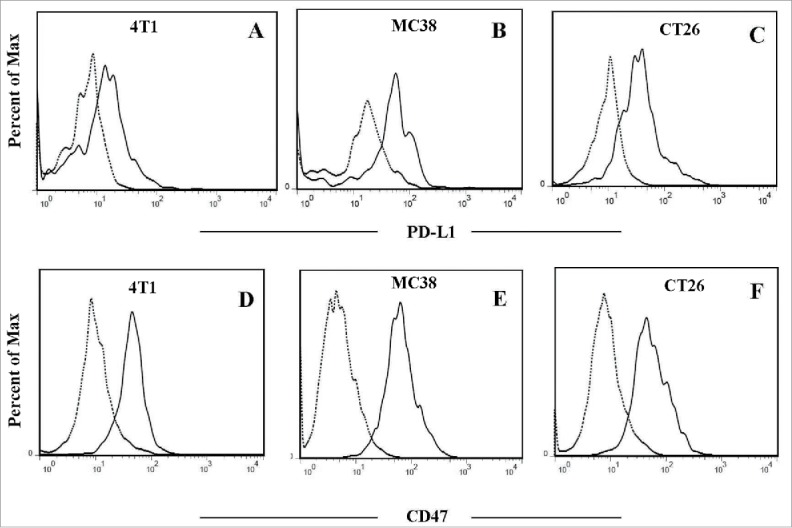
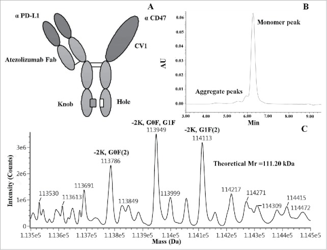
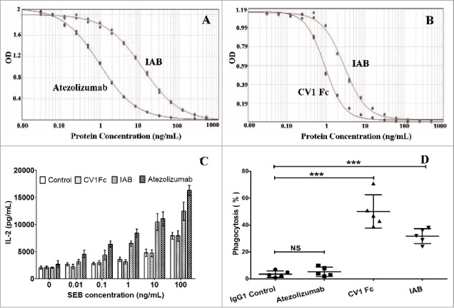
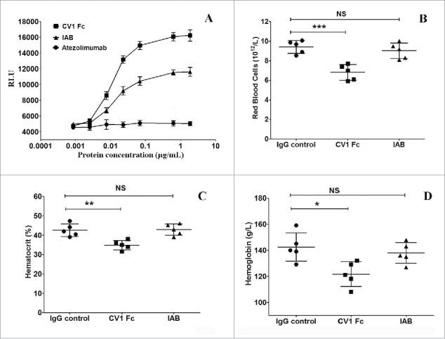
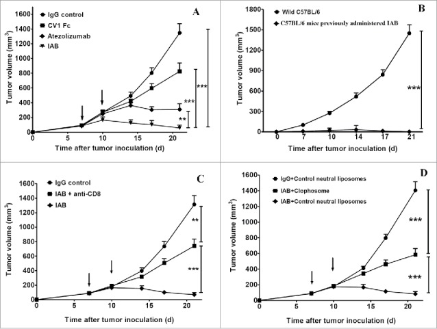
The host immune system generally serves as a barrier against tumor formation. Programmed death-ligand 1 (PD-L1) is a critical "don't find me" signal to the adaptive immune system, whereas CD47 transmits an anti-phagocytic signal, known as the "don't eat me" signal, to the innate immune system. These and similar immune checkpoints are often overexpressed on human tumors. Thus, dual targeting both innate and adaptive immune checkpoints would likely maximize anti-tumor therapeutic effect and elicit more durable responses. Herein, based on the variable region of atezolizumab and consensus variant 1 (CV1) monomer, we constructed a dual-targeting fusion protein targeting both CD47 and PD-L1 using "Knobs-into-holes" technology, denoted as IAB. It was effective in inducing phagocytosis of tumor cells, stimulating T-cell activation and mediating antibody-dependent cell-mediated cytotoxicity in vitro. No obvious sign of hematological toxicity was observed in mice administered IAB at a dose of 100 mg/kg, and IAB exhibited potent antitumor activity in an immune-competent mouse model of MC38. Additionally, the anti-tumor effect of IAB was impaired by anti-CD8 antibody or clodronate liposomes, which implied that both CD8+ T cells and macrophages were required for the anti-tumor efficacy of IAB and IAB plays an essential role in the engagement of innate and adaptive immune responses. Collectively, these results demonstrate the capacity of an elicited endogenous immune response against tumors and elucidate essential characteristics of synergistic innate and adaptive immune response, and indicate dual blockade of CD47 and PD-L1 by IAB may be a synergistic therapy that activates both innate and adaptive immune response against tumors.

Keywords: CD47; PD-L1; adaptive immunity; dual-targeting fusion protein; immune checkpoint; innate immunity.


Figures

Figure 1.
Figure 1.
Co-expression of CD47 and PD-L1 on mouse tumor cells in vivo. Representative flow cytometry analysis following staining with anti-PD-L1 or anti-CD47 antibody (solid line) and isotype antibody as negative control (dotted line). A and D:4T1 tumor cells; B and E:MC38 tumor cells;C and F:CT26 tumor cells.
Figure 2.
Figure 2.
Design and characterization of IAB. (A) The schematic diagram of IAB. (B) SEC chromatogram (SEC-UPLC) of IAB. (C) MS analysis indicates the mass of main peaks were in good agreement with expected heterodimer with post-translational modifications. -2K,G0F(2): IAB-Lysine C-TERM(2),Glycosylation G0F(2), Expected mass: 113784Da, Mass error: 9.94ppm; -2K,G0F,G1F: IAB-Lysine C-TERM(2),Glycosylation G0F,Glycosylation G1F, Expected mass: 113946Da, Mass error: 26.88ppm; -2K,G1F(2): IAB-Lysine C-TERM(2),Glycosylation G1F(2), Expected mass: 114108Da, Mass error: 48.54Da.
Figure 3.
Figure 3.
Competitive binding assay and in vitro human PBMC and murine macrophage activation. Competitive inhibition of IAB binding to PD-L1 (A) or CD47 (B) was evaluated by competitive ELISA assay (n = 2). (C) Dose increase in IL-2 secretion induced by IAB compared with CV1 and atezolizumab in SEB-stimulated PBMCs in a dose-dependent manner (n = 3). (D) IAB-mediated phagocytosis of MC38, CFSE-labeled MC38 cells were incubated with murine macrophages (RAW264.7) and indicated antibodies/proteins, the phagocytosis index was determined by flow cytometry (n = 5). Data are reported as the means ± SD, ***: p < 0.001; NS: non-significant (unpaired Student's t test).
Figure 4.
Figure 4.
IAB exhibits superior ADCC activity in vitro and minimal blood toxicity in vivo. (A) Relative ADCC activity of IAB, CV1 Fc and atezolizumab in ADCC reporter assay. (n = 3); Analysis of red blood cell (B), hematocrit (C) and hemoglobin (D) in mice intraperitoneal injected with 100 mg/Kg of IAB or parental proteins (n = 5). Data are reported as the means ± SD, *: p<0.05, **: p <0.01, ***: p <0.001; NS: non-significant (unpaired Student's t test).
Figure 5.
Figure 5.
The durable anti-tumor effect of IAB depends on both innate and adaptive immunity. (A) Anti-tumor effect: C57BL/6 (n = 5/group) mice were injected s.c. with 2 × 10⁵ MC38 cells and treated intratumorally with indicated antibodies at the dose of 10mg/Kg on day 7, and 10. (B) The re-challenge experiment: C57BL/6 mice previously administered with IAB or wild C57BL/6 (n = 5/group) mice were challenged s.c. with 1 × 10⁶ MC38 cells. (C) The CD8+ depletion experiment: MC38 tumor bearing C57BL/6 (n = 5/group) were treated intratumorally with IAB (10mg/Kg) and injected i.p.with 400 μg anti-CD8 antibody on days 7, and 10. (D) The macrophage depletion experiment: MC38 tumor bearing C57BL/6 (n = 5/group) were treated intratumorally with IAB (10mg/Kg) on days 7, and 10. 200 μl of clophosome or control liposome was administered i.p. on day 5 and every 4 days thereafter. Data are reported as means ± SEM. *: p < 0.05, **: p < 0.01,***: p < 0.001 (unpaired Student's t test).Note: For negative controls shown in Figure 5C (IgG control group) and 5D (IgG+Control neutral liposomes group), mice bearing too large or small tumor after MC38 inoculation were excluded; on day 7 after tumor inoculation, mice with measurable tumor (~100 mm3) were randomized to treatment group. The error bars represent standard error of the mean (SEM). In Figure 5C, the IgG control group mean±SEM on Days 7, 10, 14, 17 and 21 were 89±10, 185±31, 399±51, 726±87, 1297±130, respectively. In Figure 5D, the IgG control neutral liposomes group mean±SEM on Days 7, 10, 14, 17 and 21 were 92±12, 193±20, 410±35, 800±55, 1403±122, respectively.

Similar articles

Cited by

References

    1. Hanahan D, Weinberg RA. Hallmarks of cancer: the next generation. Cell. 2011;144:646–74. doi:10.1016/j.cell.2011.02.013. - DOI - PubMed
    1. Topalian SL, Drake CG, Pardoll DM. Immune checkpoint blockade: a common denominator approach to cancer therapy. Cancer Cell. 2015;27:450–61. doi:10.1016/j.ccell.2015.03.001. - DOI - PMC - PubMed
    1. Moynihan KD, Opel CF, Szeto GL, Tzeng A, Zhu EF, Engreitz JM, Williams RT, Rakhra K, Zhang MH, Rothschilds AM, et al.. Eradication of large established tumors in mice by combination immunotherapy that engages innate and adaptive immune responses. Nat Med. 2016;22:1402–10. doi:10.1038/nm.4200. - DOI - PMC - PubMed
    1. Casey SC, Tong L, Li Y, Do R, Walz S, Fitzgerald KN, Gouw AM, Baylot V, Gütgemann I, Eilers M, et al.. MYC regulates the antitumor immune response through CD47 and PD-L1. Science. 2016;352:227–31. doi:10.1126/science.aac9935. - DOI - PMC - PubMed
    1. Chen L, Han X. Anti-PD-1/PD-L1 therapy of human cancer: past, present, and future. J Clin Invest. 2015;125:3384–91. doi:10.1172/JCI80011. - DOI - PMC - PubMed

Publication types

MeSH terms