Review
. 2017 Nov 28;10(1):53.
doi: 10.1186/s13041-017-0340-9.

Oxidative stress and cellular pathologies in Parkinson's disease

Affiliations
Review

Oxidative stress and cellular pathologies in Parkinson's disease

Lesly Puspita et al. Mol Brain. .

Abstract

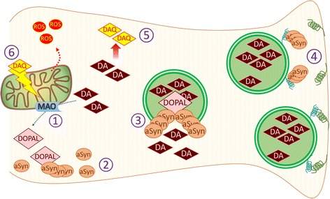
Parkinson's disease (PD) is a chronic and progressive neurodegeneration of dopamine neurons in the substantia nigra. The reason for the death of these neurons is unclear; however, studies have demonstrated the potential involvement of mitochondria, endoplasmic reticulum, α-synuclein or dopamine levels in contributing to cellular oxidative stress as well as PD symptoms. Even though those papers had separately described the individual roles of each element leading to neurodegeneration, recent publications suggest that neurodegeneration is the product of various cellular interactions. This review discusses the role of oxidative stress in mediating separate pathological events that together, ultimately result in cell death in PD. Understanding the multi-faceted relationships between these events, with oxidative stress as a common denominator underlying these processes, is needed for developing better therapeutic strategies.

Keywords: Alpha-synuclein; Dopamine neurons; Mitochondria; Oxidative stress; Parkinson’s disease; Reactive oxygen species; Unfolded protein response.


Conflict of interest statement

Ethics approval and consent to participate

Not applicable

Consent for publication

Not applicable

Competing interests

The authors declare that they have no competing interests.

Publisher’s Note

Springer Nature remains neutral with regard to jurisdictional claims in published maps and institutional affiliations.

Figures

Fig. 1
Fig. 1
Central role of chronic oxidative stress in mediating PD progression. Mitochondria depolarization, ER stress, α-synuclein accumulation and increased level of cytosolic DA are known PD phenotypes that potentially contribute to cellular oxidative stress, alone or by interacting with each other. ER regulates cytosolic calcium and thus prevents excess uptake by MCU that otherwise can stimulate ETC and ROS production in mitochondria, an essential function in neurons with intense depolarization. α-synuclein accumulation can contribute to ER stress by binding to ER chaperones and disturbing vesicle trafficking between ER and Golgi. While UPR activation also stimulate α-synuclein aggregation by unclear mechanism. Utilization of DA risks midbrain DA neurons to oxidative damage by DA metabolism. Additionally, DA has been shown to trigger α-synuclein oligomer formation and mitochondria depolarization. Altogether, these phenotypes produce oxidative environment that further amplifies the damage. Mutations that disturb the function of LRRK2, DJ-1, Parkin and PINK1 have been linked to familial cases of PD. Parkin is an E3 ubiquitin ligase and dysfunctionality of this protein results in accumulation of its substrates. Together with PINK1, Parkin is also responsible to clear up damaged mitochondria. One of DJ-1 putative role as sensor of oxidative stress may be necessary in cell protection. Single mutation in LRRK2 resulted in increased susceptibility towards oxidative stressor, even though the mechanism is less understood. Pesticide rotenone, iron and manganese also cause cellular oxidative stress by triggering mitochondria depolarization, α-synuclein oligomerization together with ROS production and UPR activation, respectively. Long term exposure of those substances has been linked with higher risk of developing sporadic PD
Fig. 2
Fig. 2
Alpha-synuclein oligomers and cytosolic DA amplify each other and synergistically contribute to oxidative stress. 1) DA anabolism enzyme, MAO, turns DA into DOPAL, that later can be converted into less reactive DOPAC by another enzyme. 2) DOPAL induces α-synuclein oligomerization and prevents fibril formation. 3) DOPAL modified α-synuclein oligomers create pore-like structure in the synaptic vesicle membrane, causing DA leakage into cytosol. 4) α-synuclein oligomers are also known to negatively regulate synaptic vesicle release by preventing SNARE formation and vesicle docking. This provides more DA vesicles that can be targeted by DOPAL-α-synuclein oligomers. 5) Oxidation of cytosolic DA produces DAQ. 6) DAQ reacts with cysteine residues of mitochondrial proteins that results in mitochondria depolarization and further ROS production

References

    1. Bellucci A, Mercuri NB, Venneri A, Faustini G, Longhena F, Pizzi M, et al. Review: Parkinson’s disease: from synaptic loss to connectome dysfunction. Neuropathol Appl Neurobiol. 2016;42:77–94. doi: 10.1111/nan.12297. - DOI - PubMed
    1. Tabbal SD, Tian LL, Karimi M, Brown CA, Loftin SK, Perlmutter JS. Low nigrostriatal reserve for motor parkinsonism in nonhuman primates. Exp Neurol. 2012;237:355–362. doi: 10.1016/j.expneurol.2012.07.008. - DOI - PMC - PubMed
    1. Valente EM, Abou-Sleiman PM, Caputo V, Muqit MM, Harvey K, Gispert S, et al. Hereditary early-onset Parkinson’s disease caused by mutations in PINK1. Science. 2004;304:1158–1160. doi: 10.1126/science.1096284. - DOI - PubMed
    1. Polymeropoulos MH, Lavedan C, Leroy E, Ide SE, Dehejia A, Dutra A, et al. Mutation in the alpha-synuclein gene identified in families with Parkinson’s disease. Science. 1997;276:2045–2047. doi: 10.1126/science.276.5321.2045. - DOI - PubMed
    1. Di Fonzo A, Rohe CF, Ferreira J, Chien HF, Vacca L, Stocchi F, et al. A frequent LRRK2 gene mutation associated with autosomal dominant Parkinson’s disease. Lancet. 2005;365:412–415. doi: 10.1016/S0140-6736(05)17829-5. - DOI - PubMed

Substances