

A magnetic resonance imaging study of early brain injury in a rat model of acute DFP intoxication
- PMID: 29183789
- PMCID: PMC5940565
- DOI: 10.1016/j.neuro.2017.11.009
A magnetic resonance imaging study of early brain injury in a rat model of acute DFP intoxication
Abstract
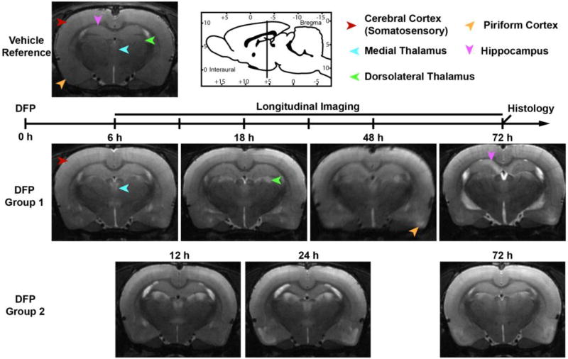
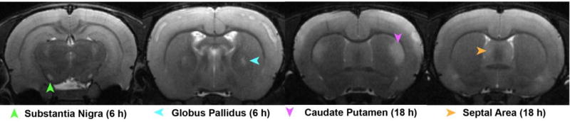
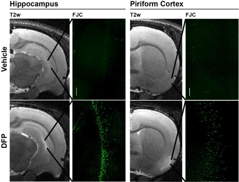
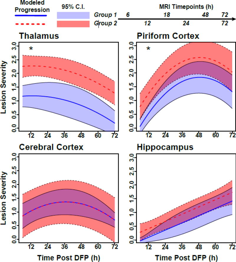
Current treatments for seizures induced by organophosphates do not protect sufficiently against progressive neurodegeneration or delayed cognitive impairment. Developing more effective therapeutic approaches has been challenging because the pathogenesis of these delayed consequences is poorly defined. Using magnetic resonance imaging (MRI), we previously reported brain lesions that persist for months in a rat model of acute intoxication with the OP, diisopropylfluorophosphate (DFP). However, the early spatiotemporal progression of these lesions remains unknown. To address this data gap, we used in vivo MRI to longitudinally monitor brain lesions during the first 3 d following acute DFP intoxication. Adult male Sprague Dawley rats acutely intoxicated with DFP (4mg/kg, sc) were MR imaged at 6, 12, 18, 24, 48, 72h post-DFP, and their brains then taken for correlative histology to assess neurodegeneration using FluoroJade C (FJC) staining. Acute DFP intoxication elicited moderate-to-severe seizure activity. T2-weighted (T2w) anatomic imaging revealed prominent lesions within the thalamus, piriform cortex, cerebral cortex, hippocampus, corpus striatum, and substantia nigra that corresponded to neurodegeneration, evident as bands of FJC positive cells. Semi-quantitative assessment of lesion severity demonstrated significant regional variation in the onset and progression of injury, and suggested that lesion severity may be modulated by isoflurane anesthesia. These results imply that the timing of therapeutic intervention for attenuating brain injury following OP intoxication may be regionally dependent, and that longitudinal assessment of OP-induced damage by MRI may be a powerful tool for assessing therapeutic response.
Keywords: In vivo imaging; Neuropathology; Organophosphate; Seizure; T2-weighted MRI.
Copyright © 2017 Elsevier B.V. All rights reserved.
Figures
References
-
- Bar-Klein G, Klee R, Brandt C, Bankstahl M, Bascunana P, Tollner K, et al. Isoflurane prevents acquired epilepsy in rat models of temporal lobe epilepsy. Ann Neurol. 2016;80:896–908. - PubMed
-
- Bareyre F, Wahl F, McIntosh TK, Stutzmann JM. Time course of cerebral edema after traumatic brain injury in rats: effects of riluzole and mannitol. J Neurotrauma. 1997;14:839–49. - PubMed
-
- Bertoglio D, Verhaeghe J, Dedeurwaerdere S, Grohn O. Neuroimaging in animal models of epilepsy. Neuroscience. 2017;358:277–99. - PubMed
-
- Bhagat YA, Obenaus A, Hamilton MG, Kendall EJ. Magnetic resonance imaging predicts neuropathology from soman-mediated seizures in the rodent. Neuroreport. 2001;12:1481–7. - PubMed
Publication types
MeSH terms
Substances
Grants and funding
LinkOut - more resources
Full Text Sources
Other Literature Sources
Miscellaneous
